. 2020 Jun 2;59(21):2032-2040.
doi: 10.1021/acs.biochem.0c00241. Epub 2020 May 19.

Orotidine 5'-Monophosphate Decarboxylase: The Operation of Active Site Chains Within and Across Protein Subunits

Affiliations

Orotidine 5'-Monophosphate Decarboxylase: The Operation of Active Site Chains Within and Across Protein Subunits

Tiago A S Brandão et al. Biochemistry. .

Abstract

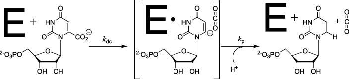
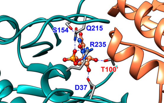
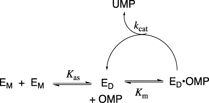
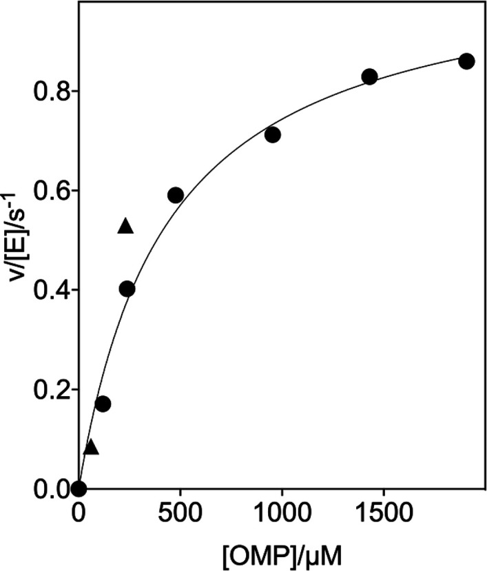
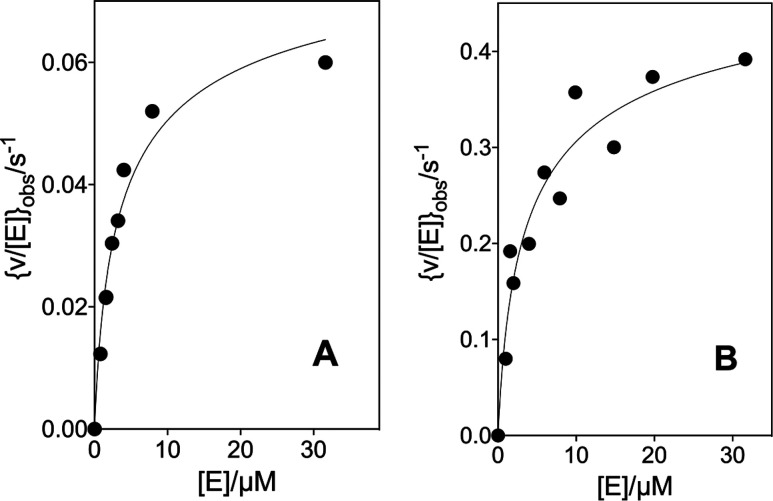
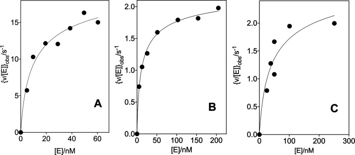
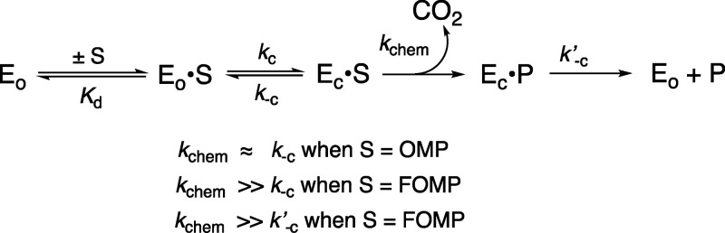
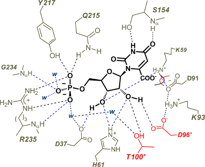
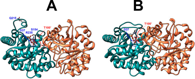
The D37 and T100' side chains of orotidine 5'-monophosphate decarboxylase (OMPDC) interact with the C-3' and C-2' ribosyl hydroxyl groups, respectively, of the bound substrate. We compare the intra-subunit interactions of D37 with the inter-subunit interactions of T100' by determining the effects of the D37G, D37A, T100'G, and T100'A substitutions on the following: (a) kcat and kcat/Km values for the OMPDC-catalyzed decarboxylations of OMP and 5-fluoroorotidine 5'-monophosphate (FOMP) and (b) the stability of dimeric OMPDC relative to the monomer. The D37G and T100'A substitutions resulted in 2 kcal mol-1 increases in ΔG for kcat/Km for the decarboxylation of OMP, while the D37A and T100'G substitutions resulted in larger 4 and 5 kcal mol-1 increases, respectively, in ΔG. The D37G and T100'A substitutions both resulted in smaller 2 kcal mol-1 decreases in ΔG for the decarboxylation of FOMP compared to that of OMP. These results show that the D37G and T100'A substitutions affect the barrier to the chemical decarboxylation step while the D37A and T100'G substitutions also affect the barrier to a slow, ligand-driven enzyme conformational change. Substrate binding induces the movement of an α-helix (G'98-S'106) toward the substrate C-2' ribosyl hydroxy bound at the main subunit. The T100'G substitution destabilizes the enzyme dimer by 3.5 kcal mol-1 compared to the monomer, which is consistent with the known destabilization of α-helices by the internal Gly side chains [Serrano, L., et al. (1992) Nature, 356, 453-455]. We propose that the T100'G substitution weakens the α-helical contacts at the dimer interface, which results in a decrease in the dimer stability and an increase in the barrier to the ligand-driven conformational change.


Conflict of interest statement

The authors declare no competing financial interest.

Figures

Scheme 1
Scheme 1. OMPDC-Catalyzed Decarboxylation of OMP to Form a Vinyl Carbanion Reaction Intermediate
Scheme 2
Scheme 2. Partitioning of the Total 31 kcal mol–1 Stabilization of the Transition State for OMPDC Decarboxylation among the Three Contributing Substrate Fragments
Scheme 3
Scheme 3. Utilization of Intrinsic Substrate Binding Energy in Order to Drive an Unfavorable Enzyme Conformational Change
Figure 1
Figure 1
A representation (PDB 3GDL) of the interactions between the OMPDC active site side chains and the tight binding inhibitor 6-azauridine 5′-monophosphate (azaUMP) at the complex to the closed form of OMPDC (EC, Scheme 3). The enzyme active site is near the subunit interface, which is shown by the blue and orange shading of the two subunits. The inhibitor complex is stabilized by the following interactions: the Q215 and R235 side chains interact directly with the substrate dianion,, the S154 side chain oxygen accepts a hydrogen bond from the pyrimidine −NH, the D37 side chain forms a hydrogen bond to the C-3′ ribosyl −OH, and the T100′ side chain from the second enzyme subunit forms a hydrogen bond to the C-2′ ribosyl −OH.,
Scheme 4
Scheme 4. Conversion of the Inactive OMPDC Monomer (EM) to the Active Dimer (ED)
Figure 2
Figure 2
Dependence of v/[E] for the decarboxylation of OMP catalyzed by the T100′G variant of OMPDC for reactions at 25 °C, pH 7.1 (30 mM MOPS), I = 0.105 (NaCl), and 32 μM OMPDC (solid circles). The solid triangles show the limiting values of v/[E] determined for reactions catalyzed by high [OMPDC] at 60 μM (Figure 3A) and 230 μM OMP (Figure 3B).
Figure 3
Figure 3
Effect of increasing concentrations of OMPDC on [v/[E]]obs for the decarboxylation of OMP catalyzed by the T100′G variant at 25 °C, pH 7.1 (30 mM MOPS), and I = 0.105 (NaCl). (A) The T100′G variant OMPDC-catalyzed decarboxylation of 60 μM OMP. (B) The T100′G variant-catalyzed decarboxylation of 230 μM OMP.
Figure 4
Figure 4
Effect of increasing concentrations of OMPDC on [v/[E]]obs for the decarboxylation of OMP catalyzed by the wild type and variant enzymes at 25 °C, pH 7.1 (30 mM MOPS), and I = 0.105 (NaCl). The solid line shows the fit of the experimental data to eq 2 where [E] is the concentration of the OMPDC monomers. (A) The wild type OMPDC-catalyzed decarboxylation reaction with 34 μM OMP. (B) The T100′A variant-catalyzed decarboxylation reaction of 99 μM OMP. (C) The D37G variant-catalyzed decarboxylation reaction of 52 μM OMP.
Scheme 5
Scheme 5. A Kinetic Scheme for OMPDC That Separates the Steps for Ligand Binding and the Enzyme Conformational Change from Open EO·S to Closed EC·S
Figure 5
Figure 5
A pancake representation of the interactions between the amino acid side chains of OMPDC and the bound substrate OMP (PDB 1DQX, but with OMP inserted into the position of the 6-hydroxyuridine 5′-monophosphate inhibitor). The D96′ and T100′ side chains from the second enzyme subunit are shaded red. The D37 side chain is hydrogen bonded to the C-3′ ribosyl hydroxyl and interacts with the substrate phosphodianion through an intervening water molecule. The interactions of the substrate phosphodianion with the Q215, Y217, and R235 side chains develop during the conformational change from the open unliganded enzyme EO to the closed Michaelis complex EC·OMP (Scheme 3).
Figure 6
Figure 6
Representations of the open (A, PDB 3GDK) and the closed or liganded (B, PDB 3GDL) forms of yeast OMPDC where the azaUMP ligand is placed at structure A at the position determined for structure B. These representations show the movements of the phosphodianion gripper loop (P202–V220) toward the pyrimidine umbrella loop (A151–T165) and R235 toward the phosphodianion, as well as the movement of the G′98–S′106 α-helix from the second subunit toward the bound substrate and the pyrimidine umbrella of the main subunit. The bridging interaction between the −CH2OH side chain of S154 and the amide side chain of Q215 at the closed enzyme is not shown.

Similar articles

Cited by

References

    1. Miller B. G.; Wolfenden R. (2002) Catalytic proficiency: the unusual case of OMP decarboxylase. Annu. Rev. Biochem. 71, 847–885. 10.1146/annurev.biochem.71.110601.135446. - DOI - PubMed
    1. Miller B. G.; Hassell A. M.; Wolfenden R.; Milburn M. V.; Short S. A. (2000) Anatomy of a proficient enzyme: the structure of orotidine 5′-monophosphate decarboxylase in the presence and absence of a potential transition state analog. Proc. Natl. Acad. Sci. U. S. A. 97, 2011–2016. 10.1073/pnas.030409797. - DOI - PMC - PubMed
    1. Goryanova B.; Amyes T. L.; Richard J. P. (2019) Role of the Carboxylate in Enzyme-Catalyzed Decarboxylation of Orotidine 5′-Monophosphate: Transition State Stabilization Dominates Over Ground State Destabilization. J. Am. Chem. Soc. 141, 13468–13478. 10.1021/jacs.9b04823. - DOI - PMC - PubMed
    1. Goryanova B.; Amyes T. L.; Gerlt J. A.; Richard J. P. (2011) OMP Decarboxylase: Phosphodianion Binding Energy Is Used To Stabilize a Vinyl Carbanion Intermediate. J. Am. Chem. Soc. 133, 6545–6548. 10.1021/ja201734z. - DOI - PMC - PubMed
    1. Tsang W.-Y.; Wood B. M.; Wong F. M.; Wu W.; Gerlt J. A.; Amyes T. L.; Richard J. P. (2012) Proton Transfer from C-6 of Uridine 5′-Monophosphate Catalyzed by Orotidine 5′-Monophosphate Decarboxylase: Formation and Stability of a Vinyl Carbanion Intermediate and the Effect of a 5-Fluoro Substituent. J. Am. Chem. Soc. 134, 14580–14594. 10.1021/ja3058474. - DOI - PMC - PubMed

MeSH terms