Review
. 2020 May 4;9(5):1131.
doi: 10.3390/cells9051131.

Lysosomal Biology and Function: Modern View of Cellular Debris Bin

Affiliations
Review

Lysosomal Biology and Function: Modern View of Cellular Debris Bin

Purvi C Trivedi et al. Cells. .

Abstract

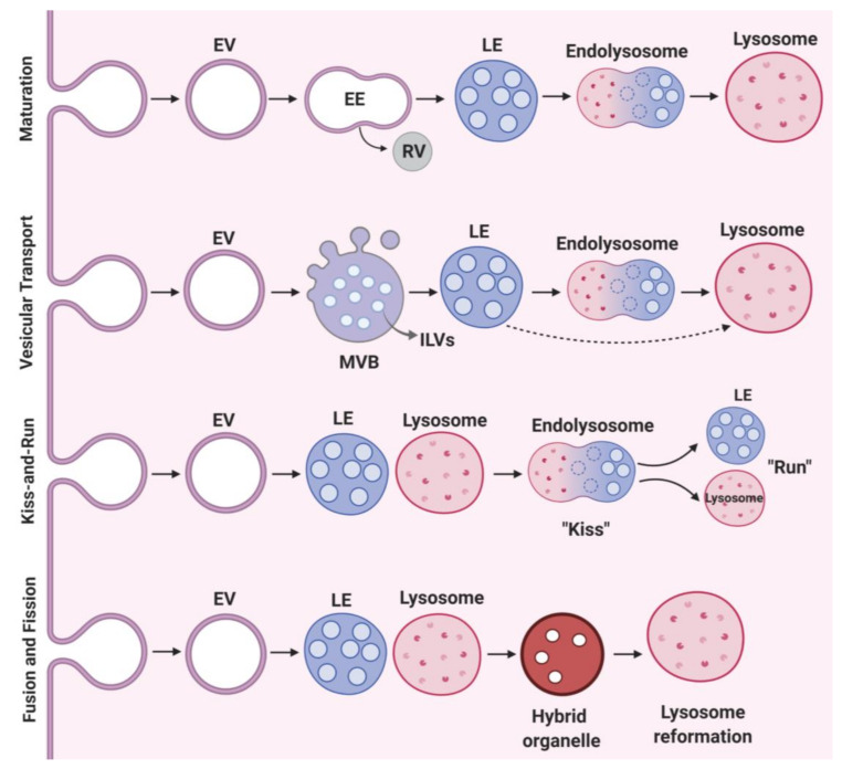
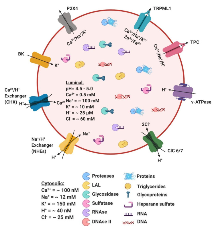
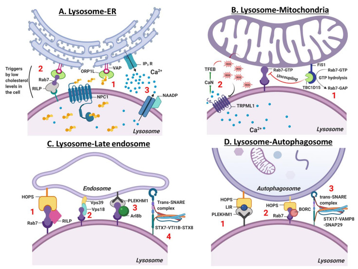
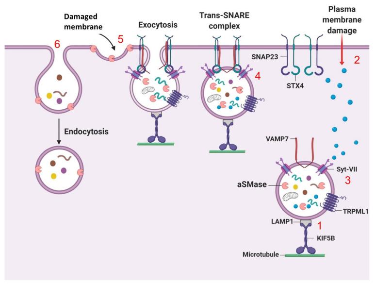
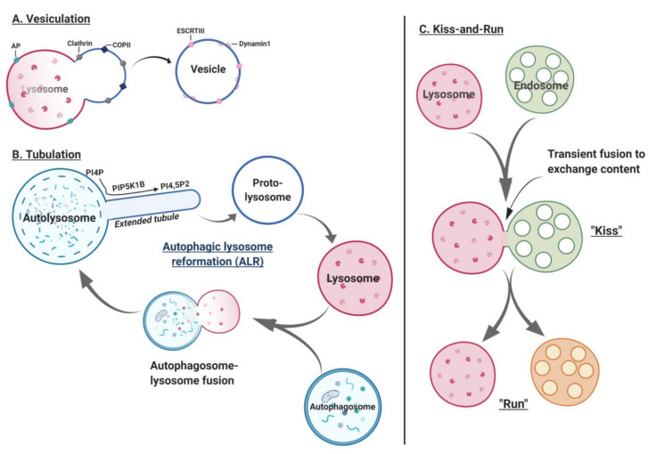
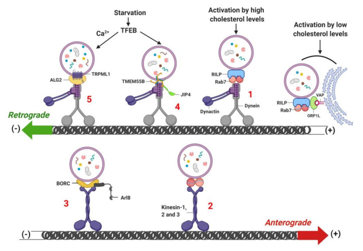
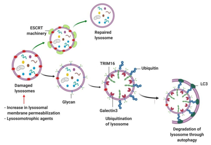
Lysosomes are the main proteolytic compartments of mammalian cells comprising of a battery of hydrolases. Lysosomes dispose and recycle extracellular or intracellular macromolecules by fusing with endosomes or autophagosomes through specific waste clearance processes such as chaperone-mediated autophagy or microautophagy. The proteolytic end product is transported out of lysosomes via transporters or vesicular membrane trafficking. Recent studies have demonstrated lysosomes as a signaling node which sense, adapt and respond to changes in substrate metabolism to maintain cellular function. Lysosomal dysfunction not only influence pathways mediating membrane trafficking that culminate in the lysosome but also govern metabolic and signaling processes regulating protein sorting and targeting. In this review, we describe the current knowledge of lysosome in influencing sorting and nutrient signaling. We further present a mechanistic overview of intra-lysosomal processes, along with extra-lysosomal processes, governing lysosomal fusion and fission, exocytosis, positioning and membrane contact site formation. This review compiles existing knowledge in the field of lysosomal biology by describing various lysosomal events necessary to maintain cellular homeostasis facilitating development of therapies maintaining lysosomal function.

Keywords: autophagy; calcium; cathepsin; endocytosis; lysosome; mannose-6-phosphate; metabolism; proton.


Conflict of interest statement

The authors declare no potential conflicts of interest.

Figures

Figure 1
Figure 1
Molecular events in lysosome biogenesis. Maturation; this model of lysosome biogenesis describes the formation of EVs (endocytic vesicles) from the plasma membrane and their progressive maturation to late endosomes and subsequently to lysosomes. The cargo targeted for recycling is carried by the TGN derived RVs (recycling vesicles), whereas cargo required for lysosomal degradation is transported by the cargo vesicle through late endosomes. Vesicular transport; requires ECV/MVBs (endosomal carrier vesicle/multi-vesicular bodies) carrying ILVs (intraluminal vesicles), which mobilize cargo from early-to-late endosomes and then to lysosomes or mobilize cargo directly from the matured late endosomes to lysosomes. Kiss-and-Run; describes contact site formation between lysosomes and late endosomes (“kiss”) followed the by cargo transfer and ensuing dissociation (“run”) of late endosomes from lysosomes. Fusion and fission; events involve heterotypic fusion of late endosome-lysosome to form a hybrid organelle and subsequently lysosome reformation.
Figure 2
Figure 2
Lysosome ion channels and transporters. The lysosomal lumen contains a battery of soluble hydrolytic enzymes that degrade proteins (proteases), triglycerides (lysosomal acid lipase; LAL), glycoproteins (glycosidases), heparan sulphate (sulfatases), and nucleic acids (nucleases, RNAase and DNAse). Lysosomal ion channels and transporters include vATPase; vacuolar H+-ATPase (proton pump), chloride channel (ClC); ClC family that exchanges cytosolic Cl for lysosomal H+ (ClC-6 and ClC-7), K+ channels (BK). Non-selective cation channels include Transient Receptor Potential Cation Channel (TRPML); permeable to Ca2+, Na+, K+, Zn2+ and Fe2+, P2X4 channel; permeable to Ca2+, Na+ and K+ and two-pore channels (TPC); permeable to H+, Ca2+ and Na+. Ion transporters include Na+/H+ exchangers (NHEs) and Ca2+/H+ exchangers (CHX).
Figure 3
Figure 3
Organellar contact site and fusion with lysosome. (A). Lysosome-ER: Lysosome-ER contact site regulates ER Ca2+ release. (1) Lysosome-ER contact site shuttles free cholesterol from the lysosome to ER via concerted action of lysosomal membrane-localized protein, NPC1 and ER membrane proteins, VAPA (vesicle-associated membrane protein (VAMP)-associated ER protein A) and VAPB and a lipid-transfer protein, ORP1L (oxysterol-binding protein-related protein 1L). (2) Low cellular cholesterol level activates ORP1L, which then triggers the formation of a contact site between ER and lysosomal membrane. ER-resident protein VAP anchors with lysosomal Rab7-RILP (Rab7-interacting protein) to initiate the movement of lysosomes to plus-end or towards the cell periphery. (3) NAADP (nicotinic acid adenine dinucleotide phosphate), a Ca2+ mobilizing entity and IP3 receptor agonist mobilizes lysosomal Ca2+ and facilitates ER Ca2+ release into the cytosol. (B). Lysosome-Mitochondria: Lysosome-mitochondria contract site mediates the bidirectional regulation of lysosome and mitochondria dynamics. (1) Mitochondria forms a contact with the lysosome via lysosomal bound Rab7-GTP. This contact site is disrupted by mitochondrial fission 1 membrane protein, FIS1 and recruits TBC1D15 (TBC1 Domain Family Member 15) which hydrolyzes Rab7-GTP to Rab7-GAP. (2) Lysosomal mucolipin channel, TRPML1 (Transient Receptor Potential Cation Channel, Mucolipin subfamily) governs mitochondria homeostasis by sensing and scavenging mitochondrial ROS (reactive oxygen species). TRPML1 is activated by ROS that induces Ca2+ release from the lysosomal lumen and activates nuclear TFEB translocation in a calcineurin-dependent manner. TFEB induces lysosome biogenesis and autophagy to clear damaged mitochondria and scavenge ROS. (C). Lysosome-Late endosome: The fusion of lysosome and late endosome occurs via tethering processes, which involves; (1) coupling of small GTPase Rab7, Rab7-interacting protein (RIPL) and vacuole protein sorting (HOPS) complex, (2) interaction of Rab7, VPS18 and VPS39 (3) Rab7, Arf-like (Ar18b) and Rab7 effector PLEKHM1 (pleckstrin homology domain-containing protein family member 1 and (4) formation of Trans-SNARE assembly composed of syntaxin-7 (STX7, Qa-SNARE), VTI1B (VPS10 tail interactor-1B, Qb-SNARE) and syntaxin-8 (STX8, Qc-SNARE). (D). Lysosome-autophagosome: The fusion of lysosome and autophagosome occurs via (1) HOPS complex and Rab7 effector protein, PLEKHM1 which binds to HOP via LC3-interacting region (LIR) (2) BLOC-1 related complex (BORC1) that recruits Rab7 and HOPS for tethering and (3) enabling trans-SNARE complex formation of STX17-VAMP8-SNAP29 to induce autophagosome-lysosome fusion.
Figure 4
Figure 4
Lysosome exocytosis-dependent plasma membrane repair. Upon damage to the PM, (1) lysosomes are translocated to the cell periphery via cytoskeleton and motor protein, Kinesin 5 subfamily member KIF5B, which is bound to LAMP1 on the lysosomal membrane. (2) Injury to the PM can lead to an influx of Ca2+ in cells, which is then (3) sensed by Ca2+ sensor, Syt-VII (synaptotagmin-VII) on the lysosomal membrane and triggers a repair mechanism. (4) Subsequently, exocytosis is initiated by the lysosomal membrane localized VAMP7 (vesicle-associated membrane protein 7) that forms a Trans-SNARE complex with syntaxin-4 and SNAP23 (synaptosome-associated protein 23 kDa) on the PM. Trans-SNARE complex brings the lysosome in a close proximity to the PM to initiate the fusion of lysosome and PM. (5) The fusion of lysosome and PM triggers an efflux of lysosomal enzyme aSMase (sphingomyelinase), which is then retain on the PM to convert sphingomyelin into ceramide, (6) leading to an inward configuration of the PM to facilitate endocytosis-dependent removal and restoration of damaged PM.
Figure 5
Figure 5
Lysosome fission. Lysosome fission involves, vesiculation process (A) is initiated by inner-coat adaptor protein-dependent cargo sorting, followed by membrane deformation mediated by clathrin and structural scaffold of COPII subcomplex. Subsequently, scission machinery including dynamin 1 and ESCRT-III catalyzes the membrane fission and dissociates vesicle from the lysosomal membrane. The process of tubulation (B) is best described by autophagic lysosome reformation (ALR), wherein proto-lysosomes derived from ALs, mature into functional lysosomes known as lysosome reformation. These matured lysosomes then subsequently fuse with autophagosomes to form ALs and initiate the cycle of lysosome reformation. The final event of lysosome fission is kiss-and-run (C), which involves the fusion of lysosomes and late endosomes. The transient interaction (“Kiss”) between lysosome and LEs allows the exchange of content, followed by separation of the lysosome (“Run”).
Figure 6
Figure 6
Retrograde- and anterograde-dependent lysosome mobilization. Lysosomes move from the cell periphery towards the perinuclear region in a retrograde manner. (1) The retrograde movement of the lysosome is initiated by RILP-Rab7-dependent recruitment and activation of dynein-dynactin, enabling the transport of lysosomes to minus-end direction. (2) While anterograde movement transport lysosomes from the perinuclear to the cell periphery, which requires kinesin-1, 2 and 3 motor proteins. (3) Anterograde movement of lysosomes also requires BLOC-1-related complex (BORC)-dependent recruitment of Arl8, which activates the kinesin proteins. (4) and (5) starvation-induced activation of TFEB increases the transcription of TRPML1 and TMEM55B to trigger lysosome mobilization in a retrograde manner via interaction with ALG2 (an EF-hand-containing protein, serve as lysosomal Ca2+ sensor) and JIP4 (C-Jun-amino-terminal kinase-interacting protein 4), respectively. Low cellular cholesterol levels restrict lysosomes at the plus end, which is mediated by the interaction between lysosomal RILP and Rab7 with VAP and ORP1L on ER membrane.
Figure 7
Figure 7
Lysosome repair by lysophagy. Damaged lysosomes can efflux luminal Ca2+ stores and activate ESCRT machinery component to repair lysosome by restoring lysosomal integrity. However, lysosomes are subjected to degradation when damaged. Severely damaged lysosomes can expose luminal glycan to the cytosol, which is sensed by galectin-3 to trigger lysosome ubiquitination via TRIM16. Following lysosome ubiquitination, lysophagy initiates lysosomal degradation.

References

    1. De Duve C. The lysosome turns fifty. Nat. Cell Biol. 2005;7:847–849. doi: 10.1038/ncb0905-847. - DOI - PubMed
    1. Bainton D.F. The discovery of lysosomes. J. Cell Biol. 1981;91:66s–76s. doi: 10.1083/jcb.91.3.66s. - DOI - PMC - PubMed
    1. Pryor P.R., Luzio J.P. Delivery of endocytosed membrane proteins to the lysosome. Biochim. Biophys. Acta. 2009;1793:615–624. doi: 10.1016/j.bbamcr.2008.12.022. - DOI - PubMed
    1. Hu Y.B., Dammer E.B., Ren R.J., Wang G. The endosomal-lysosomal system: from acidification and cargo sorting to neurodegeneration. Transl. Neurodegener. 2015;4:18. doi: 10.1186/s40035-015-0041-1. - DOI - PMC - PubMed
    1. Jegga A.G., Schneider L., Ouyang X., Zhang J. Systems biology of the autophagy-lysosomal pathway. Autophagy. 2011;7:477–489. doi: 10.4161/auto.7.5.14811. - DOI - PMC - PubMed

Publication types

LinkOut - more resources