doi: 10.5045/br.2020.2019191.
Chronic phase of chronic myeloid leukemia presenting with myeloid sarcoma in an adolescent
Affiliations
- PMID: 32375463
- PMCID: PMC7343543
- DOI: 10.5045/br.2020.2019191
Item in Clipboard
Chronic phase of chronic myeloid leukemia presenting with myeloid sarcoma in an adolescent
Blood Res.
.
No abstract available
Conflict of interest statement
No potential conflicts of interest relevant to this article were reported.
Figures

Histopathology of the thigh biopsy (A) low-power image (hematoxylin and eosin, ×100) and (B) high-power image (hematoxylin and eosin, ×400) showing medium to large cells with a cleaved nucleus and eosinophilic cytoplasm. (C) Tumor cells are diffusely positive for myeloperoxidase (immunohistochemical stain, ×400).

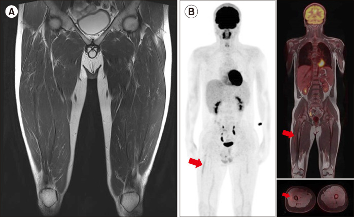
Initial thigh T2 magnetic resonance image (MRI) showing a 12×6×4 cm sized heterogeneous low signal intensity lesion in the proximal portion of the vastus lateralis muscle (A). Fluorodeoxyglucose (FDG)-positron emission tomography (PET) scan showing high uptake of FDG in the right thigh and diffused uptake in the BM of the entire skeleton (B).
After 6 months of treatment, a significant reduction in tumor size was achieved (A). Barely noticeable tumor seen on the thigh T2 MRI scan. Significant reduction in the extent and intensity of FDG uptake seen on FDG-PET scan (B).
Comment in
-
Myeloid sarcoma: "A cytological surprise".Diagn Cytopathol. 2022 Nov;50(11):538-539. doi: 10.1002/dc.25034. Epub 2022 Aug 12. Diagn Cytopathol. 2022. PMID: 35960136 No abstract available.
References
-
- Pilieri SA, Orazi A, Falini B. Myeloid sarcoma. In: Swerdlow SH, Campo E, Harris NL, et al., editors. WHO classification of tumours of haematopoietic and lymphoid tissues. 4th ed. IARC Press; Lyon, France: 2008. pp. 140–1.
-
- Dasappa L, Thanky AH, Kuntegowdanahalli LC, et al. Myeloid sarcoma as the first sign of progression of chronic myeloid leukemia in medullary chronic phase: experience from a Tertiary Cancer Centre in Southern India. Gulf J Oncolog. 2017;1:21–5. - PubMed
Publication types
LinkOut - more resources
Full Text Sources