Review
. 2020 May 6;17(1):35.
doi: 10.1186/s12987-020-00196-2.

Choroid plexus and the blood-cerebrospinal fluid barrier in disease

Affiliations
Review

Choroid plexus and the blood-cerebrospinal fluid barrier in disease

Peter Solár et al. Fluids Barriers CNS. .

Abstract

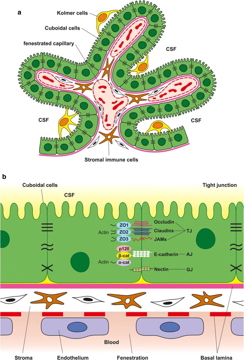
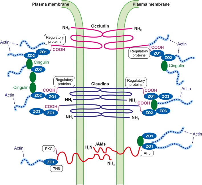
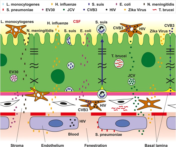
The choroid plexus (CP) forming the blood-cerebrospinal fluid (B-CSF) barrier is among the least studied structures of the central nervous system (CNS) despite its clinical importance. The CP is an epithelio-endothelial convolute comprising a highly vascularized stroma with fenestrated capillaries and a continuous lining of epithelial cells joined by apical tight junctions (TJs) that are crucial in forming the B-CSF barrier. Integrity of the CP is critical for maintaining brain homeostasis and B-CSF barrier permeability. Recent experimental and clinical research has uncovered the significance of the CP in the pathophysiology of various diseases affecting the CNS. The CP is involved in penetration of various pathogens into the CNS, as well as the development of neurodegenerative (e.g., Alzheimer´s disease) and autoimmune diseases (e.g., multiple sclerosis). Moreover, the CP was shown to be important for restoring brain homeostasis following stroke and trauma. In addition, new diagnostic methods and treatment of CP papilloma and carcinoma have recently been developed. This review describes and summarizes the current state of knowledge with regard to the roles of the CP and B-CSF barrier in the pathophysiology of various types of CNS diseases and sets up the foundation for further avenues of research.

Keywords: Autoimmune disease; Blood–cerebrospinal fluid barrier; Carcinoma; Choroid plexus; Inflammatory diseases; Neurodegenerative disease; Stroke.


Conflict of interest statement

The authors declare that they have no competing interests.

Figures

Fig. 1
Fig. 1
Schematic illustration of the anatomical organization of the CP (a) and the B-CSF barrier (b). The CP is an epithelio-endothelial convolute, comprising a highly vascularized stroma with connective tissue, and a continuous lining of epithelial cells with adhering Kolmer cells (a). The main site of the barrier (b) is at the level of the cuboidal epithelial cells that are linked by tight junctions (TJ), adherent junctions (AJ) and gap junctions (GJ). TJs are required for strong cell–cell adhesion and include transmembrane (occludin, claudins and JAMs) and cytoplasmic proteins (ZO). AJs are formed by E-cadherin, a transmembrane protein that intracellularly binds to actin through a variety of protein complexes, including catenin (p120, β-catenin, α-catenin). GJ protein complexes comprising nectin also play a role in adherence
Fig. 2
Fig. 2
Schematic illustration of the molecular interactions of TJ proteins in CP epithelial cells. TJ proteins and their complex interactions with other proteins form the connections between adjacent epithelial cells in the B-CSF barrier. Claudin, occludins, and JAMs are linked to actin filaments through ZO proteins. Cingulin, another cytoplasmic protein, is recruited to TJ through ZO. The interaction between TJ proteins and the cytoplasmic protein network is crucial for maintaining B-CSF barrier integrity (adapted from [72])
Fig. 3
Fig. 3
Schematic illustration showing the various invasion strategies of different pathogens (bacteria, viruses and parasites) through the B-CSF barrier into the CNS. Streptococus suis (S. suis) can cross the B-CSF barrier within endocytic vacuoles and there is some evidence supporting a “Trojan horse” mechanism using polymorphonuclear leukocytes. Streptococus pneumoniae (S. pneumoniae) interacts with the endothelium of the CP. Listeria monocytogenes (L. monocytogenes) has been observed invading the CNS using a “Trojan horse” mechanism inside mononuclear cells. Escherichia coli (E. coli) and Haemophilus influenzae (H. influenzae) can migrate through CP epithelial cells. Neisseria meningitidis (N. meningitidis) crosses the B-CSF barrier and forms colonies at the apical side of CP epithelial cells. Polyomavirus JC (JCV) probably forms a reservoir in CP epithelial cells. HIV has been described in endothelial and stromal cells as well as in epiplexus monocytes. Echovirus 30 (EV30) may invade and replicate in CP epithelial cells. Coxsackievirus B3 (CVB3) crosses the B-CSF barrier using myeloid cells as well as via a paracellular route through the TJs. Trypanosoma brucei (T. brucei) was found in the perivascular region of the CP and also in CP epithelial cells

References

    1. Abbott NJ, Patabendige AAK, Dolman DEM, Yusof SR, Begley DJ. Structure and function of the blood–brain barrier. Neurobiol Dis. 2010;37:13–25. doi: 10.1016/j.nbd.2009.07.030. - DOI - PubMed
    1. Hladky SB, Barrand MA. Mechanisms of fluid movement into, through and out of the brain: evaluation of the evidence. Fluids Barriers CNS. 2014;11:26. doi: 10.1186/2045-8118-11-26. - DOI - PMC - PubMed
    1. Johanson CE, Duncan JA, Stopa EG, Baird A. Enhanced prospects for drug delivery and brain targeting by the choroid plexus–CSF route. Pharm Res. 2005;22:1011–1037. doi: 10.1007/s11095-005-6039-0. - DOI - PubMed
    1. Bors L, Tóth K, Tóth EZ, Bajza Á, Csorba A, Szigeti K, et al. Age-dependent changes at the blood–brain barrier. A comparative structural and functional study in young adult and middle aged rats. Brain Res Bull. 2018;139:269–277. doi: 10.1016/j.brainresbull.2018.03.001. - DOI - PubMed
    1. Huber JD, Egleton RD, Davis TP. Molecular physiology and pathophysiology of tight junctions in the blood–brain barrier. Trends Neurosci. 2001;24:719–725. doi: 10.1016/s0166-2236(00)02004-x. - DOI - PubMed

MeSH terms

LinkOut - more resources