. 2020 Aug;1863(8):194566.
doi: 10.1016/j.bbagrm.2020.194566. Epub 2020 May 3.

The molecular basis of selective DNA binding by the BRG1 AT-hook and bromodomain

Affiliations

The molecular basis of selective DNA binding by the BRG1 AT-hook and bromodomain

Julio C Sanchez et al. Biochim Biophys Acta Gene Regul Mech. 2020 Aug.

Abstract

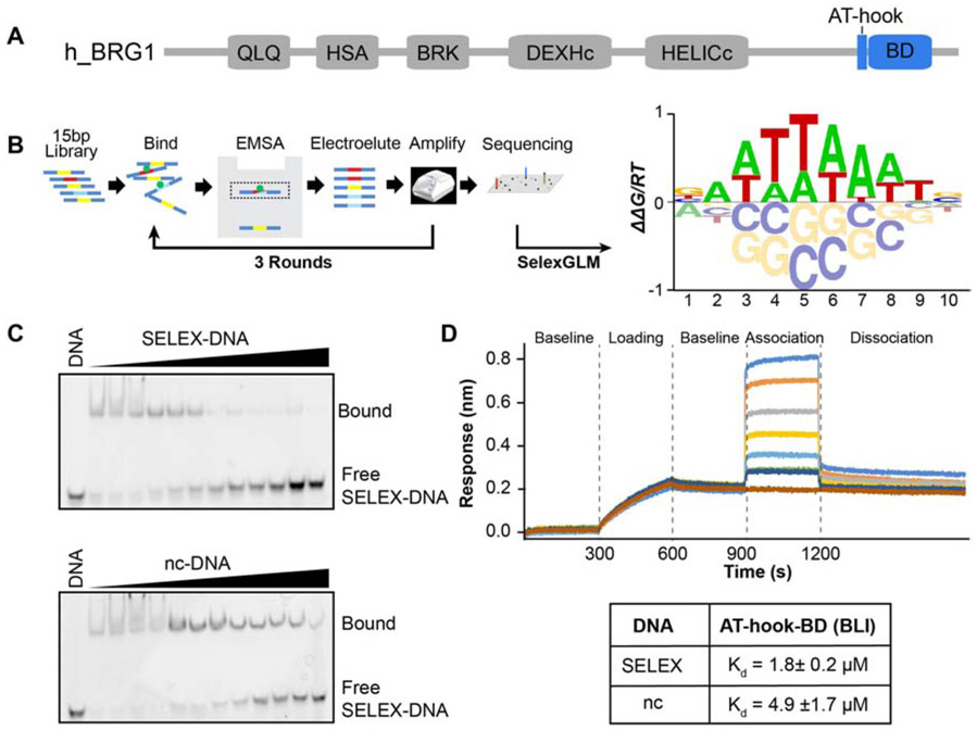
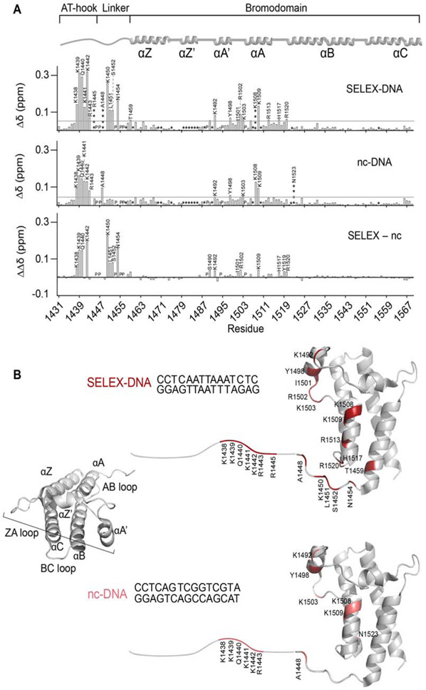
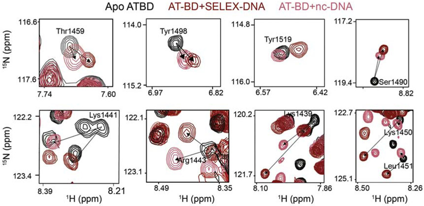
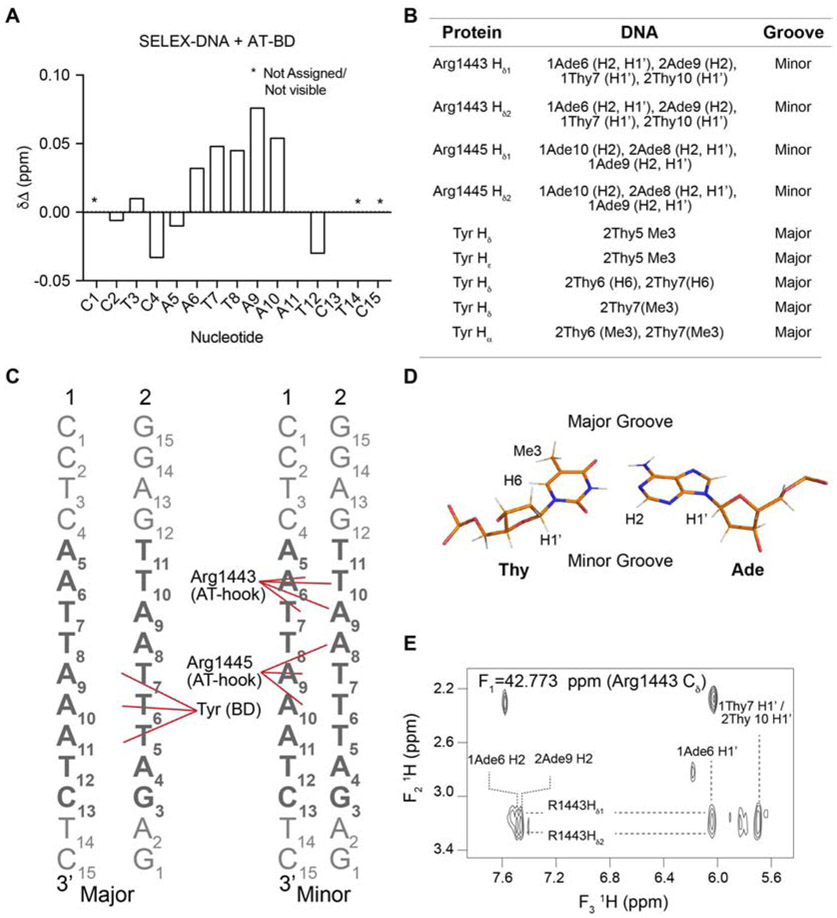
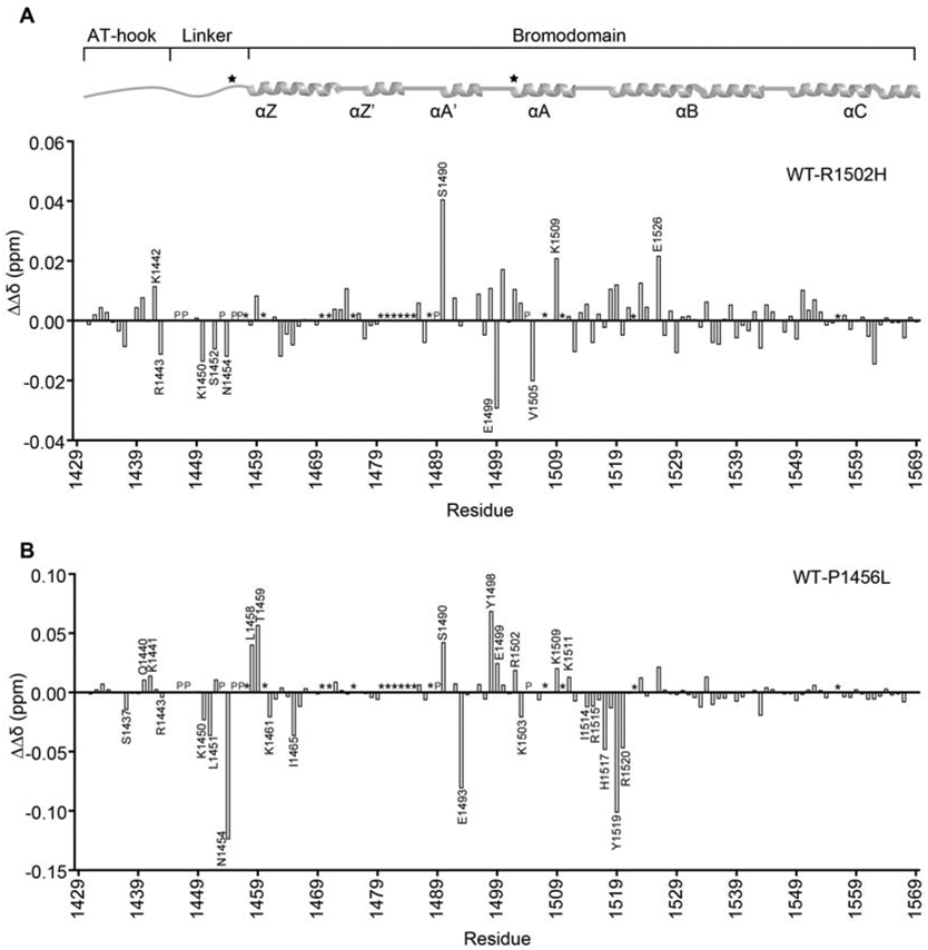
The ATP-dependent BAF chromatin remodeling complex plays a critical role in gene regulation by modulating chromatin architecture, and is frequently mutated in cancer. Indeed, subunits of the BAF complex are found to be mutated in >20% of human tumors. The mechanism by which BAF properly navigates chromatin is not fully understood, but is thought to involve a multivalent network of histone and DNA contacts. We previously identified a composite domain in the BRG1 ATPase subunit that is capable of associating with both histones and DNA in a multivalent manner. Mapping the DNA binding pocket revealed that it contains several cancer mutations. Here, we utilize SELEX-seq to investigate the DNA specificity of this composite domain and NMR spectroscopy and molecular modelling to determine the structural basis of DNA binding. Finally, we demonstrate that cancer mutations in this domain alter the mode of DNA association.


Conflict of interest statement

Declaration of competing interest The authors declare that they have no known competing financial interests or personal relationships that could have appeared to influence the work reported in this paper.

Figures

Figure 1.
Figure 1.. DNA specificity of the BRG1 ATBD.
(A) Domain architecture of the human BRG1 subunit of the BAF complex. The AT-hook and bromodomain (BD) are highlighted in blue. (B) Schematic representation of SELEX-seq followed by a construction of a position specific affinity matrix and converted into the affinity logo containing AATTAAAT. (C) Specificity was confirmed through competitive binding assays analyzed by EMSA. A 15bp double-stranded DNA containing either the SELEX determined DNA sequence (SELEX-DNA: CCTCAATTAAATCTC) or a non-consensus sequence (nc-DNA: CCTCAGTCGGTCGTA) was used. The complex of ATBD and Cy5-labeled SELEX-DNA (bound) was competed against unlabeled SELEX-DNA (top) or nc-DNA (bottom). (D) Dissociation (Kd) values were determined by BLI. Shown is a set of representative traces in a BLI binding assay (top) for 15μM, 8μM, 4μM, 2μM, 1μM, 0.5μM, and 0.25μM (from top to bottom) of protein. Calculated Kd values are in the table below, values reported are the average of 3 runs and standard deviations.
Figure 2.
Figure 2.. NMR reveals the molecular basis of enhanced binding to SELEX-DNA.
(A) Plots of normalized CSPs (Δδ) for AT-BD with SELEX-DNA (top), nc-DNA (middle), and the difference between the two (ΔΔδ, bottom). Residues perturbed more than the average plus two standard deviations (denoted by the gray line) are labeled. Where the average was calculated after trimming the top 10% of perturbations. “P” indicates a proline, “*” indicates an unassigned residue and “****” represent a signal that broadened beyond detection upon titration with ligand. The AT-BD secondary structure architecture is depicted on top of the plots for reference. (B) Residues in the AT-BD with significant CSPs upon binding to the SELEX-DNA (top) or nc-DNA (bottom) are plotted onto a cartoon representation of the previously solved structure of the BD (PDB ID 3UVD), with the AT-hook drawn in (a small break is left between the structure and the drawn-in segment).
Figure 3.
Figure 3.. NMR reveals unique binding to SELEX-DNA.
1H,15N-HSQC spectral overlays are shown for AT-BD residues saturated with either SELEX-DNA (red) or nc-DNA (salmon). Shown are resonances for residues which show a unique trajectory of CSP between the two substrates, indicating a unique binding mode. This was observed for residues in the bromodomain (Thr1459, Tyr1498, Tyr1519, Ser1490) the AT-hook (Lys1439, Lys1441, Lys1443) and the linker (Lys1450, Lys1451).
Figure 4.
Figure 4.. The AT-BD spans the major and minor groove.
(A) CSPs (Δδ) of the imino resonances of the SELEX-DNA saturated with AT-BD. “*” indicates an unassigned or not visible resonance. (B) Table of Intermolecular NOEs observed between the 13C-AT-BD and 12C-SELEX-DNA. (C) Schematic of AT-hook and BD contacts with SELEX-DNA (strands 1 and 2 labeled) in the major (left) and minor (right) grooves. Contacts are highlighted by red lines. The core SELEX determined sequence is in bold. (D) structure of an AT base pair with hydrogens forming NOEs labeled, as well as the major and minor groove sides. (E) Representative NOE cross-peaks between AT-BD and SELEX-DNA indicate that these protons are <6Å from each other in space.
Figure 5.
Figure 5.. Molecular model of the AT-BD/SELEX-DNA complex.
(A) Model of AT-BD/SELEX-DNA complex. The AT-hook is highlighted in blue, the αA in deep sky blue, the ZA loop in ice blue and the linker in cyan. The DNA and the rest of the BRG1 BD are in gray. In this binding mode (mode 1) the AT-hook is positioned in the minor groove and the ZA loop the major groove, consistent with NOE data. (B) A zoom in of the linker shows a significant bend induced at in the polypeptide backbone at Pro1456. (C) A zoom in shows the AT-hook arginines (1443 and 1445) inserted into the minor groove. Carbons are colored in cyan, oxygens in red and nitrogens in blue. (D) The minimum distance between AT-BD elements and SELEX-DNA. (E) The number of hydrogen bonds between AT-BD elements and SELEX-DNA in the percentage of frames during the 500 ns simulations.
Figure 6.
Figure 6.. Cancer mutations alter the mode of DNA binding.
Differences in the magnitude of CSPs (ΔΔδ) for binding of AT-BD to SELEX-DNA were computed between wild-type (WT) and the R1502H (A) or P1456L (B) mutants. Residues which show substantially different binding are marked. “P” denotes proline and “*” denotes unassigned residues. The AT-BD secondary structure architecture is depicted on top of the plots for reference with mutation sites marked with a star.

References

    1. Luger K, Mäder AW, Richmond RK, Sargent DF, Richmond TJ, Crystal structure of the nucleosome core particle at 2.8 Å resolution, Nature. 389 (1997) 251–260. doi:10.1038/38444. - DOI - PubMed
    1. Wu JI, Lessard J, Crabtree GR, Understanding the Words of Chromatin Regulation, Cell. 136 (2009) 200–206. doi:10.1016/j.cell.2009.01.009. - DOI - PMC - PubMed
    1. Alpsoy A, Dykhuizen EC, Glioma tumor suppressor candidate region gene 1 (GLTSCR1) and its paralog GLTSCR1-like form SWI/SNF chromatin remodeling subcomplexes, J Biol Chem. 293 (2018) 3892–3903. doi:10.1074/jbc.ra117.001065. - DOI - PMC - PubMed
    1. Gatchalian J, Malik S, Ho J, Lee D-S, Kelso TWR, Shokhirev MN, Dixon JR, Hargreaves DC, A non-canonical BRD9-containing BAF chromatin remodeling complex regulates naive pluripotency in mouse embryonic stem cells., Nature Communications. 9 (2018) 5139. doi:10.1038/s41467-018-07528-9. - DOI - PMC - PubMed
    1. Alfert A, Moreno N, Kerl K, The BAF complex in development and disease, Epigenet Chromatin. 12 (2019) 19. doi:10.1186/s13072-019-0264-y. - DOI - PMC - PubMed

Publication types

MeSH terms

LinkOut - more resources