. 2020;10(3):658-671.e1.
doi: 10.1016/j.jcmgh.2020.04.015. Epub 2020 May 4.

Norepinephrine Has Dual Effects on Human Colonic Contractions Through Distinct Subtypes of Alpha 1 Adrenoceptors

Affiliations

Norepinephrine Has Dual Effects on Human Colonic Contractions Through Distinct Subtypes of Alpha 1 Adrenoceptors

Masaaki Kurahashi et al. Cell Mol Gastroenterol Hepatol. 2020.

Abstract

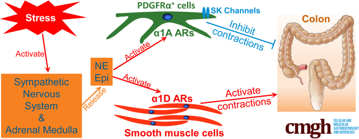
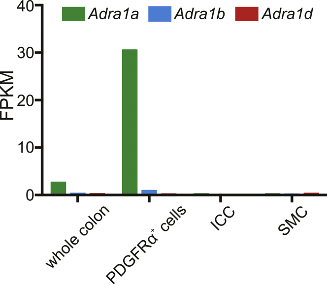
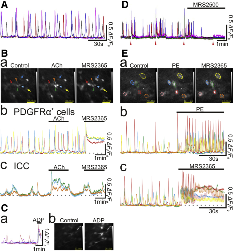
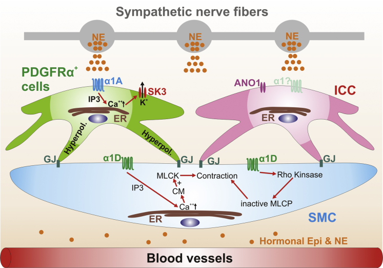
Background & aims: Colonic musculature contain smooth muscle cells (SMC), interstitial cells of Cajal (ICC), and platelet-derived growth factor receptor α+ cells (PDGFRα+ cells), which are electrically coupled and operate together as the SIP syncytium. PDGFRα+ cells have enriched expression of small conductance Ca2+-activated K+ (SK) channels. Purinergic enteric neural input activates SK channels in PDGFRα+ cells, hyperpolarizes SMC, and inhibits colonic contractions. Recently we discovered that PDGFRα+ cells in mouse colon have enriched expression of α1A adrenoceptors (ARs), which coupled to activation of SK channels and inhibited colonic motility, and α1A ARs were principal targets for sympathetic regulation of colonic motility. Here we investigated whether PDGFRα+ cells in human colon express α1A ARs and share the roles as targets for sympathetic regulation of colonic motility.

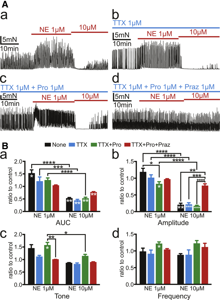
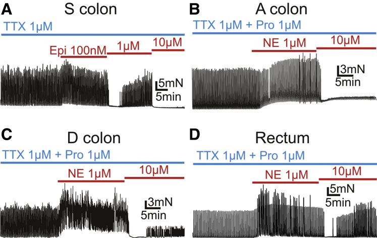
Methods: Isometric tension recording, intracellular recording, and Ca2+ imaging were performed on muscles of the human colon. Responses to α1 ARs agonists or electric field stimulation with AR antagonists and neuroleptic reagents were studied.

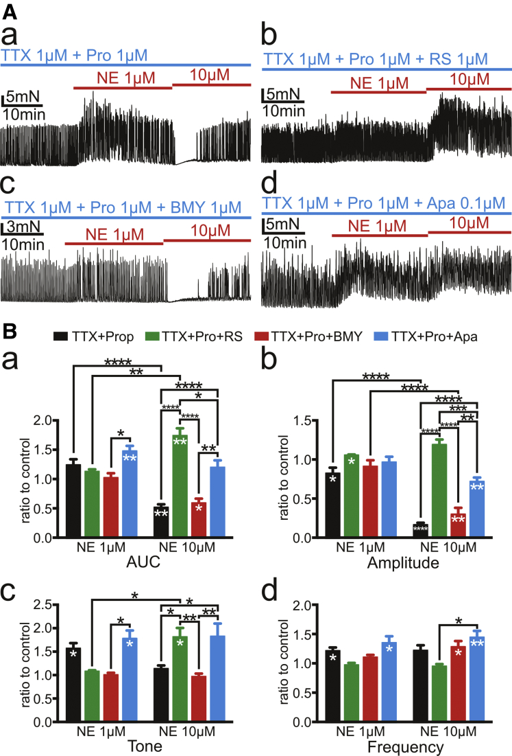
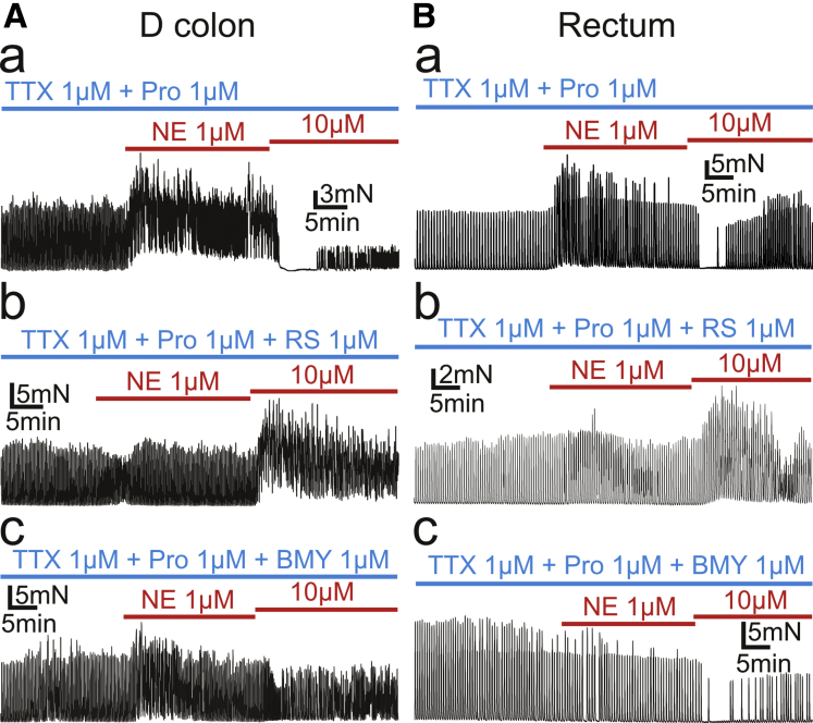
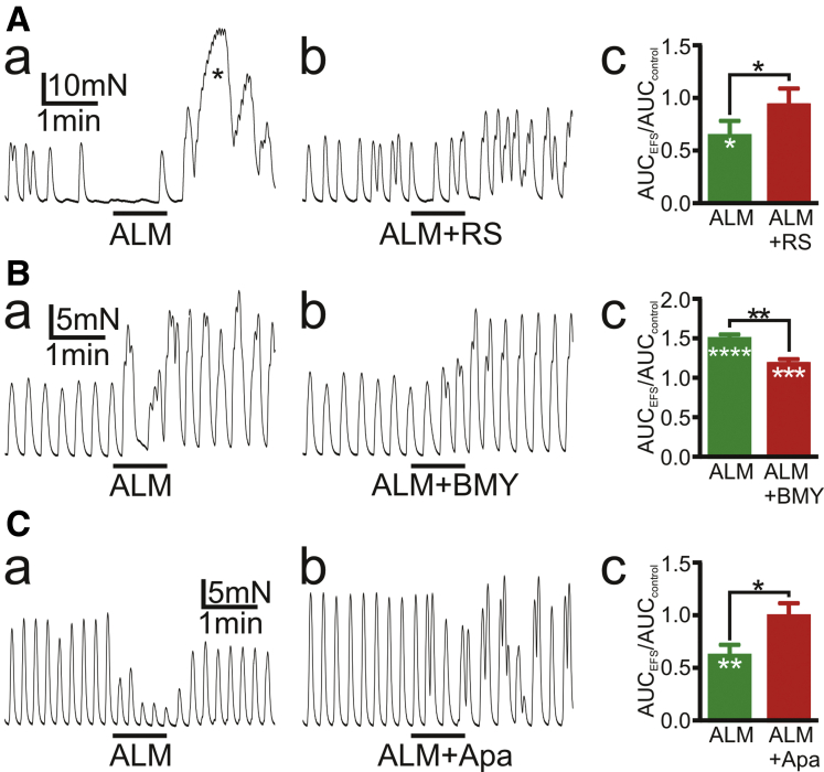
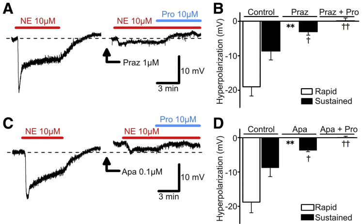
Results: Exogenous or endogenous norepinephrine released from nerve fibers inhibited colonic contractions through binding to α1A ARs or enhanced colonic contractions by acting on α1D ARs. Inhibitory responses were blocked by apamin, an antagonist of SK channels. Phenylephrine, α1 AR agonists, or norepinephrine increased intracellular [Ca2+] in PDGFRα+ cells, but not in ICC, and hyperpolarized SMCs by binding to α1 ARs expressed by PDGFRα+ cells.

Conclusions: Human colonic contractions are inhibited by α1A ARs expressed in PDGFRα+ cells and activated by α1D ARs expressed in SMC.

Keywords: Colonic Motility; PDGFRα(+) Cells; SIP Syncytium; Sympathetic Nervous System; α1 Adrenoceptor.


Figures

None
Graphical abstract
Figure 1
Figure 1
Bar graph depicting expression profile of the genes of adrenergic receptor α1 (AR α1) family created from transcriptome data of SIP syncytium of mouse colon that we published in 2015–2017., , Fragments per kilobase of transcript per million (FPKM) of AR α1A, α1B, and α1D are 30.41, 0.80, and 0.07 in PDGFRα+ cells, 0.11, 0.00, and 0.00 in ICC, and 0.13, 0.06, and 0.21 in SMC, respectively.
Figure 2
Figure 2
Tension recordings of CM strips of human sigmoid colon. (A) NE 1 μmol/L activated SPCs and NE 10 μmol/L inhibited them (a) regardless of the presence of TTX 1 μmol/L (b). Propranolol 1 μmol/L (Pro) enhanced the elevation of tone by NE but reduced the increments of amplitude of contractions by NE (c). Prazosin (Praz) 1 μmol/L blocked responses of muscle strips to NE (d). (B) Summary of 4 parameters, AUC, amplitudes, tone, and frequency of SPCs for 10 minutes after applying NE 1 μmol/L and 10 μmol/L is shown by ratio to controls for 10 minutes before applying NE in the same recordings. Black asterisks (∗) indicate statistically significant differences between the values connected by black lines, and white asterisks indicate statistically significant differences of the values against controls. The numbers of asterisks indicate the following: ∗.05 > P ≥ .01; ∗∗.01 > P ≥ .001; ∗∗∗.001 > P ≥ .0001; ∗∗∗∗.0001 > P.
Figure 3
Figure 3
Tension recordings of CM strips of sigmoid colon (S colon) (A), ascending colon (A colon) (B), descending colon (D colon) (C), and rectum (D). (A) Epinephrine (Epi) 100 nmol/L activated the spontaneous contractions of muscle strips, but Epi 1 μmol/L and 10 μmol/L inhibited them in dose-dependent manner under existence of TTX 1 μmol/L. (B–D) A and D colon and rectum also showed similar responses to NE to sigmoid colon, in which NE 1 μmol/L activated tonic contractions of muscle strips and NE 10 μmol/L inhibited amplitude of contractions under existence of TTX 1 μmol/L and propranolol (Pro) 10 μmol/L. In (B), A colon looked to have the tonic contraction increased in dose-dependent manner, while it had the amplitude of contractions inhibited by NE 10 μmol/L. In (D), rectum looked to have the amplitude of contractions increased as well as the tonic contractions by NE 1 μmol/L.
Figure 4
Figure 4
Tension recordings of CM strips of sigmoid colon in the presence of TTX 1 μmol/L and propranolol (Pro) 1 μmol/L. (A) RS100329 (RS) 1 μmol/L and apamin (Apa) 0.1 μmol/L blocked inhibitory effects on amplitude of contractions by NE 10 μmol/L and revealed excitatory effects on tone by that (b and d). BMY7378 (BMY) 1 μmol/L did not block inhibitory effect on amplitude of contractions of NE 10 μmol/L but inhibited excitatory effects of NE 1 μmol/L in tone (c). (B) Summary of 4 parameters of responses of SPCs for 10 minutes after applying NE 1 μmol/L and 10 μmol/L is displayed using ratio to the controls as described in Figure 2. Black asterisks (∗) indicate statistically significant difference between the values connected by black line, and white asterisks indicate statistically significant difference of the values against controls. The numbers of asterisks indicate the following: ∗.05 > P ≥ .01; ∗∗.01 > P ≥ .001; ∗∗∗.001 > P ≥ .0001; ∗∗∗∗.0001 > P.
Figure 5
Figure 5
Tension recordings of CM muscle strips of D colon (A) and rectum (B) were performed. Both D colon and rectum showed similar responses to NE under the presence of RS100329 (RS) 1 μmol/L or BMY7378 (BMY) 1 μmol/L to S colon, in which RS inhibited inhibitory effects by NE 10 μmol/L and revealed excitatory effects by that (Ab and Bb), and BMY inhibited excitatory effects by NE 1 μmol/L (Ac and Bc).
Figure 6
Figure 6
Tension recordings of CM strips of sigmoid colon.Black bars represent EFS with 50-millisecond duration and 100 V at 5 Hz for 1 minute. Responses of SPCs to EFS in the presence of antagonists of main neurotransmitters, atropine 1 μmol/L, L-NNA 100 μmol/L, and MRS2500 500 nmol/L (ALM), were recorded. EFS induced inhibitory effects (Aa and Ca) or excitatory effects (Ba) on SPCs. Inhibitory effects of EFS on SPCs were attenuated by RS100329 (RS) 1 μmol/L (Ab) or apamin (Apa) 0.1 μmol/L (Cb). In (A), EFS evoked rebound excitation immediately after EFS as indicated by asterisk ∗ in (Aa), which were inhibited by RS. Excitatory effects of EFS on SPCs (Ba) were inhibited by BMY7378 (BMY) 1 μmol/L (Bb). Ac, Bc, and Cc depict the summary of AUC during EFS for 1 minute divided by AUC of control SPCs for 1 minute. Black asterisks (∗) indicate statistically significant difference between the values connected by black line, and white asterisks indicate statistically significant difference of the values against controls. AUC values (means ± standard error) (mN·min) were (Ac) ALM, 4.37 ± 0.37; ALM + RS, 5.96 ± 0.95; (Bc) ALM, 9.91 ± 0.31; ALM + BMY, 7.64 ± 0.29; and (Cc) ALM, 3.10 ± 0.56; ALM + apamin, 6.86 ± 1.04. The numbers of asterisks indicate the following: ∗.05 > P ≥ .01; ∗∗.01 > P ≥ .001; ∗∗∗.001 > P ≥ .0001; ∗∗∗∗.0001 > P.
Figure 7
Figure 7
Ca2+ imaging in human colonic muscles. (A) In a muscle preparation, 4 PDGFRα+ cells generated developed asynchronous spontaneous Ca2+ transients independently from each other. (B) In a preparation where PDGFRα+ cells generated asynchronous spontaneous Ca2+ transients (b), ICC exhibited synchronous spontaneous Ca2+ transients within their cluster (c). PDGFRα+ cells responded to MRS2365 100 nmol/L but not ACh 1 μmol/L (arrows in a and b), whereas ICC responded to ACh 1 μmol/L but not MRS2365 100 nmol/L (arrowheads in a and c). Graph of Ca2+ signals picked in each of cells pointed by color arrows or arrowheads in (a) were depicted in (b) and (c) in the same color as that of arrow or arrowhead. (C) In a preparation where PDGFRα+ cells generated asynchronous spontaneous Ca2+ transients, ADP 100 μmol/L evoked increases in basal Ca2+ level (a and b). (D) In a preparation where PDGFRα+ cells generated asynchronous spontaneous Ca2+ transients, EFS (20 Hz for 1 second) triggered synchronous increases in basal Ca2+ level (red arrowheads in D). MRS2500 500 nmol/L largely suppressed EFS-induced Ca2+ transients and also prevented generation of spontaneous Ca2+ transients. (E) In a preparation where PE 10 μmol/L caused increases in basal Ca2+ level associated with superimposed Ca2+ oscillations in several cells (middle panel in a and b), MRS2365 100 nmol/L evoked sustained increases in basal Ca2+ level in the same cells (right panel in a and c). Graph of Ca2+ signals picked in each of color circles in (a) were depicted in (b) and (c) in the same color as that of the circle.
Figure 8
Figure 8
Effects of NE on membrane potentials of human S colon circular SMCs. Application of NE 10 μmol/L induced a two-phase hyperpolarization, a rapid component followed by a sustained component. (A) NE-induced two-phase hyperpolarization was changed to small, sustained hyperpolarization by pretreatment of prazosin (Praz) 1 μmol/L. Residual hyperpolarization was inhibited by propranolol (Pro) 10 μmol/L. (B) Summarized bar graphs showing effects of Praz and Pro on NE-induced two-phase hyperpolarization. ∗∗P < .01, significant difference from control responses of rapid component. P < .05, significant difference from control responses of sustained component. ††P < .01, significant difference from sustained responses in presence of Praz alone. (C) Apamin (Apa) 0.1 μmol/L inhibited two-phase hyperpolarization induced by NE, resulting in sustained hyperpolarization, which was inhibited by Pro 10 μmol/L. (D) Summary showing effects of Apa and Pro on NE-induced two-phase hyperpolarization. ∗∗P < .01, significant difference from control responses of rapid component. P < .05, significant difference from control responses of sustained component. ††P < .01, significant difference from sustained responses in presence of Apa alone. Resting membrane potentials were A, −46 mV; C, −49 mV. A and C were recorded from different tissues. Each record in a given set of two was obtained from the same impalement.
Figure 9
Figure 9
Schematic diagram of the new concept based on this study. ARs are expressed on PDGFRα+ cells. Neuronal or hormonal NE or Epi, via binding to and activating α1A ARs in PDGFRα+ cells, opens SK3 channels through increasing intracellular [Ca2+] by inositol triphosphate (IP3) and hyperpolarize (Hyperpol) them. Hyperpolarization of PDGFRα+ cells is propagated to SMC via gap junctions (GJ) and inhibits contractions of them. α1D ARs are expressed by SMC. Neuronal or hormonal NE or Epi, via binding to and activating α1D ARs on SMC, can make myosin light chain kinase (MLCK) activated and SMC contract through activation of calmodulin (CM) via increase of intracellular [Ca2+] by IP3 or can activate Rho kinase pathway and inactivate myosin light chain phosphatase (MLCP), which leads to contractions of SMC. ICC might not express α1 ARs. ANO1, anoctamin-1, Ca2+ -activated Cl channels. Altogether, neuronal or hormonal NE or Epi can inhibit human colonic contractions via α1A AR-SK channel signaling pathway in PDGFRα+ cells and excite them via α1D AR on SMC.

Similar articles

Cited by

References

    1. Komuro T., Seki K., Horiguchi K. Ultrastructural characterization of the interstitial cells of Cajal. Arch Histol Cytol. 1999;62:295–316. - PubMed
    1. Sanders K.M., Koh S.D., Ro S., Ward S.M. Regulation of gastrointestinal motility-insights from smooth muscle biology. Nat Rev Gastroenterol Hepatol. 2012;9:633–645. - PMC - PubMed
    1. Sanders K.M., Kito Y., Hwang S.J., Ward S.M. Regulation of gastrointestinal smooth muscle function by interstitial cells. Physiology. 2016;31:316–326. - PMC - PubMed
    1. Iino S., Horiguchi K., Horiguchi S., Nojyo Y. c-Kit-negative fibroblast-like cells express platelet-derived growth factor receptor alpha in the murine gastrointestinal musculature. Histochem Cell Biol. 2009;131:691–702. - PubMed
    1. Kurahashi M., Zheng H., Dwyer L., Ward S.M., Koh S.D., Sanders K.M. A functional role for the 'fibroblast-like cells' in gastrointestinal smooth muscle. J Physiol. 2011;589:697–710. - PMC - PubMed

Publication types

MeSH terms

Substances