

[T1-weighted magnetic resonance imaging contrast agents and their theranostic nanoprobes]
- PMID: 32376585
- PMCID: PMC7167323
- DOI: 10.12122/j.issn.1673-4254.2020.03.24
[T1-weighted magnetic resonance imaging contrast agents and their theranostic nanoprobes]
Abstract
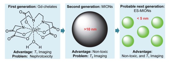
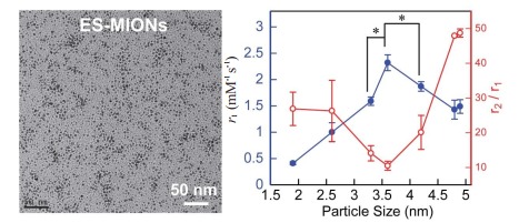
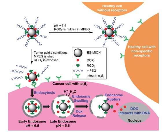
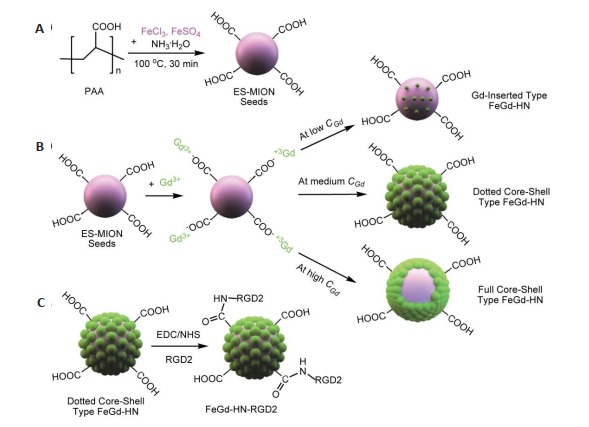
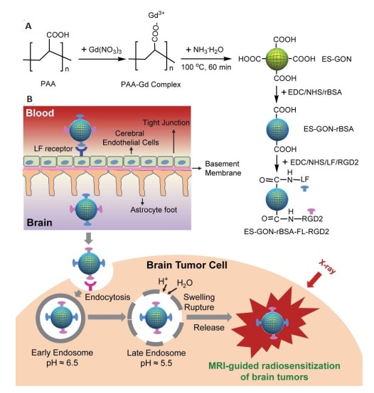
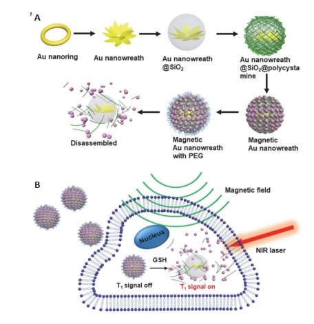
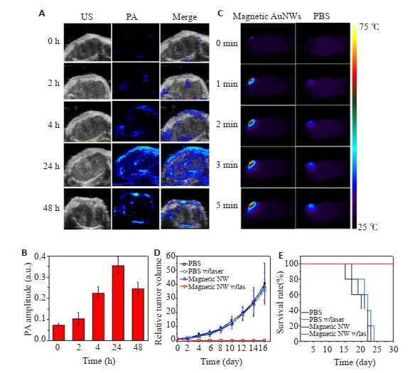
Magnetic resonance imaging (MRI) is an important imaging modality for clinical disease diagnosis, and nearly 50% of clinical MRI examinations require contrast agents to enhance the diagnostic sensitivity. This review provides a summary of the major MRI contrast agents and their classification, and the advantages and limits of the commercially available MRI contrast agents, and elaborates on the exceedingly small magnetic iron oxide nanoparticles (ES-MIONs), dotted core-shell iron and gadolinium hybrid nanoparticles (FeGd-HN) and exceedingly small gadolinium oxide nanoparticles (ES-GON). These nanoparticles can greatly improve the efficiency of T1-weighted MRI due to their high r1 value and low r2/r1 ratio, and are expected to be translated into clinical contrast agents for T1-weighted MRI. The authors also review the diagnostic and therapeutic integration system that combines MRI contrast agents with various tumor therapies, such as MRI-guided ferroptosis therapy, radiosensitization therapy, and photothermal therapy, which allow efficient treatment as well as real-time monitoring of tumors and serve as potential cancer therapy strategies. The possible future research directions in the field of MRI-based multifunctional diagnostic and therapeutic formulations are also discussed.
MRI已经成为临床疾病诊断的重要手段之一,而40%~50%的临床MRI检查需要使用造影剂来提高MRI的灵敏度,因此,MRI造影剂也成为重要的临床诊断药物。本文主要介绍了MRI造影剂及其分类,市售MRI造影剂的优缺点,并对极小磁性氧化铁纳米粒子、点式核壳型铁钆复合纳米粒子和极小氧化钆纳米粒子进行了评述,这三者因其r1值很高且r2/r1比值很低,T1-加权MRI成像的效果很好,有望转化为临床医用T1-加权MRI造影剂。本文对将MRI造影剂与各种肿瘤治疗策略相结合的诊断治疗一体化体系进行了评述,如MRI指导的铁凋亡治疗、MRI指导的放射增敏治疗和MRI指导的光热治疗,它们实现了对肿瘤的高效治疗和实时监控,有望成为可行的癌症治疗策略。最后对MRI基多功能诊疗制剂这一领域的未来可能研究方向进行了展望。
Keywords: dotted coreshell iron and gadolinium hybrid nanoparticles; exceedingly small gadolinium oxide nanoparticles; exceedingly small magnetic iron oxide nanoparticles; magnetic resonance imaging contrast agents; tumor theranostics.
Figures
Similar articles
-
Multifunctional Theranostic Nanoparticles Based on Exceedingly Small Magnetic Iron Oxide Nanoparticles for T1-Weighted Magnetic Resonance Imaging and Chemotherapy.ACS Nano. 2017 Nov 28;11(11):10992-11004. doi: 10.1021/acsnano.7b04924. Epub 2017 Oct 19. ACS Nano. 2017. PMID: 29039917
-
Exceedingly Small Magnetic Iron Oxide Nanoparticles for T1 -Weighted Magnetic Resonance Imaging and Imaging-Guided Therapy of Tumors.Small. 2023 Dec;19(49):e2302856. doi: 10.1002/smll.202302856. Epub 2023 Aug 18. Small. 2023. PMID: 37596716 Review.
-
Exceedingly Small Gadolinium Oxide Nanoparticles with Remarkable Relaxivities for Magnetic Resonance Imaging of Tumors.Small. 2019 Oct;15(41):e1903422. doi: 10.1002/smll.201903422. Epub 2019 Aug 25. Small. 2019. PMID: 31448577
-
Biodegradable and biocompatible exceedingly small magnetic iron oxide nanoparticles for T1-weighted magnetic resonance imaging of tumors.J Nanobiotechnology. 2022 Jul 30;20(1):350. doi: 10.1186/s12951-022-01562-y. J Nanobiotechnology. 2022. PMID: 35908057 Free PMC article.
-
Paramagnetic and Superparamagnetic Inorganic Nanoparticles for T1-Weighted Magnetic Resonance Imaging.Curr Med Chem. 2018;25(25):2970-2986. doi: 10.2174/0929867324666170314124616. Curr Med Chem. 2018. PMID: 28292235 Review.
Cited by
-
VHPKQHR Peptide Modified Ultrasmall Paramagnetic Iron Oxide Nanoparticles Targeting Rheumatoid Arthritis for T1-Weighted Magnetic Resonance Imaging.Front Bioeng Biotechnol. 2022 Feb 28;10:821256. doi: 10.3389/fbioe.2022.821256. eCollection 2022. Front Bioeng Biotechnol. 2022. PMID: 35295653 Free PMC article.
-
[Mn2+-doped Prussian blue nanoparticles for T1-T2 dual-mode magnetic resonance imaging and photothermal therapy in vitro].Nan Fang Yi Ke Da Xue Xue Bao. 2021 Jun 20;41(6):909-915. doi: 10.12122/j.issn.1673-4254.2021.06.14. Nan Fang Yi Ke Da Xue Xue Bao. 2021. PMID: 34238744 Free PMC article. Chinese.
References
-
- Lauterbur PC. Image formation by induced local interactions: examples employing nuclear magnetic resonance. Nature. 1973;242(5394):190–1. doi: 10.1038/242190a0. [Lauterbur PC. Image formation by induced local interactions: examples employing nuclear magnetic resonance[J]. Nature, 1973, 242(5394): 190-1.] - DOI - PubMed
-
- Denis HC. Paramagnetic contrast media for magnetic resonance imaging of the mediastinum and lungs. J Thorac Imaging. 1985;1(1):74–8. doi: 10.1097/00005382-198512000-00010. [Denis HC. Paramagnetic contrast media for magnetic resonance imaging of the mediastinum and lungs[J]. J Thorac Imaging, 1985, 1 (1): 74-8.] - DOI - PubMed
-
- Shellock FG, Kanal E. Safety of magnetic resonance imaging contrast agents. J Magn Reson Imaging. 1999;10(3):477–84. doi: 10.1002/(SICI)1522-2586(199909)10:3<477::AID-JMRI33>3.0.CO;2-E. [Shellock FG, Kanal E. Safety of magnetic resonance imaging contrast agents[J]. J Magn Reson Imaging, 1999, 10(3): 477-84.] - DOI - PubMed
-
- 李 坤成. 磁共振成像对比剂的临床应用. 北京: 人民卫生出版社; 2016. pp. 20–4. [李坤成.磁共振成像对比剂的临床应用[M].北京:人民卫生出版社, 2016: 20-4.]
-
- Zou ZT, Lin M. Nephrogenic systemic fibrosis: review of 408 biopsy-confirmed cases. http://d.old.wanfangdata.com.cn/OAPaper/oai_doaj-articles_6c75c58348afe6.... Cardiovasc Imaging. 2011;56(1):65–73. [Zou ZT, Lin M. Nephrogenic systemic fibrosis: review of 408 biopsy-confirmed cases[J]. Cardiovasc Imaging, 2011, 56(1): 65- 73.] - PMC - PubMed
Publication types
MeSH terms
Substances
LinkOut - more resources
Full Text Sources
Medical