. 2020 Jul 1;80(13):2804-2817.
doi: 10.1158/0008-5472.CAN-19-1523. Epub 2020 May 6.

Pattern of Invasion in Human Pancreatic Cancer Organoids Is Associated with Loss of SMAD4 and Clinical Outcome

Affiliations

Pattern of Invasion in Human Pancreatic Cancer Organoids Is Associated with Loss of SMAD4 and Clinical Outcome

Wenjie Huang et al. Cancer Res. .

Abstract

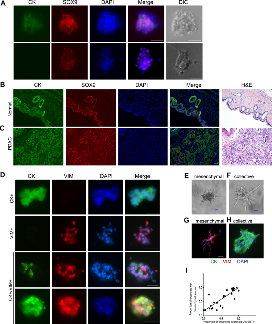
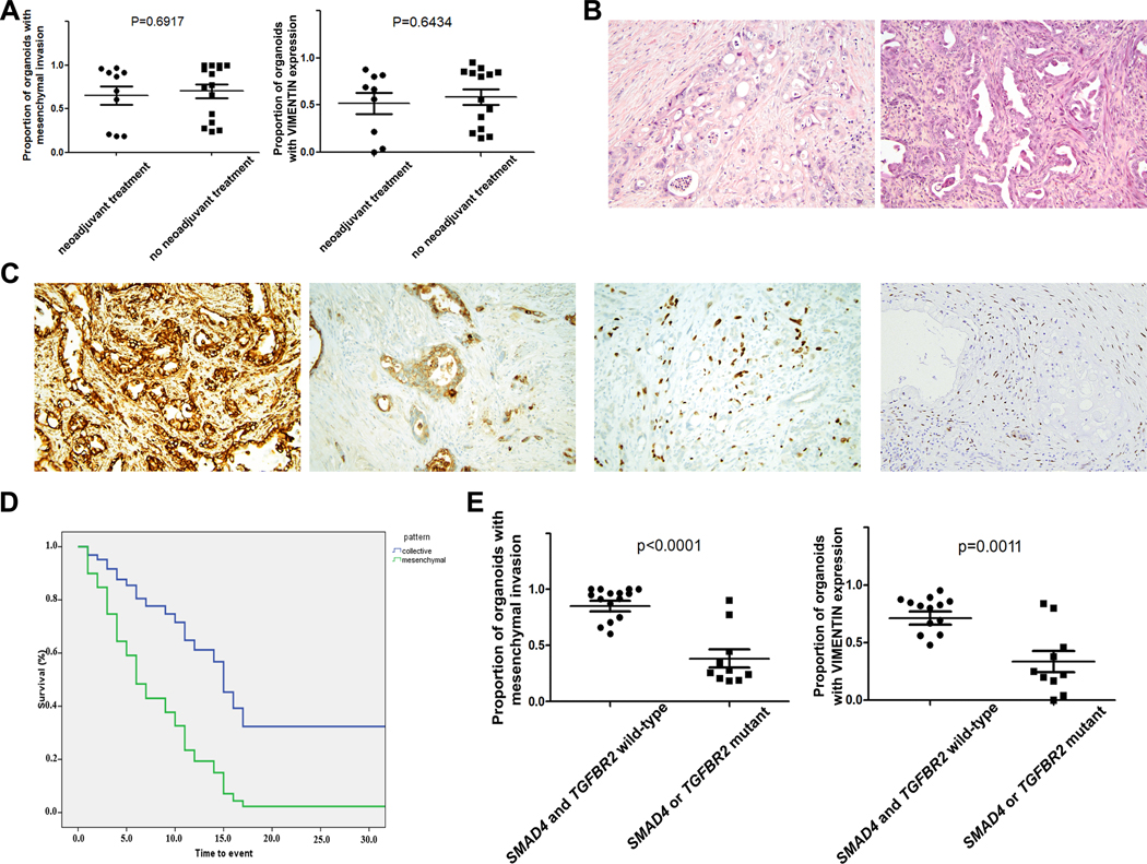
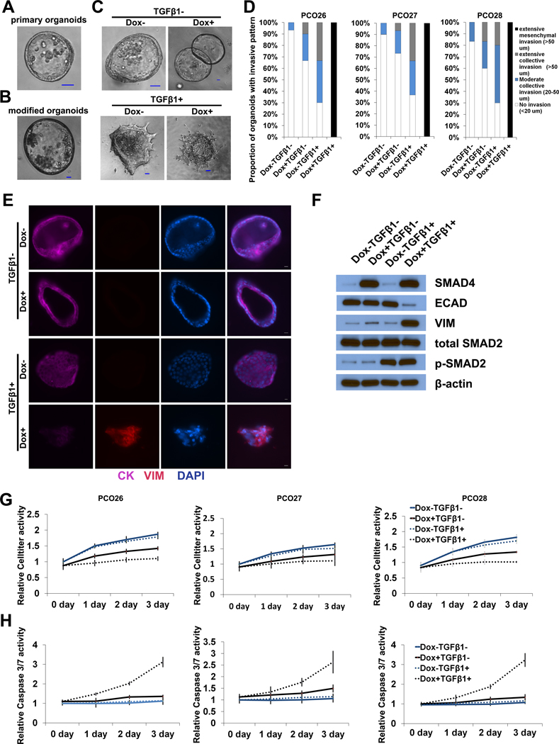
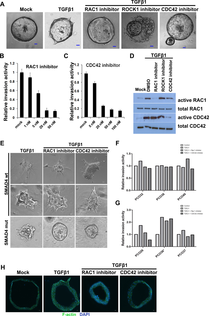
Pancreatic ductal adenocarcinoma (PDAC) is an aggressive malignancy characterized by extensive local invasion and systemic spread. In this study, we employed a three-dimensional organoid model of human pancreatic cancer to characterize the molecular alterations critical for invasion. Time-lapse microscopy was used to observe invasion in organoids from 25 surgically resected human PDAC samples in collagen I. Subsequent lentiviral modification and small-molecule inhibitors were used to investigate the molecular programs underlying invasion in PDAC organoids. When cultured in collagen I, PDAC organoids exhibited two distinct, morphologically defined invasive phenotypes, mesenchymal and collective. Each individual PDAC gave rise to organoids with a predominant phenotype, and PDAC that generated organoids with predominantly mesenchymal invasion showed a worse prognosis. Collective invasion predominated in organoids from cancers with somatic mutations in the driver gene SMAD4 (or its signaling partner TGFBR2). Reexpression of SMAD4 abrogated the collective invasion phenotype in SMAD4-mutant PDAC organoids, indicating that SMAD4 loss is required for collective invasion in PDAC organoids. Surprisingly, invasion in passaged SMAD4-mutant PDAC organoids required exogenous TGFβ, suggesting that invasion in SMAD4-mutant organoids is mediated through noncanonical TGFβ signaling. The Rho-like GTPases RAC1 and CDC42 acted as potential mediators of TGFβ-stimulated invasion in SMAD4-mutant PDAC organoids, as inhibition of these GTPases suppressed collective invasion in our model. These data suggest that PDAC utilizes different invasion programs depending on SMAD4 status, with collective invasion uniquely present in PDAC with SMAD4 loss. SIGNIFICANCE: Organoid models of PDAC highlight the importance of SMAD4 loss in invasion, demonstrating that invasion programs in SMAD4-mutant and SMAD4 wild-type tumors are different in both morphology and molecular mechanism.


Conflict of interest statement

Conflict of Interest: LDW served as a consultant for Personal Genome Diagnostics and receives research funding from Applied Materials. AJE has pending patent applications on molecular markers for cancer invasion and antibody-based strategies to treat cancer, and his wife is an employee of ImmunoCore. The other authors report no conflict of interest.

Figures

Figure 1.
Figure 1.. Expression of cytokeratin, vimentin, and SOX9 in human PDAC organoids and pancreatic tissue.
(A) Immunofluorescence co-staining of PDAC organoids for cytokeratin (green) and SOX9 (red) reveals that even cytokeratin-negative organoids express SOX9. Scale bars, 25 um. Images show two representative organoids (rows). (B-C) Immunofluorescence co-staining of normal pancreas and PDAC tissue with cytokeratin (green) and SOX9 (red). Scale bars, 50 um. (B) Representative normal pancreas sample shows SOX9 staining limited to ductal epithelium. (C) Representative PDAC sample shows SOX9 localization in malignant cells, but the stroma remains negative. (D) Immunofluorescence co-staining of PDAC organoids for cytokeratin (green) and vimentin (red) demonstrates variable expression patterns, including organoids expressing each marker alone, as well as organoids expressing both cytokeratin and vimentin. Scale bars, 25 um. (E-F) PDAC organoids invade with different phenotypes in collagen I gels: (E) mesenchymal invasion (F) collective invasion. (G-H) Immunofluorescence co-staining of PDAC organoids for cytokeratin (green) and vimentin (red) after 48 hours in culture in collagen I gels shows that organoids with mesenchymal invasion express vimentin, while those with collective invasion express cytokeratin. (I) Correlation of vimentin expression and mesenchymal invasion in PDAC organoids cultured in collagen I gel.
Figure 2.
Figure 2.. Pattern of invasion is associated with clinical outcome and SMAD4 mutation.
(A) There was no difference in pattern of invasion or VIM expression between patients who did or did not receive neoadjuvant chemotherapy prior to surgical resection. (B) Representative images of primary tumor morphology (H&E) showing predominantly glandular architecture: PCO18 (left) had predominantly collective invasion in our organoid model, while PCO5 (right) had predominantly mesenchymal invasion. (C) Multiple features of mesenchymal differentiation can be identified by immunohistochemistry in primary PDACs that give rise to mesenchymally invading organoids, including VIM expression (far left), ECAD loss (middle left), P40 expression (middle right), and GATA6 loss (far right). (D) Kaplan-Meier analysis of 25 resected PDAC patients shows increased risk of death associated with mesenchymal invasion (green) compared to collective invasion (blue) in our organoid culture model (HR=15.277; 95%CI: 2.000–116.689; p=0.009). (F) Collective invasion and lack of VIM expression in our organoid model were strongly associated with mutation in SMAD4 or TGFBR2 (p<0.001 and p=0.0011, t test).
Figure 3.
Figure 3.. Re-expression of SMAD4 leads to mesenchymal invasion in presence of TGFβ.
(A-B) Representative images showing morphology of unmodified (A) and lentivirally transduced (B) SMAD4-mutant PDAC organoids cultured in Matrigel. (C) Representative images of modified SMAD4-mutant PDAC organoids after treatment with doxycycline (Dox) and/or TGFβ and culture in collagen I. (D) Extent of invasion of modified SMAD4-mutant PDAC organoids after treatment with doxycycline (Dox) and/or TGFβ while cultured in collagen I. PCO26, PCO27, and PCO28 represent the organoids derived from 3 independent SMAD4-mutant PDACs. (E) Immunofluorescence co-staining of modified SMAD4-mutant PDAC organoids after treatment with doxycycline (Dox) and/or TGFβ while cultured in collagen I: cytokeratin (purple) and vimentin (red). (F) Expression of epithelial and mesenchymal markers in modified SMAD4-mutant PDAC organoids after treatment with doxycycline (Dox) and/or TGFβ as assayed by Western blot. (G-H) Effect of doxycycline (Dox) and/or TGFβ treatment on proliferation and apoptosis in modified SMAD4-mutant PDAC organoids: (G) proliferation as assessed by Celltiter assay. (H) apoptosis as assessed by caspase 3/7 activity. PCO26, PCO27, and PCO28 represent the organoids derived from 3 independent SMAD4-mutant PDACs.
Figure 4.
Figure 4.. RAC1 and CDC42 activation is required for TGFβ-induced collective invasion in SMAD4-mutant PDAC organoids.
(A) Representative images of organoids from a SMAD4-mutant PDAC cultured in collagen I and treated with TGFβ and/or RAC1, ROCK1, or CDC42 inhibitors. (B-C) Relative invasion of organoids from a SMAD4-mutant PDAC treated with different doses of RAC1 inhibitor (B) or CDC42 inhibitor (C). (D) Inhibition of RAC1 and CDC42 in PDAC organoids was confirmed by active GTPase pulldown followed by Western blot. (E) Representative images of organoids from additional SMAD4-wildtype (top) and SMAD4-mutant (bottom) PDACs cultured in collagen I and treated with TGFβ and/or RAC1 or CDC42 inhibitors. We identified a single SMAD4-wildtype PDAC (PCO36) with collective invasion in our organoid model (middle panels). Only SMAD4-mutant organoids decreased invasion after treatment with RAC1 or CDC42 inhibitors. (F) Impact of TGFβ and/or RAC1 or CDC42 inhibition on invasion in organoids with mesenchymal invasion. PDAC organoids with mesenchymal invasion (all SMAD4-wildtype) had little to no decrease in invasion after RAC1/CDC42 inhibition. (G) Impact of TGFβ and/or RAC1 or CDC42 inhibition on invasion in organoids with collective invasion. All organoids with collective invasion showed increased invasion after TGFβ treatment, but only those with SMAD4 mutations had decreased invasion after RAC1/CDC42 inhibition. *PCO36 is SMAD4-wildtype, while PCO29 and PCO37 are SMAD4-mutant. (H) Phalloidin labeling of F-actin in PDAC organoids in collagen I gels after treatment with TGFβ and/or RAC1 or CDC42 inhibitors.

References

    1. Siegel RL, Miller KD, Jemal A. Cancer Statistics, 2017. CA Cancer J Clin 2017;67:7–30. - PubMed
    1. Rahib L, Smith BD, Aizenberg R, Rosenzweig AB, Fleshman JM, Matrisian LM. Projecting cancer incidence and deaths to 2030: the unexpected burden of thyroid, liver, and pancreas cancers in the United States. Cancer Res 2014;74:2913–2921. - PubMed
    1. Jones S, Zhang X, Parsons DW, Lin JC, Leary RJ, Angenendt P, Mankoo P, et al. Core signaling pathways in human pancreatic cancers revealed by global genomic analyses. Science 2008;321:1801–1806. - PMC - PubMed
    1. Waddell N, Pajic M, Patch AM, Chang DK, Kassahn KS, Bailey P, Johns AL, et al. Whole genomes redefine the mutational landscape of pancreatic cancer. Nature 2015;518:495–501. - PMC - PubMed
    1. Biankin AV, Waddell N, Kassahn KS, Gingras MC, Muthuswamy LB, Johns AL, Miller DK, et al. Pancreatic cancer genomes reveal aberrations in axon guidance pathway genes. Nature 2012;491:399–405. - PMC - PubMed

Publication types

MeSH terms