. 2020 Aug;19(8):1263-1280.
doi: 10.1074/mcp.RA120.002029. Epub 2020 May 6.

Proteomics of Campylobacter jejuni Growth in Deoxycholate Reveals Cj0025c as a Cystine Transport Protein Required for Wild-type Human Infection Phenotypes

Affiliations

Proteomics of Campylobacter jejuni Growth in Deoxycholate Reveals Cj0025c as a Cystine Transport Protein Required for Wild-type Human Infection Phenotypes

Lok Man et al. Mol Cell Proteomics. 2020 Aug.

Abstract

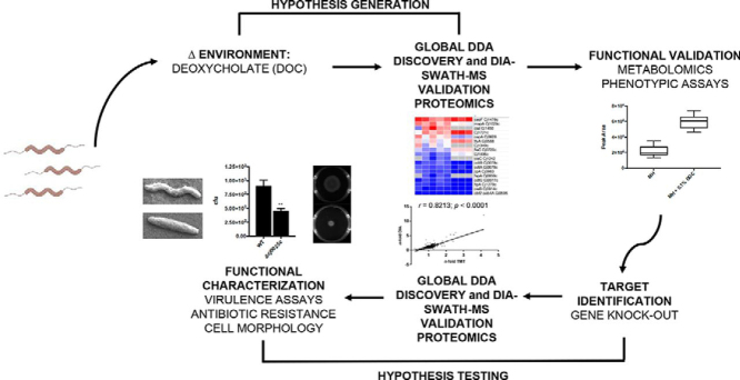
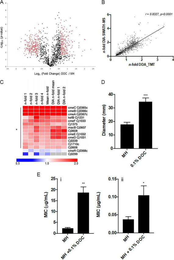
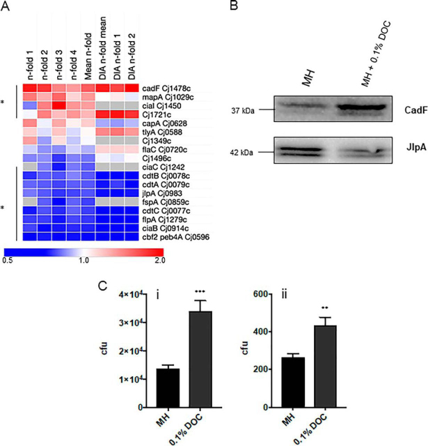
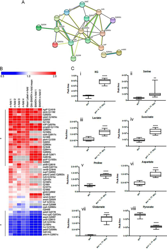
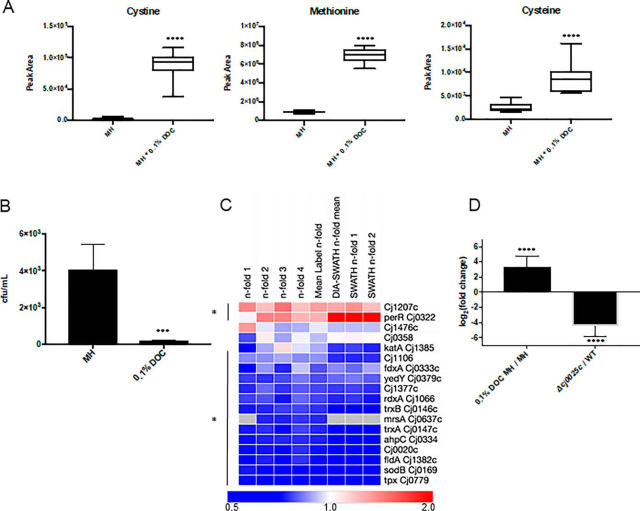
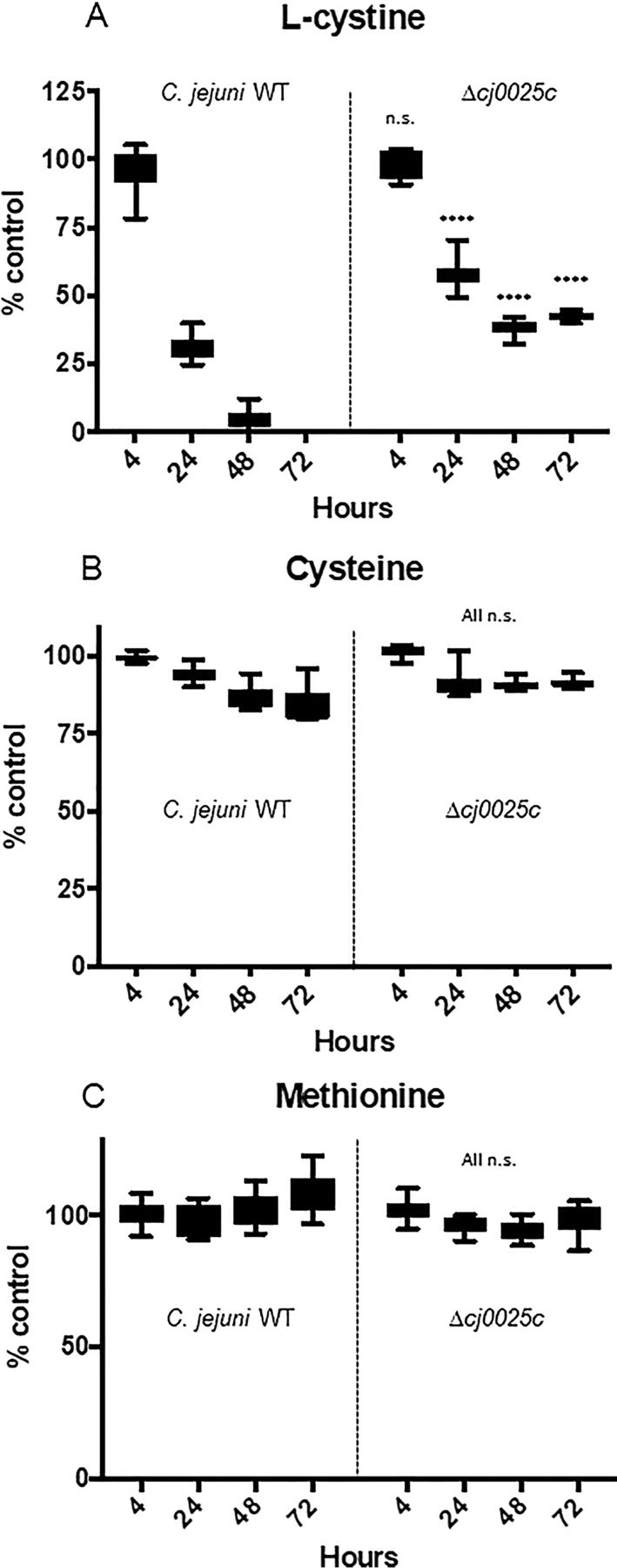
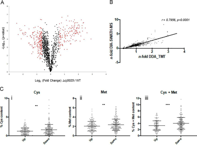
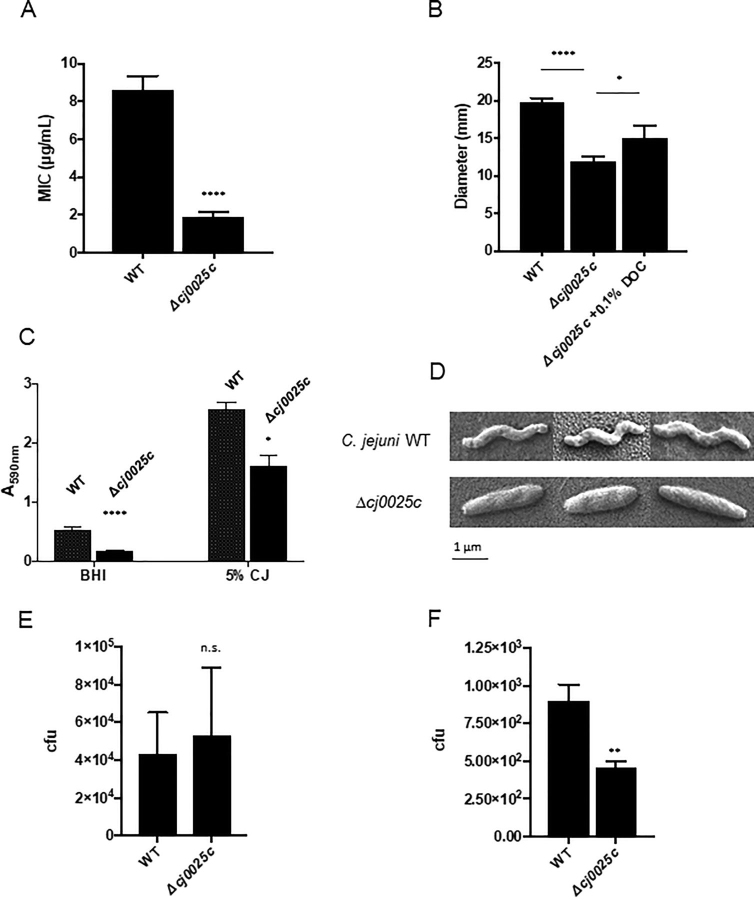
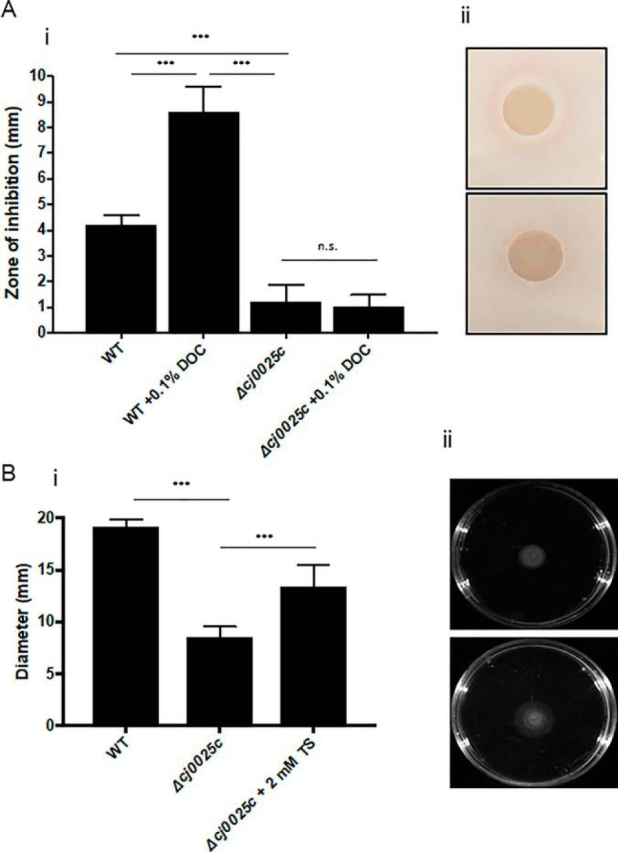
Campylobacter jejuni is a major cause of food-borne gastroenteritis. Proteomics by label-based two-dimensional liquid chromatography coupled to tandem mass spectrometry (LC-MS/MS) identified proteins associated with growth in 0.1% sodium deoxycholate (DOC, a component of gut bile salts), and system-wide validation was performed by data-independent acquisition (DIA-SWATH-MS). LC-MS/MS quantified 1326 proteins (∼82% of the predicted C. jejuni proteome), of which 1104 were validated in additional biological replicates by DIA-SWATH-MS. DOC resulted in a profound proteome shift with 512 proteins showing significantly altered abundance. Induced proteins were associated with flagellar motility and antibiotic resistance; and these correlated with increased DOC motility and resistance to polymyxin B and ciprofloxacin. DOC also increased human Caco-2 cell adherence and invasion. Abundances of proteins involved in nutrient transport were altered by DOC and aligned with intracellular changes to their respective carbon sources. DOC increased intracellular levels of sulfur-containing amino acids (cysteine and methionine) and the dipeptide cystine (Cys-Cys), which also correlated with reduced resistance to oxidative stress. A DOC induced transport protein was Cj0025c, which has sequence similarity to bacterial Cys-Cys transporters. Deletion of cj0025ccj0025c) resulted in proteome changes consistent with sulfur starvation, as well as attenuated invasion, reduced motility, atypical morphology, increased antimicrobial susceptibility and poor biofilm formation. Targeted metabolomics showed Δcj0025c could use known C. jejuni amino and organic acid substrates commensurate with wild-type. Medium Cys-Cys levels however, were maintained in Δcj0025c relative to wild-type. A toxic Cys-Cys mimic (selenocystine) inhibited wild-type growth, but not Δcj0025c Provision of an alternate sulfur source (2 mm thiosulfate) restored Δcj0025c motility. Our data confirm that Cj0025c is a Cys-Cys transporter that we have named TcyP consistent with the nomenclature of homologous proteins in other species.

Keywords: Bacteria; Bacterial pathogenesis; Campylobacter jejuni; Cystine; Mass Spectrometry; Metabolomics; Nutrient transport; Pathogens; Sulfur; Virulence.


Conflict of interest statement

Conflict of interest—Authors declare no competing interests.

Figures

None
Graphical abstract
Fig. 1
Fig. 1
DOC induces a large proteome shift in C. jejuni NCTC11168 and influences phenotypes associated with virulence.A, Volcano plot for cells grown in 0.1% DOC compared with control; x axis represents averaged Log2(DOC/control), y axis represents log10(p value). Significantly differentially abundant proteins are highlighted in red (p < 0.05); B, Correlation plot based on n-fold changes observed in the discovery set (n-fold DDA_TMT) and validation set (n-fold DIA-SWATH-MS), Pearson correlation r = 0.8213 (p < 0.0001) determined from 1104 aligned proteins; C, Heat map of C. jejuni proteins associated with antibiotic resistance and efflux ordered by largest mean n-fold change (label-based discovery; top). Data from each of 4 label-based replicates (n-fold 1–4) and DIA-SWATH-MS validation (‘DIA’ mean of 2 biological replicates [DIA n-fold 1–2]) are shown. Values are gray where the protein was not identified by DIA-SWATH-MS (* denotes proteins significantly altered in abundance); D, Increased motility associated with growth in 0.1% DOC based on colony diameter in mm on semi-solid agar (*** p < 0.001); E, Antibiotic resistance assays for (i) polymyxin B, (ii) ciprofloxacin (** p < 0.005, * p < 0.05).
Fig. 2
Fig. 2
C. jejuni NCTC11168 growth in DOC influences abundance of virulence-associated proteins and virulence phenotypes.A, Heat map of known C. jejuni virulence-associated proteins ordered by largest mean n-fold change (label-based discovery; top). Data from each of 4 label-based replicates (n-fold 1–4) and DIA-SWATH-MS validation (“DIA” mean of 2 biological replicates [DIA n-fold 1–2]) are shown. Values are gray where the protein was not identified in a biological replicate and/or by DIA-SWATH MS (* denotes proteins significantly altered in abundance); B, Western blotting using (upper) anti-CadF and (lower) anti-JlpA serum confirming changes in their DOC abundance (JlpA appears as two major bands as it contains a single glycosylation site in NCTC11168 (addition of 1406 Da (18)) (loading controls are shown in Suppl. Fig. S3); C, (i) Adherence to and (ii) invasion of Caco-2 cells for C. jejuni passaged in 0.1% DOC (*** p < 0.001, ** p < 0.005).
Fig. 3
Fig. 3
C. jejuni NCTC11168 growth in DOC is associated with changes in nutrient acquisition proteins that correlate with abundance of intracellular nutrients.A, STRINGdb cluster of nutrient transport proteins significantly elevated in abundance during growth in 0.1% DOC; B, Heat map of C. jejuni nutrient transport proteins ordered by largest mean n-fold change (label-based discovery; top). Data from each of 4 label-based replicates (n-fold 1–4) and DIA-SWATH-MS validation (“DIA” mean of 2 biological replicates [DIA n-fold 1–2]) are shown. Values are gray where the protein was not identified in a biological replicate and/or by DIA-SWATH MS (* denotes proteins significantly altered in abundance); C, (i-viii) Comparative intracellular abundances of known C. jejuni carbon sources following growth in MH medium with or without 0.1% DOC and measured by targeted LC-MS/MS metabolomics (**** p < 0.0001, ** p < 0.005).
Fig. 4
Fig. 4
Increased intracellular sulfur-containing amino acids in C. jejuni NCTC11168 grown in DOC correlate with reduced resistance to oxidative stress and increased abundance of the Cj0025c putative sodium:dicarboxylate transporter.A, Comparative intracellular abundances of sulfur-containing amino acids in MH medium with or without 0.1% DOC measured by targeted LC-MS/MS metabolomics (**** p < 0.0001); B, Growth in 0.1% DOC reduces resistance against oxidative stress (cells were exposed to 5 mm H2O2 for 30 min, serial dilutions were plated onto MH agar for CFU enumeration; *** p < 0.001); C, Heat map of C. jejuni antioxidant proteins ordered by largest mean n-fold change (label-based discovery; top). Data from each of 4 label-based replicates (n-fold 1–4) and DIA-SWATH-MS validation (“DIA” mean of 2 biological replicates [DIA n-fold 1–2]) are shown. Values are gray where the protein was not identified in a biological replicate and/or by DIA-SWATH MS (* denotes proteins significantly altered in abundance); D, qPCR showing increased cj0025c gene expression in 0.1% DOC and removal of cj0025c expression in Δcj0025c C. jejuni.
Fig. 5
Fig. 5
Culture supernatant levels of sulfur-containing amino acids for C. jejuni NCTC11168 WT and Δcj0025c grown in MCLMAN medium across 0–72 h growth. (A) l-cystine; (B) cysteine; (C) methionine (**** p < 0.0001, n.s. not significant). Data expressed at each time point as a % of the uninoculated control (% control). Uninoculated controls were incubated identically and measurements for each amino acid taken at the same time-points, no significant changes were observed (data not shown).
Fig. 6
Fig. 6
Deletion of cj0025c alters the C. jejuni NCTC11168 proteome consistent with sulfur starvation.A, Volcano plot for Δcj0025c compared with WT C. jejuni; x axis represents averaged Log2cj0025c/WT), y axis represents log10(p value). Significantly differentially abundant proteins are highlighted in red (p < 0.05); B, Correlation plot based on n-fold changes observed in the discovery set (n-fold DDA_TMT) and validation set (n-fold DIA-SWATH-MS), Pearson correlation r = 0.7956 (p < 0.0001) determined from 983 aligned proteins; C, % sulfur-containing amino acid content for proteins significantly increased (Up) and decreased (Down) in abundance in Δcj0025c compared with WT; (i) cysteine, (ii) methionine, (iii) cysteine + methionine (** p < 0.005, *** p < 0.001).
Fig. 7
Fig. 7
Deletion of cj0025c results in altered C. jejuni NCTC11168 phenotypes associated with virulence.A, Polymyxin B resistance assay (**** p < 0.0001); B, Motility assay based on colony diameter in mm on semi-solid agar (**** p < 0.0001, * p < 0.05); C, Biofilm assays in (left) BHI medium and (right) BHI medium supplemented with 5% chicken exudate (‘juice’ [CJ]; **** p < 0.0001, * p < 0.05); D, Cell morphology assayed by S.E.; E, C. jejuni Caco-2 cell adherence and (F) invasion (** p < 0.005, n.s. not significant).
Fig. 8
Fig. 8
Cj0025c is a TcyP-like Cys-Cys transport protein.A, Selenocystine inhibition assay (i) Se-Cys-Cys inhibits WT C. jejuni and 0.1% DOC increases inhibition consistent with increased abundance of Cj0025c under 0.1% DOC growth (Fig. 3B), whereas Δcj0025c is only slightly inhibited and 0.1% DOC does not influence this phenotype (*** p < 0.001, n.s. not significant), (ii) representative Se-Cys-Cys inhibition plates (upper) C. jejuni WT and (lower) Δcj0025c; B, Provision of an alternative sulfur source (2 mm thiosulfate [TS]) (i) restores Δcj0025c motility toward WT levels, (ii) representative motility plate assay (upper) Δcj0025c, (lower) Δcj0025c + 2 mm TS (*** p < 0.001).

Similar articles

Cited by

References

    1. Epps S.V.R., Harvey R.B., Hume M.E., Phillips T.D., Anderson R.C., Nisbet D.J. Foodborne Campylobacter: infections, metabolism, pathogenesis and reservoirs. Int. J. Environ. Res. Public Health. 2013;10:6292–6304. - PMC - PubMed
    1. Hermans D., Pasmans F., Heyndrickx M., Van Immerseel F., Martel A., Van Deun K., Haesebrouck F. A tolerogenic mucosal immune response leads to persistent Campylobacter jejuni colonization in the chicken gut. Crit. Rev. Microbiol. 2012;38:17–29. - PubMed
    1. Humphrey S., Chaloner G., Kemmett K., Davidson N., Williams N., Kipar A., Humphrey T., Wigley P. Campylobacter jejuni is not merely a commensal in commercial broiler chickens and affects bird welfare. mBio. 2014;5:e01364–14. - PMC - PubMed
    1. Awad W.A., Hess C., Hess M. Re-thinking chicken-Campylobacter jejuni interaction: a review. Avian Pathol. 2018;47:352–363. - PubMed
    1. Black R.E., Levine M.M., Clements M.L., Hughes T.P., Blaser M.J. Experimental Campylobacter jejuni infection in humans. J. Infect. Dis. 1988;157:472–479. - PubMed

Uncited reference

    1. Neal-McKinney J.M., Konkel M.E. The Campylobacter jejuni CiaC virulence protein is secreted from the flagellum and delivered to the cytosol of host cells. Front. Cell. Infect. Microbiol. 2012;20:31. - PMC - PubMed

Publication types

MeSH terms