Multicenter Study
. 2020 Jul 15;26(14):3662-3670.
doi: 10.1158/1078-0432.CCR-20-0073. Epub 2020 May 6.

Medium-throughput Drug Screening of Patient-derived Organoids from Colorectal Peritoneal Metastases to Direct Personalized Therapy

Affiliations
Multicenter Study

Medium-throughput Drug Screening of Patient-derived Organoids from Colorectal Peritoneal Metastases to Direct Personalized Therapy

Vignesh Narasimhan et al. Clin Cancer Res. .

Abstract

Purpose: Patients with colorectal cancer with peritoneal metastases (CRPMs) have limited treatment options and the lowest colorectal cancer survival rates. We aimed to determine whether organoid testing could help guide precision treatment for patients with CRPMs, as the clinical utility of prospective, functional drug screening including nonstandard agents is unknown.

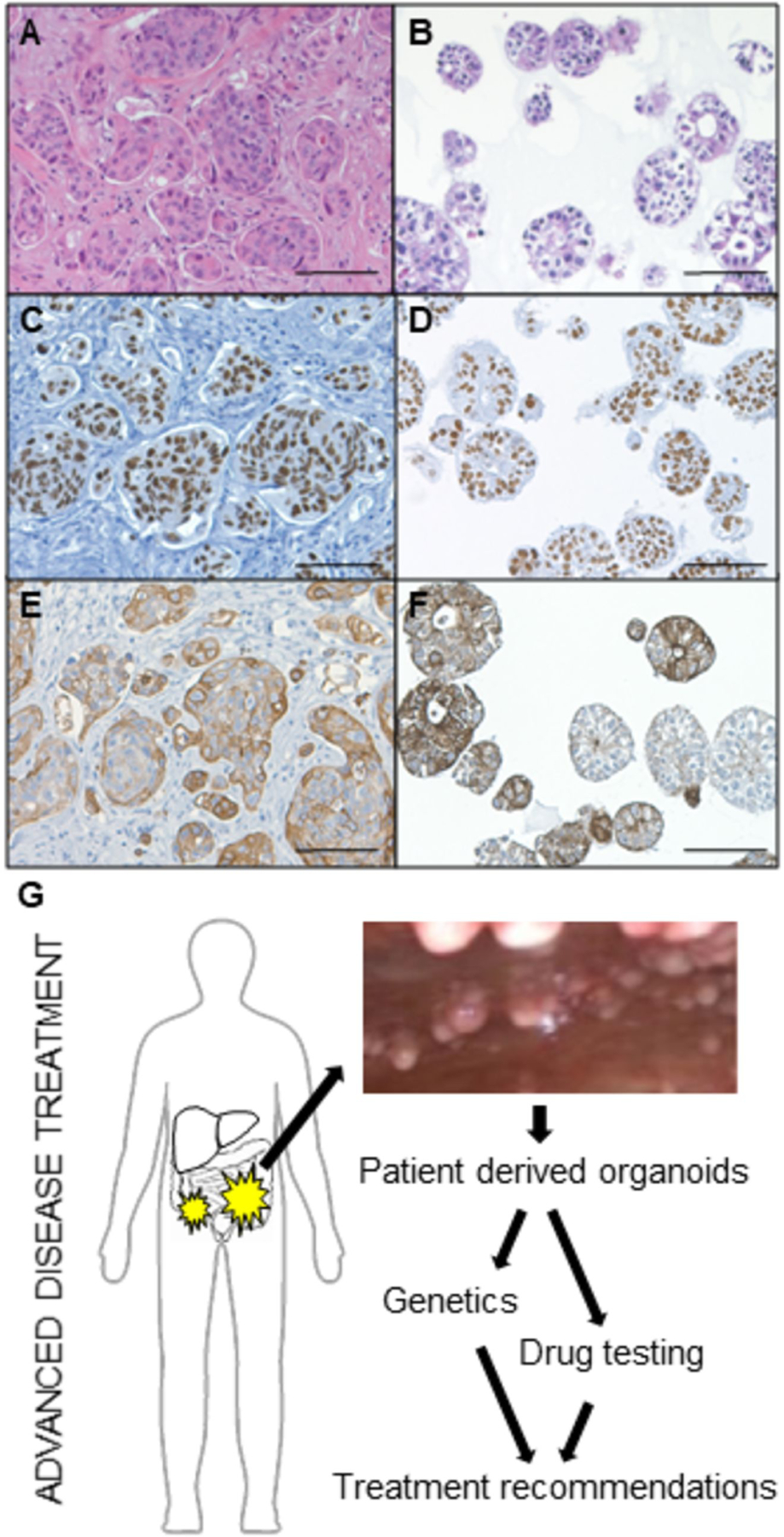
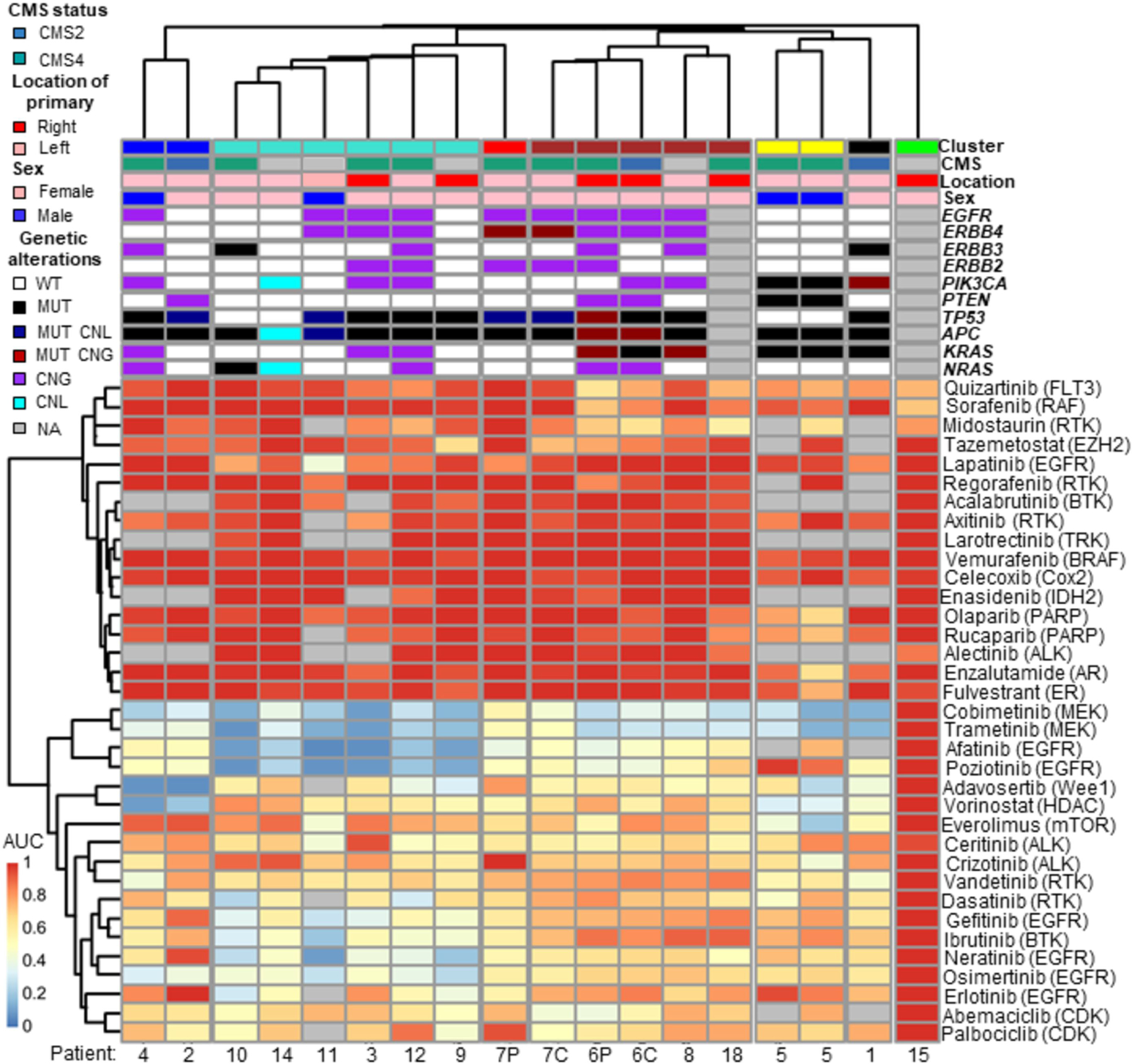
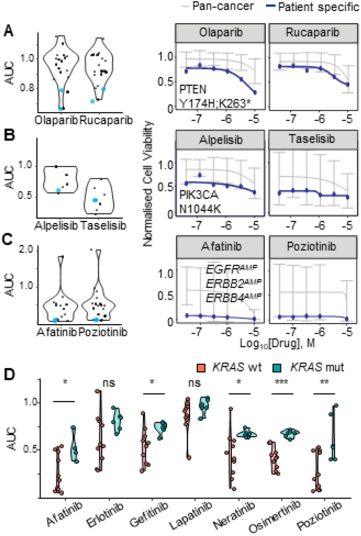
Experimental design: CRPM organoids (peritonoids) isolated from patients underwent parallel next-generation sequencing and medium-throughput drug panel testing ex vivo to identify specific drug sensitivities for each patient. We measured the utility of such a service including: success of peritonoid generation, time to cultivate peritonoids, reproducibility of the medium-throughput drug testing, and documented changes to clinical therapy as a result of the testing.

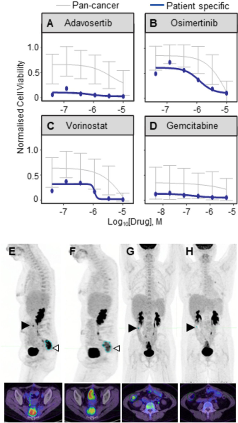
Results: Peritonoids were successfully generated and validated from 68% (19/28) of patients undergoing standard care. Genomic and drug profiling was completed within 8 weeks and a formal report ranking drug sensitivities was provided to the medical oncology team upon failure of standard care treatment. This resulted in a treatment change for two patients, one of whom had a partial response despite previously progressing on multiple rounds of standard care chemotherapy. The barrier to implementing this technology in Australia is the need for drug access and funding for off-label indications.

Conclusions: Our approach is feasible, reproducible, and can guide novel therapeutic choices in this poor prognosis cohort, where new treatment options are urgently needed. This platform is relevant to many solid organ malignancies.


Conflict of interest statement

Potential conflicts of interest Outside of the research in this manuscript, TP reports grants from Amgen; MM from Ipsen Australia Pty Ltd, Merck Serono Australia and Amgen Australia Pty Ltd. RGR receives research support from Merck Serono Australia, Invion, Australia and Fisher and Paykel Healthcare, New Zealand. CG has patents pending regarding systems and methods for personalised cancer treatment and drug development. All other authors declare no competing interests.

Figures

Figure 1.
Figure 1.. Peritonoids are representative of the human tumors from which they are derived.
Peritonoids (B, D, F) resemble the tumor cell morphology of the native tumor (A, C, E) and express intestinal epithelial markers (A-B H&E, C-D CDX2, E-F CK20). Scale bar 100μm. (G) Schema depicting our precision medicine screening platform to guide patient-specific treatment for patients with worst prognosis CRC. Peritonoids and peripheral blood mononuclear cells undergo next-generation whole exome sequencing (WES) to identify genetic alterations found in the tumor and germline of each patient. This is combined with medium-throughput drug panel testing to identify specific drug sensitivities for each patient. Results are presented to the medical oncology team to provide treatment change options should patients exhaust standard care chemotherapy.
Figure 2.
Figure 2.. Medium-throughput drug screening of peritonoids reveals shared and specific drug sensitivities to targeted agents.
Clinical and molecular features summarised (top), non-supervised clustering of normalised dose response AUC data from ex vivo medium-throughput drug testing of peritonoids depicted below (red 100% viable cells to blue 0% viable cells, normalised to vehicle alone). Matched samples: C, tumoroid derived from primary colorectal cancer; P, peritonoid derived from CRPM. CMS, consensus molecular subtype, WT, wild type; MUT, mutant; CNG, copy number gain; CNL, copy number loss; NA, not available
Figure 3.
Figure 3.. Concordant patient specific genomic alterations and peritonoid drug responses.
Peritonoid dose response AUC data displayed as a violin plot for inhibitors of: (A) PARP; (B) PI3K; (C-D) EGFR. (A-C) Blue data points indicate the peritonoid with dose response curves displayed on right: (A) Patient 5 PTEN Y174H,K263; (B) Patient 1 PIK3CA N1044K; (C) Patient 3 EGFRAMP, ERBB2AMP, ERBB4AMP. Blue line on dose response curves is patient specific response, grey line indicates average response for previously screened cancer organoid and cell lines. AUC, area under the curve, AMP, copy number gain.
Figure 4.
Figure 4.. Peritonoid-guided drug choice for chemo-refractory disease.
Disease in 43 year old patient 2 worsened despite standard care surgery and 5 rounds of chemo(radio)therapy, including EGFRi treatment. The medical oncology team considered candidate alternate therapies based on peritonoid testing results. Peritonoid dose response curves (in blue) for (A) Wee1 inhibitor Adavosertib, (B) Osimertinib, (C) Vorinostat and (D) Gemcitabine. In grey is average cell viability from prior SEngine testing of cancer cell and organoid lines. Error bars denote st dev. FDG-PET-CT scan of patient immediately prior to treatment change (E, G) and 3 months after change to Gemcitabine (F, H). (E, F) Sagittal and (G, H) frontal images. Position of the transverse axial slice (bottom) is indicated by open arrowhead in top image. Pelvic hotspot marked in blue (E, F) has reduced from 15 to 9.7 and right abdomen lesion (closed arrow, serosal deposit on bowel) is no longer visible.

References

    1. Siegel R, Miller K, Jemal A. Cancer statistics, 2019. CA Cancer J Clin 2019;69(1):7–34 doi 10.3322/caac.21551. - DOI - PubMed
    1. Simkens GA, Rovers KP, Nienhuijs SW, de Hingh IH. Patient selection for cytoreductive surgery and HIPEC for the treatment of peritoneal metastases from colorectal cancer. Cancer Manag Res 2017;9:259–66 doi 10.2147/CMAR.S119569. - DOI - PMC - PubMed
    1. Franko J, Shi Q, Goldman C, Pockaj B, Nelson G, Goldberg R, et al.Treatment of colorectal peritoneal carcinomatosis with systemic chemotherapy: a pooled analysis of north central cancer treatment group phase III trials N9741 and N9841. J Clin Oncol 2012;30(3):263–7 doi 10.1200/JCO.2011.37.1039. - DOI - PMC - PubMed
    1. Franko J, Shi Q, Meyers JP, Maughan TS, Adams RA, Seymour MT, et al.Prognosis of patients with peritoneal metastatic colorectal cancer given systemic therapy: an analysis of individual patient data from prospective randomised trials from the Analysis and Research in Cancers of the Digestive System (ARCAD) database. Lancet Oncol 2016;17(12):1709–19 doi 10.1016/S1470-2045(16)30500-9. - DOI - PubMed
    1. Sugarbaker PH. Colorectal cancer: prevention and management of metastatic disease. Biomed Res Int 2014;2014:782890 doi 10.1155/2014/782890. - DOI - PMC - PubMed

Publication types

MeSH terms

Substances