. 2020 Jun 26;295(26):8746-8758.
doi: 10.1074/jbc.RA119.012339. Epub 2020 May 6.

A C3-specific nanobody that blocks all three activation pathways in the human and murine complement system

Affiliations

A C3-specific nanobody that blocks all three activation pathways in the human and murine complement system

Henrik Pedersen et al. J Biol Chem. .

Abstract

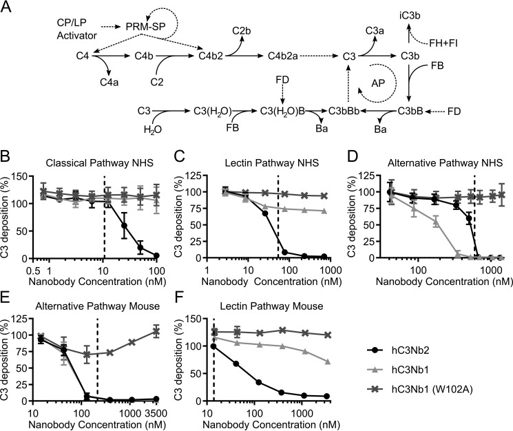
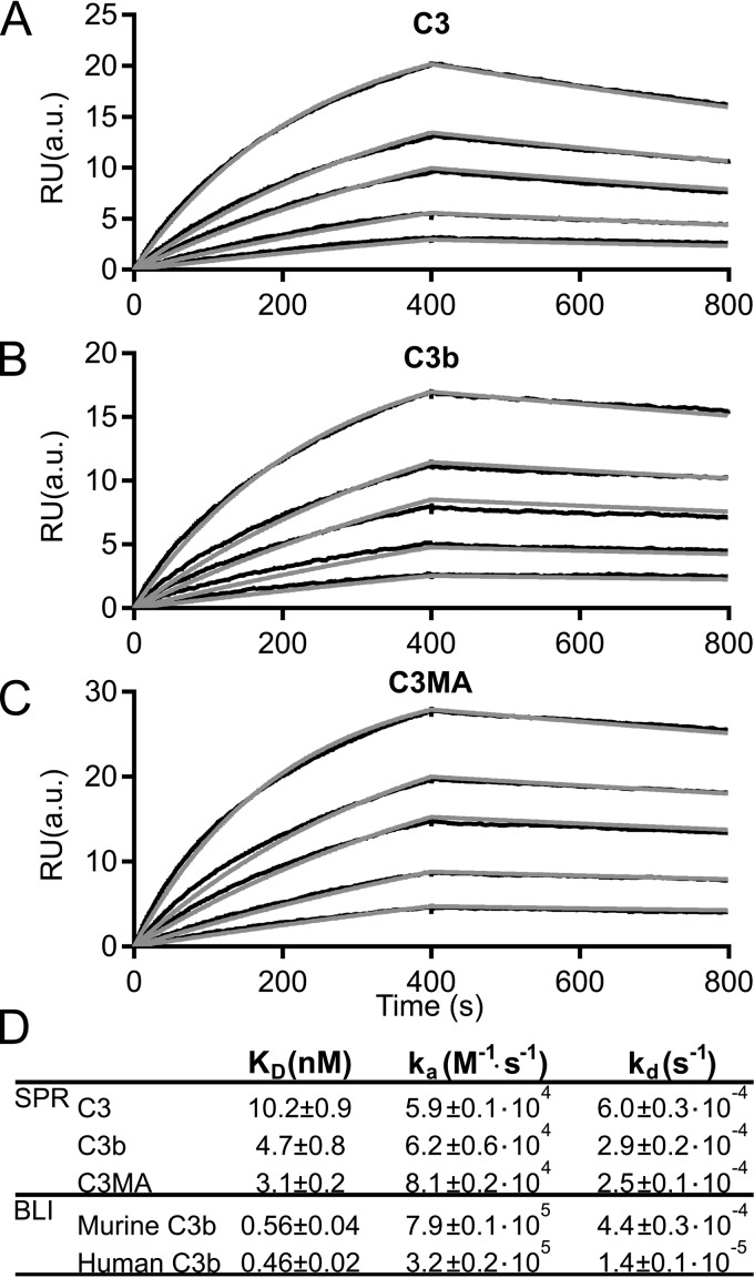
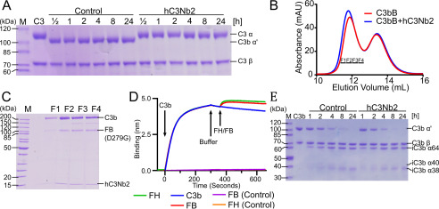
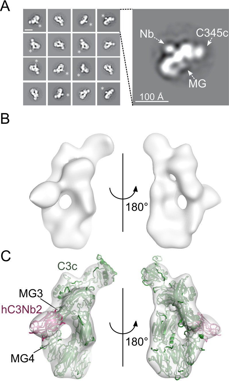
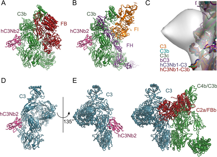
The complement system is a tightly controlled proteolytic cascade in the innate immune system, which tags intruding pathogens and dying host cells for clearance. An essential protein in this process is complement component C3. Uncontrolled complement activation has been implicated in several human diseases and disorders and has spurred the development of therapeutic approaches that modulate the complement system. Here, using purified proteins and several biochemical assays and surface plasmon resonance, we report that our nanobody, hC3Nb2, inhibits C3 deposition by all complement pathways. We observe that the hC3Nb2 nanobody binds human native C3 and its degradation products with low nanomolar affinity and does not interfere with the endogenous regulation of C3b deposition mediated by Factors H and I. Using negative stain EM analysis and functional assays, we demonstrate that hC3Nb2 inhibits the substrate-convertase interaction by binding to the MG3 and MG4 domains of C3 and C3b. Furthermore, we notice that hC3Nb2 is cross-reactive and inhibits the lectin and alternative pathway in murine serum. We conclude that hC3Nb2 is a potent, general, and versatile inhibitor of the human and murine complement cascades. Its cross-reactivity suggests that this nanobody may be valuable for analysis of complement activation within animal models of both acute and chronic diseases.

Keywords: C3 convertase; antibody; complement system; inhibitor; innate immunity; nanobody; single-domain antibody; single-domain antibody (sdAb nanobody); structural biology.


Conflict of interest statement

Conflict of interest—G. R. A. has a collaboration with Alexion pharmaceuticals.

Figures

Figure 1.
Figure 1.
The hC3Nb2 nanobody inhibits all complement activation pathways in human and mouse serum. A, the activation pathways of complement. PRMs of the CP and LP bind the activating surface. This binding activates the PRM-associated serine proteases (PRM-SP) that cleave C4 and the C2 in the proconvertase C4b2. This results in generation of the CP C3 convertase, C4b2a. This convertase cleaves C3 into C3a and C3b, and C3b may then associate with FB. FD cleaves the C3b-bound FB, resulting in the formation of the AP C3 convertase, C3bBb. C3 can spontaneously hydrolyze, and the resulting hydrolysis product C3(H2O) is a functional homolog of C3b and can associate with FB to, upon activation by FD, form the fluid-phase AP C3 convertase, C3(H2O)Bb. Dashed lines, proteolytic activation. Solid lines, conversions. B, hC3Nb2 is a potent classical pathway inhibitor of C3 fragment deposition onto a surface of heat-aggregated IgG in 0.2% NHS. C, as in B but on a mannan surface, showing that hC3Nb2 also inhibits C3 fragment deposition through the lectin pathway in 5% NHS. D, hC3Nb2 likewise suppresses C3 fragment deposition by the alternative pathway onto a surface of zymosan in 11% NHS. E, inhibition by nanobodies of the alternative pathway in 5% mouse serum (i.e. C3 fragment deposition onto zymosan-coated surfaces). F, inhibition of the LP (i.e. deposition onto a mannan surface) in 0.3% mouse serum. B–F display the C3 deposition at the indicated nanobody concentrations. The C3 deposition was normalized to the C3 deposition obtained without added nanobodies (100% deposition). The effect of hC3Nb2 (black) was compared with the strong alternative pathway inhibitor hC3Nb1 (light gray) and the inactive hC3Nb1 (W102A) mutant (dark gray). The data for hC3Nb1 and hC3Nb1 (W102A) in C and E were published previously (20). Dashed lines, final C3 concentration at the given serum dilution, assuming a C3 concentration of 5.3 µm in undiluted human serum (55) and 4.2 µm in undiluted mouse serum (56). Average and S.D. (error bars) are shown for n = 3 experiments in B and D and n = 2 experiments in C, E, and F.
Figure 2.
Figure 2.
The hC3Nb2 inhibits classical pathway-mediated hemolysis. The classical pathway was activated on sheep erythrocytes by anti-sheep erythrocyte antibodies. The activated sheep erythrocytes were incubated in 7.5% NHS (A) or FB-depleted serum (B). The effects of hC3Nb2 (black) were compared with those of the alternative pathway inhibitor hC3Nb1 (light gray) and its inactive hC3Nb1 (W102A) mutant (dark gray). Lysis, as measured as absorption at 405 nm in the supernatants, was normalized to lysis by H2O (100%), whereas erythrocytes incubated in PBS were defined as 0% lysis. Dashed lines, putative C3 concentration in 7.5% serum. Average and S.D. (error bars) are shown for n = 3 experiments in A and n = 2 experiments in B.
Figure 3.
Figure 3.
The hC3Nb2 binds multiple functional states of C3 with nanomolar affinity. hC3Nb2 was immobilized through a biotinylated C-terminal AviTag on a streptavidin-coated surface plasmon resonance flow sensor. The analyte was injected over the hC3Nb2-coated sensor at concentrations of 5, 10, 20, 30, or 60 nm C3 (A), C3b (B), or C3MA (C). D, summary of the SPR and BLI binding and rate constants. Binding and rate constants from SPR were determined using the BiaCore T200 evaluation software; n = 3 for C3 and C3b, n = 2 for C3MA. BLI binding curves are presented in Fig. S1. Binding and rate constants from BLI-based experiments were determined as described under “Experimental procedures”; n = 2 for murine C3b, n = 2 for human C3b. RU, response units. a.u., arbitrary units.
Figure 4.
Figure 4.
The hC3Nb2 nanobody inhibits C3 cleavage but allows endogenous regulation by FI degradation. A, time course experiment where C3 degradation by CVFBb at 37 °C was monitored by SDS-PAGE in the presence or absence of hC3Nb2. Nanobody binding to C3 completely prevents cleavage revealing inhibition at the substrate level. B and C, proconvertase assembly assay. C3b was mixed with a 1.5-fold molar excess of the inactive, stabilized FB (D279G/S699A) and subjected to size-exclusion chromatography in either the presence or absence of a 2-fold molar excess of hC3Nb2. Nonreduced SDS-PAGE analysis of the fractions from B marked by bars reveals that FB and hC3Nb2 do not compete for binding to C3b. D, competition assay where immobilized hC3Nb2 on BLI sensors was dipped in 50 nm C3b. The sensors were subsequently transferred to FB (D279G) or FH. In addition, the binding of FB (D279G) and FH alone were analyzed for nonspecific background binding. Sensorgrams for FB (D279G) and FH are indistinguishable. E, a FH-mediated FI cleavage assay reveals that hC3Nb2 does not interfere with C3b degradation. The C3b was incubated with 0.2% (w/w) FH and 1% (w/w) FI for the indicated time at 37 °C in either the presence or absence of a 1.2-fold molar excess of hC3Nb2.
Figure 5.
Figure 5.
Mapping of the hC3Nb2 binding site on C3c. A, negative stain EM 2D class averages of the hC3Nb2:C3c complex. *, extra density, which was ascribed to the hC3Nb2 in the 2D class averages. In the expanded view of one 2D class to the right, the MG ring and the C-terminal C345c domain are marked by arrows, whereas the likely location of hC3Nb2 is marked Nb. The scale bar in the top left 2D class is 100 Å. B, a 3D EM reconstruction based on the hC3Nb2:C3c particles. C, fitting of the C3c (PDB entry 2A74) and the hC3Nb1 (PDB entry 6EHG) into the 3D reconstruction. The locations of the MG3 and MG4 domains are marked.
Figure 6.
Figure 6.
A structure-based model for the inhibitory mechanism. A docking of hC3Nb2 onto the structures of C3bB (PDB entry 2XWJ) (A) and C3b:miniFH:FI (PDB entry 5O32) (B) using the hC3Nb2:C3c envelope is consistent with the interpretation that hC3Nb2 does not compete with either FB or FH. C, a superposition of the known structures at the MG3-MG4 interface suggests that the epitope of hC3Nb2 is conserved in C3 (PDB entries 2A73 and 2B39), C3b (PDB entry 5FO7), C3c (PDB entry 2A74), hC3Nb1:C3 (PDB entry 6RU5), and hC3Nb1:C3b (PDB entry 6EHG). D, mapping of hC3Nb2 onto native C3 (PDB entry 2A73) by comparison with C3c. E, a comparison between the model of the hC3Nb2:C3 complex in D and the general model of the convertase-substrate complexes implies that hC3Nb2 inhibits C3 cleavage by all convertases by preventing substrate recognition.

References

    1. Merle N. S., Noe R., Halbwachs-Mecarelli L., Fremeaux-Bacchi V., and Roumenina L. T. (2015) Complement system part II: role in immunity. Front. Immunol. 6, 257 10.3389/fimmu.2015.00257 - DOI - PMC - PubMed
    1. Stephan A. H., Barres B. A., and Stevens B. (2012) The complement system: an unexpected role in synaptic pruning during development and disease. Annu. Rev. Neurosci. 35, 369–389 10.1146/annurev-neuro-061010-113810 - DOI - PubMed
    1. Kojouharova M., Reid K., and Gadjeva M. (2010) New insights into the molecular mechanisms of classical complement activation. Mol. Immunol. 47, 2154–2160 10.1016/j.molimm.2010.05.011 - DOI - PubMed
    1. Kjaer T. R., Thiel S., and Andersen G. R. (2013) Toward a structure-based comprehension of the lectin pathway of complement. Mol. Immunol. 56, 222–231 10.1016/j.molimm.2013.05.220 - DOI - PubMed
    1. Bajic G., Degn S. E., Thiel S., and Andersen G. R. (2015) Complement activation, regulation, and molecular basis for complement-related diseases. EMBO J. 34, 2735–2757 10.15252/embj.201591881 - DOI - PMC - PubMed

Publication types