. 2020 Jun 19;295(25):8575-8588.
doi: 10.1074/jbc.RA119.011979. Epub 2020 May 6.

The post-synaptic scaffolding protein tamalin regulates ligand-mediated trafficking of metabotropic glutamate receptors

Affiliations

The post-synaptic scaffolding protein tamalin regulates ligand-mediated trafficking of metabotropic glutamate receptors

Saurabh Pandey et al. J Biol Chem. .

Abstract

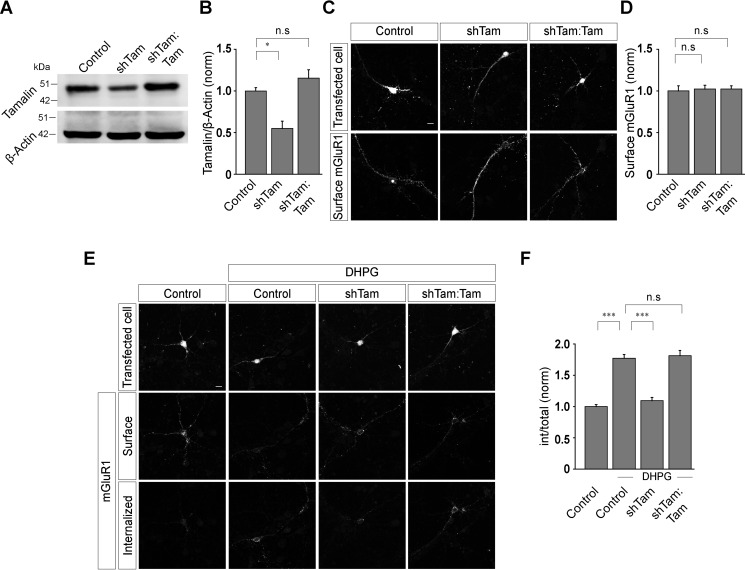
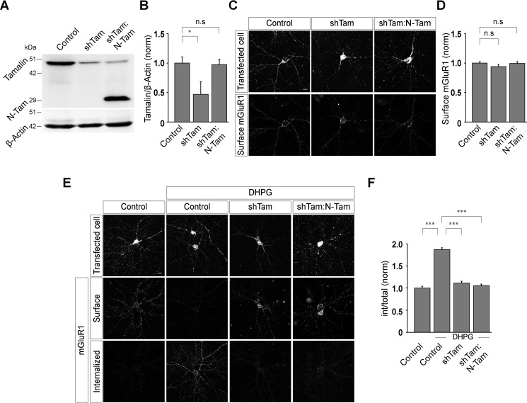
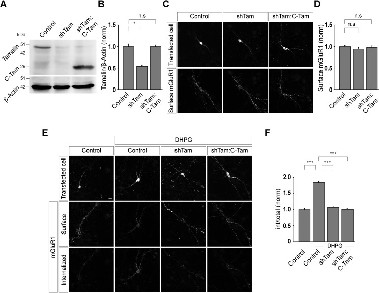
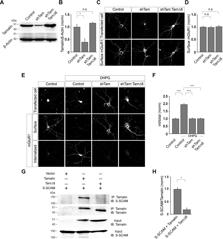
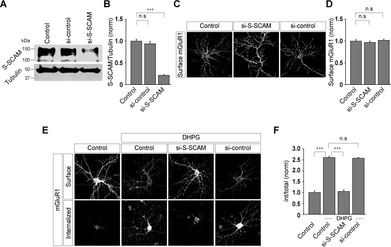
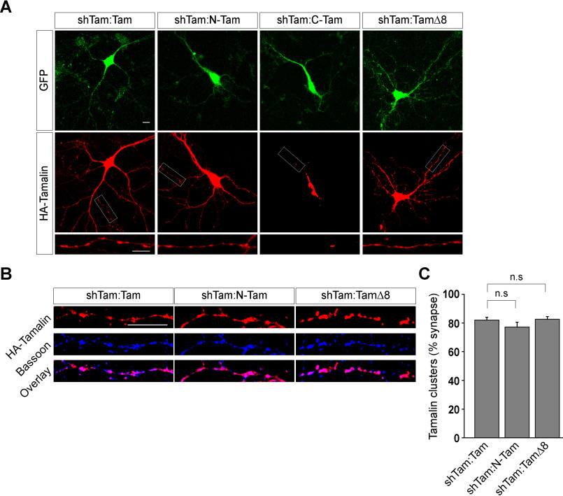
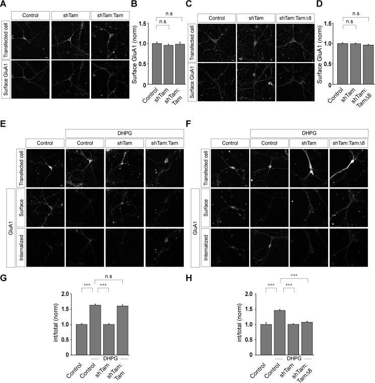
Group I metabotropic glutamate receptors (mGluRs) play important roles in various neuronal functions and have also been implicated in multiple neuropsychiatric disorders like fragile X syndrome, autism, and others. mGluR trafficking not only plays important roles in controlling the spatiotemporal localization of these receptors in the cell but also regulates the activity of these receptors. Despite this obvious significance, the cellular machineries that control the trafficking of group I metabotropic glutamate receptors in the central nervous system have not been studied in detail. The post-synaptic scaffolding protein tamalin has been shown to interact with group I mGluRs and also with many other proteins involved in protein trafficking in neurons. Using a molecular replacement approach in mouse hippocampal neurons, we show here that tamalin plays a critical role in the ligand-dependent internalization of mGluR1 and mGluR5, members of the group I mGluR family. Specifically, knockdown of endogenous tamalin inhibited the ligand-dependent internalization of these two receptors. Both N-terminal and C-terminal regions of tamalin played critical roles in mGluR1 endocytosis. Furthermore, we found that tamalin regulates mGluR1 internalization by interacting with S-SCAM, a protein that has been implicated in vesicular trafficking. Finally, we demonstrate that tamalin plays a critical role in mGluR-mediated internalization of α-amino-3-hydroxy-5-methyl-4-isoxazolepropionic acid receptors, a process believed to be the cellular correlate for mGluR-dependent synaptic plasticity. Taken together, these findings reveal a mechanistic role of tamalin in the trafficking of group I mGluRs and suggest its physiological implications in the brain.

Keywords: G-protein coupled receptor; S-SCAM; endocytosis; internalization; metabotropic glutamate receptors; neurotransmitter receptors; synaptic plasticity; tamalin; trafficking.


Conflict of interest statement

Conflict of interest—The authors declare that they have no conflicts of interest with the contents of this article.

Figures

Figure 1.
Figure 1.
Knockdown of endogenous tamalin inhibits the ligand-mediated endocytosis of mGluR1. A and B, Western blotting (A) and quantitation of the Western blots (B), showing the efficient knock-down of endogenous tamalin by shTam and replacement of the endogenous tamalin with full-length tamalin. C and D, Representative images (C) and quantitation of surface Myc-mGluR1 (D) showing that knockdown of endogenous tamalin with shTam and expression of the WT tamalin replacement construct had no effect on the surface expression of Myc-mGluR1 (n values: control, 35; shTam, 39; shTam:Tam, 38). E, Representative examples of surface and internalized Myc-mGluR1, 30 min after application of 100 μm R,S-DHPG in control cells, shTam-expressing cells, and shTam- and WT-tamalin-expressing cells. F, Quantitation of the endocytosis index suggested that knockdown of endogenous tamalin inhibited the R,S-DHPG-mediated internalization of Myc-mGluR1 and expression of WT tamalin rescued the normal trafficking of the receptor (n values: control, 35; control + DHPG, 36; shTam + DHPG, 38; shTam:Tam + DHPG, 39). The results are presented as means ± S.E. from three independent experiments. Scale bar, 10 μm. ***, p < 0.001; *, p < 0.05; n.s, p > 0.05.
Figure 2.
Figure 2.
N-terminal domain of tamalin alone is not sufficient for the ligand-mediated endocytosis of mGluR1. A and B, Western blotting (A) and quantitation of the Western blots (B), showing the effective knockdown of endogenous tamalin by shTam and expression of the N-Tam replacement construct. C and D, Representative images (C) and quantitation (D) showing that expression of shTam and shTam:N-Tam had no effect on the surface expression of Myc-mGluR1 (n values: control, 44; shTam, 43; shTam:N-Tam, 42). E and F, Representative cells (E) and quantitation (F) of 100 μm R,S-DHPG-induced internalization of Myc-mGluR1 suggested that knockdown of endogenous tamalin led to the inhibition of ligand-mediated endocytosis of Myc-mGluR1, and expression of the N-Tam replacement construct did not rescue the normal endocytosis of the receptor (n values: control, 42; control + DHPG, 43; shTam + DHPG, 42; shTam:N-Tam + DHPG, 44). The results are presented as means ± S.E. from three independent experiments. Scale bar, 10 μm. ***, p < 0.001; *, p < 0.05; n.s, p > 0.05.
Figure 3.
Figure 3.
The C-terminal domain of tamalin alone is not sufficient for the ligand-mediated endocytosis of mGluR1. A and B, Acute knockdown of endogenous tamalin and replacement of endogenous tamalin with C-Tam, as shown by Western blotting (A) and quantitation of the Western blots (B). C and D, Representative images (C) and quantitation (D) showing no effect on the surface expression of Myc-mGluR1 in shTam and shTam:C-Tam-expressing neurons (n values: control, 44; shTam, 43; shTam:C-Tam, 44). E and F, Representative images (E) and quantitation (F) suggested that knockdown of the endogenous tamalin inhibited the R,S-DHPG-mediated endocytosis of Myc-mGluR1, and replacement of the endogenous tamalin with C-Tam failed to rescue the normal internalization of the receptor (n values: control, 44; control + DHPG, 44; shTam + DHPG, 42; shTam:C-Tam + DHPG, 42). The results are presented as means ± S.E. from three independent experiments. Scale bar, 10 μm. ***, p < 0.001; *, p < 0.05; n.s, p > 0.05.
Figure 4.
Figure 4.
The last 8 amino acids of tamalin play a critical role in the ligand-mediated internalization of mGluR1. A and B, Western blotting (A) and quantitation of the Western blots (B), showing the knockdown of the endogenous tamalin by shTam and expression of the TamΔ8 replacement construct. C and D, Representative images (C) and quantitation (D) of the surface-localized Myc-mGluR1 suggested that acute knockdown of the endogenous tamalin by shTam and replacement of the endogenous tamalin with TamΔ8 had no effect on the surface expression of Myc-mGluR1 (n values: control, 42; shTam, 42; shTam + TamΔ8; 43). E and F, Representative cells (E) and quantitation (F) suggested that knockdown of the endogenous tamalin resulted in the inhibition of the R,S-DHPG-mediated endocytosis of Myc-mGluR1, and the TamΔ8 replacement construct did not rescue the normal internalization of the receptor (n values: control, 42; control + DHPG, 45; shTam + DHPG, 42; shTam:TamΔ8 + DHPG, 48). G, Coimmunoprecipitation assays demonstrated that deletion of the last 8 amino acids of tamalin disrupted binding of S-SCAM to tamalin. H, Quantitation of the coimmunoprecipitation assays. All the results are presented as means ± S.E. from three independent experiments. Scale bar, 10 μm. ***, p < 0.001; *, p < 0.05; n.s, p > 0.05.
Figure 5.
Figure 5.
Knockdown of S-SCAM inhibits the ligand-mediated internalization of mGluR1. A and B, Western blotting analyses (A) and quantitation of the Western blots (B), showing the efficient knockdown of endogenous S-SCAM by si-S-SCAM. C and D, Representative images (C) and quantitation (D) suggested that acute knockdown of endogenous S-SCAM had no effect on the surface expression of Myc-mGluR1 (n values: control, 36; si-S-SCAM, 35; si-control, 36). E and F, Representative cells (E) and quantitation of the endocytosis index (F) showing that knockdown of the endogenous S-SCAM led to the inhibition in the R,S-DHPG-mediated internalization of Myc-mGluR1, whereas in control cells and si-control-transfected cells, the receptor internalized normally (n values: control, 36; control + DHPG, 38; si-S-SCAM + DHPG, 37; si-control + DHPG, 37). The results are presented as means ± S.E. from three independent experiments. Scale bar, 10 μm. ***, p < 0.001; n.s, p > 0.05.
Figure 6.
Figure 6.
Synaptic localization of tamalin constructs. A, Representative images showing that WT tamalin is expressed throughout the hippocampal neuron and targeted to the dendrites. N-Tam and TamΔ8 expression was also observed throughout the hippocampal neuron, and they were also seen to be localized in the dendrites, similar to the WT tamalin. In contrast, C-Tam did not target properly to the dendrites of the neuron. B, Representative images showing colocalization of Bassoon, an active zone synaptic marker with various forms of tamalin (WT, N-Tam, and TamΔ8). All these constructs were found to colocalize with Bassoon, suggesting that they were targeted to the synapse. C, Quantitation also suggested that all the above forms of tamalin were localized at the synapse to a similar extent (n values: shTam:Tam, 34; shTam:N-Tam, 34; shTam:TamΔ8, 33). Scale bar, 10 μm. n.s, p > 0.05.
Figure 7.
Figure 7.
Tamalin regulates mGluR-mediated AMPAR endocytosis. A and B, Representative images (A) and quantitation (B) of surface AMPARs (GluA1-containing receptors) suggested that knockdown of the endogenous tamalin had no effect on the surface expression of these receptors (n values: control, 46; shTam, 45; shTam:Tam, 44). C and D, Representative images (C) and quantitation (D) of the surface AMPARs suggested that knockdown of the endogenous tamalin and replacement of the endogenous tamalin with TamΔ8 had no effect on the surface expression of the GluA1-containing receptors (n values: control, 28; shTam, 30; shTam:TamΔ8, 31). E and G, Representative images (E) and quantitation (G) of mGluR-mediated AMPAR endocytosis suggested that in control cells, the receptors internalized when they were stimulated with 100 μm R,S-DHPG for 5 min. In contrast, application of 100 μm R,S-DHPG did not cause endocytosis of GluA1-containing receptors in tamalin knockdown cells. Expression of WT tamalin rescued the R,S-DHPG-mediated endocytosis of the receptor (n values: control, 46; control + DHPG, 45; shTam + DHPG, 49; shTam:Tam + DHPG, 44). F and H, The inhibition of the mGluR-mediated AMPAR endocytosis due to the knockdown of the endogenous tamalin was not rescued by replacing the endogenous tamalin with TamΔ8, as observed from representative images (F) and quantitation (H) (n values: control, 28; control + DHPG, 24; shTam + DHPG, 30; shTam:TamΔ8 + DHPG, 27). The results are presented as means ± S.E. from three independent experiments. Scale bar, 10 μm. ***, p < 0.001; n.s, p > 0.05.

Similar articles

Cited by

References

    1. Pin J. P., and Duvoisin R. (1995) The metabotropic glutamate receptors: structure and functions. Neuropharmacology 34, 1–26 10.1016/0028-3908(94)00129-G - DOI - PubMed
    1. Dhami G. K., and Ferguson S. S. (2006) Regulation of metabotropic glutamate receptor signaling, desensitization and endocytosis. Pharmacol. Ther. 111, 260–271 10.1016/j.pharmthera.2005.01.008 - DOI - PubMed
    1. Bhattacharyya S. (2016) Inside story of group I metabotropic glutamate receptors (mGluRs). Int. J. Biochem. Cell Biol. 77, 205–212 10.1016/j.biocel.2016.03.003 - DOI - PubMed
    1. Mahato P. K., Ramsakha N., Ojha P., Gulia R., Sharma R., and Bhattacharyya S. (2018) Group I metabotropic glutamate receptors (mGluRs): ins and outs. Adv. Exp. Med. Biol. 1112, 163–175 10.1007/978-981-13-3065-0_12 - DOI - PubMed
    1. Conn P. J., and Pin J. P. (1997) Pharmacology and functions of metabotropic glutamate receptors. Annu. Rev. Pharmacol. Toxicol. 37, 205–237 10.1146/annurev.pharmtox.37.1.205 - DOI - PubMed

Publication types

MeSH terms