. 2020 Jul 3;295(27):8914-8927.
doi: 10.1074/jbc.RA119.011955. Epub 2020 May 6.

Copper stabilizes antiparallel β-sheet fibrils of the amyloid β40 (Aβ40)-Iowa variant

Affiliations

Copper stabilizes antiparallel β-sheet fibrils of the amyloid β40 (Aβ40)-Iowa variant

Elliot J Crooks et al. J Biol Chem. .

Abstract

Cerebral amyloid angiopathy (CAA) is a vascular disorder that primarily involves deposition of the 40-residue-long β-amyloid peptide (Aβ40) in and along small blood vessels of the brain. CAA is often associated with Alzheimer's disease (AD), which is characterized by amyloid plaques in the brain parenchyma enriched in the Aβ42 peptide. Several recent studies have suggested a structural origin that underlies the differences between the vascular amyloid deposits in CAA and the parenchymal plaques in AD. We previously have found that amyloid fibrils in vascular amyloid contain antiparallel β-sheet, whereas previous studies by other researchers have reported parallel β-sheet in fibrils from parenchymal amyloid. Using X-ray fluorescence microscopy, here we found that copper strongly co-localizes with vascular amyloid in human sporadic CAA and familial Iowa-type CAA brains compared with control brain blood vessels lacking amyloid deposits. We show that binding of Cu(II) ions to antiparallel fibrils can block the conversion of these fibrils to the more stable parallel, in-register conformation and enhances their ability to serve as templates for seeded growth. These results provide an explanation for how thermodynamically less stable antiparallel fibrils may form amyloid in or on cerebral vessels by using Cu(II) as a structural cofactor.

Keywords: Abeta40-Iowa; Alzheimer disease; Alzheimer's disease; Aβ40-Iowa; Fourier transform IR (FTIR); NMR spectroscopy; X-ray fluorescence microscopy (XFM); amyloid-beta (AB); cerebral amyloid angiopathy (CAA,); copper; fibril; metal ions; neurodegeneration; vascular disease.


Conflict of interest statement

Conflict of interest—The authors declare no competing financial interests.

Figures

Figure 1.
Figure 1.
X-ray fluorescence microscopy of copper localization in vascular amyloid. Thioflavin S staining (A–C) and copper XFM images (D–F) of vessels in human control, sporadic CAA, and Iowa-type familial CAA. Scale bars are 20 μm. The relative copper content (G) was determined by normalizing to the protein content at each pixel and shows that copper is significantly elevated (*, p < 0.05) over the controls using a Student's paired t test with a two-tailed distribution. The protein content at each pixel was determined by integrating the amide II protein peak using FTIR microspectroscopy (see “Materials and methods”).
Figure 2.
Figure 2.
Rapid fibril formation of Aβ40-Iowa. A, thioflavin T fluorescence of Aβ40-Iowa (100 μm) in the presence and absence of Cu(II) at 24 °C. Copper was added to monomeric Aβ40-Iowa at Cu(II):Aβ molar ratios of 1:10, 1:5, and 1:2. The thioflavin T fluorescence of Aβ40-WT in the absence of copper under the same conditions is shown for comparison (dashed line). B–D, single touch AFM images of the Aβ40-Iowa peptides after incubation of 10 (B), 30 (C), and 60 (D) min at room temperature reveal oligomers associating into protofibrils and fibrils. The scale bars for the AFM figures are 50 (B), 50 (C), and 400 nm (D).
Figure 3.
Figure 3.
Distinguishing anti-parallel and parallel fibrils. A, amide I and II regions of Aβ40-WT fibrils with (black) and without (red) LAGG 13C labeling after 3 weeks of incubation at 37 °C with strong agitation (200 rpm). The amide I region (1600-1700 cm−1) is sensitive to the secondary structure with the observed band between ∼1626 and 1634 cm−1 being characteristic of β-sheet. B, amide I and II regions of Aβ40-Iowa fibrils with (black) and without (red) LAGG 13C labeling formed at room temperature for 24 h under quiescent conditions.
Figure 4.
Figure 4.
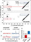
Distinguishing antiparallel and parallel fibrils. A, region of the 2D NMR spectrum of parallel Aβ40-WT fibrils. The blue box indicates the position of a cross-peak expected to arise between the [2-13C]Ala-30 and [3-13C]Ala-30 diagonal resonances. Cross-peaks marked with an asterisk are artifacts arising from MAS side bands. B, region of the 2D NMR spectrum of anti-parallel Aβ40-Iowa fibrils. The red box indicates the position of a cross-peak expected to arise between the [1-13C]Val-36 and [2-13C]Ala-30 diagonal resonances. C, rows are shown through the diagonal resonances as in A. The regions shown correspond to the colored boxes. D, rows are shown through the diagonal resonances as in B. E, integration of the cross-peaks diagnostic of parallel and anti-parallel structure. The total intensity for the regions of containing the parallel and anti-parallel cross-peaks was normalized to 1. The intensities of the parallel cross-peak (n = 2) in C, and the anti-parallel cross-peak (n = 3) in D were signficant (p < 0.05) relative to the noise using a Student's paired t test with a two-tailed distribution.
Figure 5.
Figure 5.
FTIR spectroscopy and TEM of fibril formation of Aβ40-Iowa. A, evolution of Aβ40-Iowa FTIR spectra as a function of time and temperature. B, expansion of the A. The amide I region of Aβ40-Iowa (black) is shown after incubation for 24 h at room temperature under quiescent conditions. The temperature was then increased to 37 °C and the sample was shaken at 170 rpm. FTIR spectra were obtained after 24 (orange), 48 (red), 72 (green), and 96 h (blue). These time points are designated 24 + 24 h, 24 + 48 h, 24 + 72 h, and 24 + 96 h, respectively. B is an expansion of A. C, influence of Cu(II) on the evolution of Aβ40-Iowa at 37 °C. D is an expansion of the C. Cu(II) was added after 24 h of room temperature incubation and the sample was then further incubated at 37 °C with shaking. FTIR spectra were obtained after Cu(II) addition (black), 24 + 24 h (orange), 24 + 48 h (red), 24 + 72 h (green), and 24 + 96 h (blue). TEM images obtained after 24 h of room temperature incubation (E), after 24 + 96 h without copper (F), and after 24 + 96 h with Cu(II) (G and H) show the presence of fibrils. The scale bars in (E–H) are 100 nm.
Figure 6.
Figure 6.
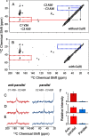
Two-dimensional 13C solid-state NMR of Aβ40-Iowa. Two-dimensional 13C DARR NMR spectrum of Aβ40-Iowa fibrils obtained after 96 h of incubation at room temperature under quiescent conditions without Cu(II) (A) and with Cu(II) (B). The fibrils were formed from an equimolar mixture of two 13C-labeled peptides; one containing [2-13C]Ala-30 and [1-13C]Val-36, and one containing [3-13C]Ala-30. The blue boxes indicate the position of cross-peaks expected to arise between the [2-13C]Ala-30 and [3-13C]Ala-30 diagonal resonances, diagnostic of parallel, in-register fibril structure, whereas the red boxes indicate the position of cross-peaks expected to arise between the [1-13C]Val-36 and [2-13C]Ala-30 diagonal resonances, diagnostic of anti-parallel fibril structure. Cross-peaks marked with an asterisk are artifacts arising from MAS side bands. C, rows are shown through the diagonal resonances of [2-13C]Ala-30 (red) and [3-13C]Ala-30 (blue) of the 2D NMR spectrum after the initial 24 h of incubation without Cu(II). These rows are duplicate experiments of those shown in Fig. 4D indicating the reproducibility of these measurements. The regions shown correspond to the colored boxes. D, rows are shown through the diagonal resonances as in A after incubation of the Aβ40-Iowa sample for 24 + 96 h. E, rows are shown through the diagonal resonances as in B after incubation of the Aβ40-Iowa sample for 24 + 96 h with Cu(II) added after the first 24 h incubation step at room temperature. F, integration of the cross-peaks for parallel and anti-parallel structure. The total intensity for the regions of containing the parallel and anti-parallel cross-peaks was normalized to 1. The intensities of the anti-parallel cross-peak (n = 3) in C, the anti-parallel and parallel cross-peaks (n = 3) in D, and the anti-parallel cross-peak (n = 2) in E were significant (p < 0.05) relative to the noise using a Student's paired t test with a two-tailed distribution.
Figure 7.
Figure 7.
Influence of Cu(II) on Aβ fibril growth from Aβ40-Iowa seeds with Aβ40-Iowa monomer. A and B, thioflavin T fluorescence of Aβ40-Iowa monomer (100 μm) in the presence of Aβ40-Iowa seeds with (A) or without (B) bound Cu(II). The seeds were added at levels of 5, 10, 15, or 20% (w/w) relative to the monomer concentration and the mixtures was incubated at room temperature under quiescent conditions. Seeded growth was observed under all conditions in A, but only with 15 and 20% seeds in B.
Figure 8.
Figure 8.
Copper binding to anti-parallel and parallel fibrils of Aβ40-Iowa. A, binding of Cu(II) by antiparallel Aβ40-Iowa fibrils. The copper-Aβ solution was prepared by adding Cu(II) in a 1:2 Cu:Aβ ratio to Aβ40-Iowa incubating at room temperature for 24 h, and then incubating the solution at 37 °C for an additional 96 h. Fibrils were pelleted by centrifugation. The amount of Cu(II) in the pellet and supernatant was determined using a photometric assay with TETD. The amount of Aβ40-Iowa peptide was determined by absorption at 280 nm. B, binding of Cu(II) by parallel Aβ40-Iowa fibrils, prepared as described under “Materials and methods” and analyzed as described above. C, control experiment showing copper distribution between the supernatant and pellet in the absence of amyloid peptide. Cu(II) ions partition equally between the supernatant (53.6%) and pellet (46.4%). Pairwise comparison was achieved via a unidirectional Wilcoxon rank sum test. Asterisk (*) indicates p < 0.05. n.s. indicates p > 0.05. D, FTIR spectra of anti-parallel (red) and parallel (black) Aβ40-Iowa fibrils having LAGG 13C labeling.
Figure 9.
Figure 9.
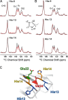
Copper coordination by histidine in anti-parallel Aβ40-Iowa fibrils. Solid-state 13C MAS NMR spectra are shown for Aβ40-Iowa uniform-13C-labeled at His-6, His-13, and His-14 with (red) and without (black) Cu(II). Anti-parallel fibrils (A) were formed by incubation at room temperature for 24 h. Copper was added after 24 h of room temperature incubation at a molar ratio of 1:2 copper:Aβ and the buffered (10 mm sodium phosphate buffer) solution was then incubated at 37 °C for an additional 96 h with shaking (50 rpm) before lyophilizing for NMR measurements. B, copper coordination by histidine in parallel Aβ40-Iowa fibrils. Parallel fibrils of Aβ40-Iowa were formed by incubation at 37 °C with 200 rpm shaking for 2 weeks (10 mm sodium phosphate buffer, 50 mm NaCl). Cu(II) was added after the 2-week period and then incubated for an additional 24 h. C, structure of the anti-parallel fibril of Aβ40-Iowa constructed by adding the N-terminal residues Asp-1–His-14 to the solid-state NMR coordinates (PDB code 2LNQ) from Gln-15–Val-40. The structure shows the relative locations of His-13 and His-14 on one monomer, and Glu-22 and Val-24 on the adjacent antiparallel monomer, after energy minimization and MD simulations. The β-strand between Lys-16 and Glu-22 continues through to at least His-13 and His-14. This geometry places His-14 next to Glu-22 on the adjacent monomer in a solvent-exposed position, whereas His-13 is oriented toward the fibril interior. The flexible N terminus allows His-6 to form a copper-binding site with His-14 and Glu-22.

References

    1. Selkoe D. J., and Hardy J. (2016) The amyloid hypothesis of Alzheimer's disease at 25 years. EMBO Mol. Med. 8, 595–608 10.15252/emmm.201606210 - DOI - PMC - PubMed
    1. Thal D. R., Griffin W. S., de Vos R. A., and Ghebremedhin E. (2008) Cerebral amyloid angiopathy and its relationship to Alzheimer's disease. Acta Neuropathol. 115, 599–609 10.1007/s00401-008-0366-2 - DOI - PubMed
    1. Qi-Takahara Y., Morishima-Kawashima M., Tanimura Y., Dolios G., Hirotani N., Horikoshi Y., Kametani F., Maeda M., Saido T. C., Wang R., and Ihara Y. (2005) Longer forms of amyloid β protein: implications for the mechanism of intramembrane cleavage by γ-secretase. J. Neurosci. 25, 436–445 10.1523/JNEUROSCI.1575-04.2005 - DOI - PMC - PubMed
    1. Selkoe D. J. (2004) Alzheimer disease: mechanistic understanding predicts novel therapies. Ann. Internal Med. 140, 627–638 10.7326/0003-4819-140-8-200404200-00047 - DOI - PubMed
    1. Castaño E. M., Prelli F., Soto C., Beavis R., Matsubara E., Shoji M., and Frangione B. (1996) The length of amyloid-β in hereditary cerebral hemorrhage with amyloidosis, Dutch type: implications for the role of amyloid-β 1-42 in Alzheimer's disease. J. Biol. Chem. 271, 32185–32191 10.1074/jbc.271.50.32185 - DOI - PubMed

Publication types

MeSH terms

Associated data

LinkOut - more resources