. 2020 Jun 19;295(25):8550-8559.
doi: 10.1074/jbc.RA120.013794. Epub 2020 May 6.

Androgen attenuates the inactivating phospho-Ser-127 modification of yes-associated protein 1 (YAP1) and promotes YAP1 nuclear abundance and activity

Affiliations

Androgen attenuates the inactivating phospho-Ser-127 modification of yes-associated protein 1 (YAP1) and promotes YAP1 nuclear abundance and activity

Bekir Cinar et al. J Biol Chem. .

Abstract

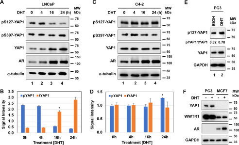
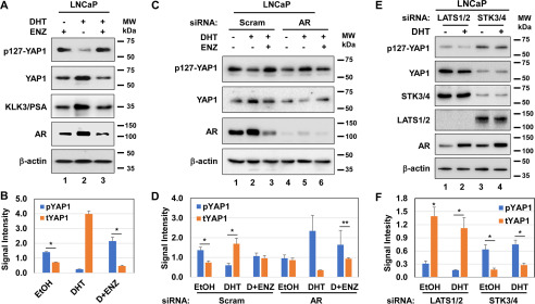
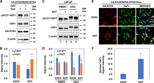
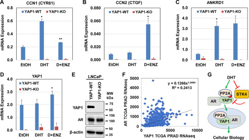
The transcriptional coactivator YAP1 (yes-associated protein 1) regulates cell proliferation, cell-cell interactions, organ size, and tumorigenesis. Post-transcriptional modifications and nuclear translocation of YAP1 are crucial for its nuclear activity. The objective of this study was to elucidate the mechanism by which the steroid hormone androgen regulates YAP1 nuclear entry and functions in several human prostate cancer cell lines. We demonstrate that androgen exposure suppresses the inactivating post-translational modification phospho-Ser-127 in YAP1, coinciding with increased YAP1 nuclear accumulation and activity. Pharmacological and genetic experiments revealed that intact androgen receptor signaling is necessary for androgen's inactivating effect on phospho-Ser-127 levels and increased YAP1 nuclear entry. We also found that androgen exposure antagonizes Ser/Thr kinase 4 (STK4/MST1) signaling, stimulates the activity of protein phosphatase 2A, and thereby attenuates the phospho-Ser-127 modification and promotes YAP1 nuclear localization. Results from quantitative RT-PCR and CRISPR/Cas9-aided gene knockout experiments indicated that androgen differentially regulates YAP1-dependent gene expression. Furthermore, an unbiased computational analysis of the prostate cancer data from The Cancer Genome Atlas revealed that YAP1 and androgen receptor transcript levels correlate with each other in prostate cancer tissues. These findings indicate that androgen regulates YAP1 nuclear localization and its transcriptional activity through the androgen receptor-STK4/MST1-protein phosphatase 2A axis, which may have important implications for human diseases such as prostate cancer.

Keywords: Hippo pathway; Hippo/MST1/STK4; YAP1; YAP1 nuclear localization; androgen; androgen receptor; androgen/AR signaling; androgens; cell signaling; gene transcription; nuclear translocation; phosphorylation; post-translational modification; protein phosphatase PP2A; protein phosphorylation; protein serine/threonine phosphatase (PSP); protein–protein interaction; signal transduction; yes-associated protein (YAP).


Conflict of interest statement

Conflict of interest—The authors declare no competing conflicts of interest with the content of this article.

Figures

Figure 1.
Figure 1.
Expression of YAP1, WWTR1, and AR in prostate cancer cell models. A, total proteins were isolated from cells at 80% confluence in steady-state growth conditions and analyzed by Western blotting for YAP1, WWTR1, or AR protein abundance. β-Actin was used as a loading control in immunoblots. The membranes were blotted with the protein-specific antibody. B, quantification of YAP1 protein was normalized to the β-actin. ImageJ software was used to quantify YAP1 and β-actin protein blots. C, expression of YAP1 transcripts in the select cell lines. Total RNA was isolated from cells at 80% confluence in steady-state conditions 24 h after cell seeding and analyzed by quantitative RT-PCR for the abundance of YAP1 transcripts, *,**, p < 0.01. YAP1 mRNA expression was normalized to 18S rRNA transcript. The data ± S.D. are from three independent experiments. MW, molecular mass.
Figure 2.
Figure 2.
Steroid hormone androgen regulates phospho–Ser-127 and YAP1 protein levels in cultures. A–D, Western blotting and quantification of phospho–Ser-127 and total YAP1 proteins in androgen-dependent LNCaP (*, p < 0.01) and androgen-independent C4-2 (*, p > 0.05) cells, respectively. The cells were exposed to DHT (10 nm) at varying times. The data in B and C were normalized to α-tubulin. The phospho–Ser-127 blot was included as a control. E and F, Western blotting analysis of YAP1 and WWTR1 proteins in the PC3 prostate cancer and MCF7 breast cancer cell lines that were also treated with vehicle (ethanol) or DHT overnight (16–18 h). DHT treatment was conducted in 5% CSS-fed growth conditions. ImageJ software was used to quantify the intensity of phospho–Ser-127, total YAP, and α-tubulin protein bands. YAP1 and AR blots were included as a positive control in F. The α-tubulin or GAPDH protein blot was incorporated as a loading control. The membranes were blotted with the protein-specific antibody. The data ± S.D. are representative of two independent experiments. MW, molecular mass.
Figure 3.
Figure 3.
AR and STK4/3 signaling is necessary for the inhibition of phospho–Ser-127 by androgen. A and B, Western blotting and quantification of phospho–Ser-127 and total YAP1 proteins, respectively. *,**, p < 0.01. KLK3/prostate specific antigen (PSA) blot was included as a positive control to assess the activity of DHT. C and D, Western blotting and quantification of phospho–Ser-127 and total YAP1 protein in LNCaP cells after transient transfection with the pool of scrambled (Scram) or the AR gene-specific siRNA for 36 h, followed by treatment with or without DHT and ENZ overnight in 5% CSS-fed conditions. E and F, Western blotting and quantification of phospho–Ser-127 and total YAP1 in LNCaP cells transfected with Scram siRNA or the LATS1/2 and STK4/3 (MST1/2) gene-specific siRNA for 36 h, followed by treatment with or without DHT overnight in 5% CSS-fed growth conditions. β-Actin was used as a loading control in immunoblots. The membranes were blotted with the protein-specific antibody. ImageJ software was used to quantify the intensity of the phospho–Ser-127 and the total YAP1 signal. The data ± S.D. are representative of three independent experiments. MW, molecular mass.
Figure 4.
Figure 4.
Androgen negatively regulates the Hippo/STK4 signaling. A and B, Western blotting and quantification of phospho–Ser-127 and total YAP1 proteins in C4-2/HA-STK4/MST1 cells that express tetracycline- or Dox-inducible HA-STK4 protein (45), respectively. C and D, Western blotting and quantification of phospho–Ser-127 and total YAP1 protein in LNCaP cells that were exposed to DMSO (vehicle) or OKA followed by treatment with or without DHT overnight in 5% CSS-fed growth condition. β-Actin was used as a loading control in immunoblots. E, immunofluorescence analysis of ectopic HA-STK4 (red), and native YAP1 (green) proteins and nuclei (DAPI, blue) in Dox-treated C4-2/HA-STK4 cells. Scale bar, 20 μm. Micrographs are the representation of multiple images. F, quantification of nuclear YAP1 protein by ImageJ from multiple images and nuclear YAP1 signals were normalized to the total number of cells subjected to image quantification. The ectopic expression of HA-STK4/MST1 protein was assessed using an HA tag antibody. The cells were treated with EtOH or DHT (10 nm) and Dox (1 μg/ml) in serum-depleted conditions overnight before analysis. *,**, p < 0.05. MW, molecular mass.
Figure 5.
Figure 5.
Androgen promotes YAP1 nuclear localization. A and B, coimmunofluorescence imaging of YAP1 and AR proteins in LNCaP cells. The cells were treated with or without DHT (10 nm) and ENZ (20 μm) overnight in 5% CSS-fed growth conditions. Images of AR (Alexa 647, red), YAP1 (Alexa 488, green), and nuclei (DAPI, blue) were acquired using confocal microscopy. Scale bar, 20 μm. The micrographs are the representation of multiple images. B, ImageJ software was used to quantify the intensity of the nuclear AR and YAP1 protein from multiple images. The data ± S.D. normalized to the total cell number from multiple images of three independent experiments. *,**, p < 0.0001. C, coimmunoprecipitation and Western blotting analysis of the cytoplasmic and nuclear YAP1, PP2A C, and AR proteins in LNCaP cells after treatment with or without DHT overnight in 5% CSS-fed growth condition. Topoisomerase I (TOPI) was used as a nuclear marker. The blots are representative of three independent experiments. MW, molecular mass.
Figure 6.
Figure 6.
Androgen signaling regulates the transcriptional activity of YAP1. A–C, quantitative RT-PCR analysis of YAP1 and the YAP1 target gene expression in YAP1-WT and YAP1-KO LNCaP cell models after treatment with EtOH, DHT, and DHT plus ENZ overnight in 5% CSS-fed growth condition. D and E, quantitative RT-PCR and Western blotting analysis YAP1 transcripts and protein expression in YAP1-WT and YAP1-KO LNCaP cell models, respectively. The data ± S.D. are from three independent experiments. *,**, p < 0.01. F, coexpression and correlation analysis of AR and YAP1 mRNA levels in TCGA Pan-Cancer prostate adenocarcinoma data set (PRAD) from 493 patients (46). Pearson correlation = 0.45, p = 1.57e−14). The data were accessed using the cBioPortal website (RRID:SCR_014555). G, a model illustrates the regulation of YAP1 nuclear localization by androgen. In this model, androgen antagonizes the Hippo/STK4/MST1 in a way that attenuates phospho-YAP and induces the YAP1 nuclear localization. Androgen also enhanced YAP1 nuclear localization by promoting protein–protein interaction between YAP1, PP2A, and AR, leading to cellular biology. MW, molecular mass.

References

    1. Chen Y. A., Lu C. Y., Cheng T. Y., Pan S. H., Chen H. F., and Chang N. S. (2019) WW domain–containing proteins YAP and TAZ in the Hippo pathway as key regulators in stemness maintenance, tissue homeostasis, and tumorigenesis. Front. Oncol. 9, 60 10.3389/fonc.2019.00060 - DOI - PMC - PubMed
    1. Ma S., Meng Z., Chen R., and Guan K. L. (2019) The Hippo pathway: biology and pathophysiology. Annu. Rev. Biochem. 88, 577–604 10.1146/annurev-biochem-013118-111829 - DOI - PubMed
    1. Zhao B., Li L., Lei Q., and Guan K. L. (2010) The Hippo–YAP pathway in organ size control and tumorigenesis: an updated version. Genes Dev. 24, 862–874 10.1101/gad.1909210 - DOI - PMC - PubMed
    1. Liu A. M., Wong K. F., Jiang X., Qiao Y., and Luk J. M. (2012) Regulators of mammalian Hippo pathway in cancer. Biochim. Biophys. Acta 1826, 357–364 10.1016/j.bbcan.2012.05.006 - DOI - PubMed
    1. Yu F. X., and Guan K. L. (2013) The Hippo pathway: regulators and regulations. Genes Dev. 27, 355–371 10.1101/gad.210773.112 - DOI - PMC - PubMed

Publication types

MeSH terms