. 2020 May 6;5(3):e00190-20.
doi: 10.1128/mSphere.00190-20.

Extracellular Vesicles from Aspergillus flavus Induce M1 Polarization In Vitro

Affiliations

Extracellular Vesicles from Aspergillus flavus Induce M1 Polarization In Vitro

Verônica S Brauer et al. mSphere. .

Abstract

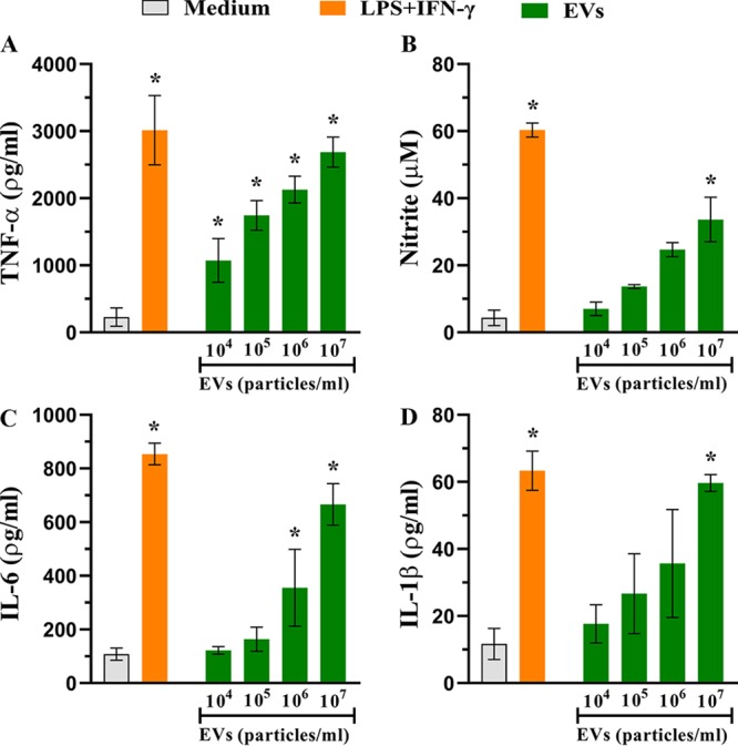
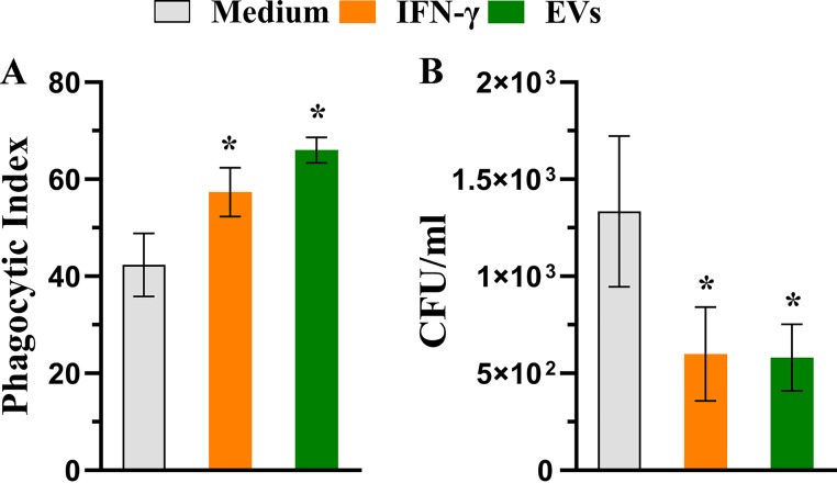
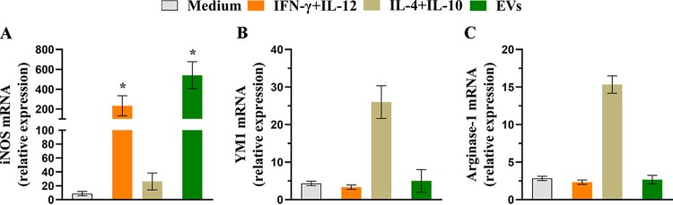
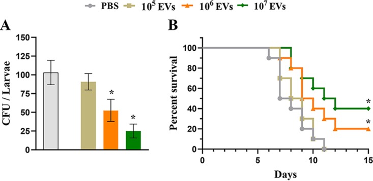
Aspergillus flavus, a ubiquitous and saprophytic fungus, is the second most common cause of aspergillosis worldwide. Several mechanisms contribute to the establishment of the fungal infection. Extracellular vesicles (EVs) have been described as "virulence factor delivery bags" in several fungal species, demonstrating a crucial role during the infection. In this study, we evaluated production of A. flavus EVs and their immunomodulatory functions. We verified that A. flavus EVs induce macrophages to produce inflammatory mediators, such as nitric oxide, tumor necrosis factor alpha (TNF-α), interleukin-6 (IL-6), and IL-1β. Furthermore, the A. flavus EVs enhance phagocytosis and killing by macrophages and induce M1 macrophage polarization in vitro In addition, a prior inoculation of A. flavus EVs in Galleria mellonella larvae resulted in a protective effect against the fungal infection. Our findings suggest that A. flavus EVs are biologically active and affect the interaction between A. flavus and host immune cells, priming the innate immune system to eliminate the fungal infection. Collectively, our results suggest that A. flavus EVs play a crucial role in aspergillosis.IMPORTANCE Immunocompromised patients are susceptible to several fungal infections. The genus Aspergillus can cause increased morbidity and mortality. Developing new therapies is essential to understand the fungal biology mechanisms. Fungal EVs carry important virulence factors, thus playing pivotal roles in fungal pathophysiology. No study to date has reported EV production by Aspergillus flavus, a fungus considered to be the second most common cause of aspergillosis and relevant food contaminator found worldwide. In this study, we produced A. flavus EVs and evaluated the in vitro immunomodulatory effects of EVs on bone marrow-derived macrophages (BMDMs) and in vivo effects in a Galleria mellonella model.

Keywords: Aspergillus flavus; Galleria mellonella; cytokine; extracellular vesicles; killing; macrophage; phagocytosis; polarization.


Figures

FIG 1
FIG 1
Extracellular vesicles (EVs) produced by Aspergillus flavus. Nanoparticle-tracking analysis of EVs isolated from A. flavus culture supernatant was performed using NanoSight NS300. (A) Representative histogram depicting the particle-size distribution and concentration of EV profiles from A. flavus (EVs × 108 particles/ml). (B) Representative graphic of EV average sizes from 12 independent experiments. (C) Screenshot from the video recorded by NanoSight NS300, presenting EV distribution.
FIG 2
FIG 2
EVs from A. flavus induce the production of inflammatory mediators by bone marrow-derived macrophages (BMDMs). BMDMs obtained from C57BL/6 mice were cultured with different concentrations of EVs (104 to 107 particles/ml) for 48 h at 37°C. As positive and negative controls, the BMDMs were treated with lipopolysaccharide (LPS, 1 μg/ml) plus gamma interferon (IFN-γ) (2 ng/ml) or medium only as indicated. The culture supernatant was used to quantify the levels of (A) tumor necrosis factor alpha (TNF-α), (B) nitrite, (C) interleukin-6 (IL-6), and (D) IL-1β using enzyme-linked immunosorbent assay (ELISA). Data represent results from three independent experiments. One-way ANOVA and Bonferroni’s multiple-comparison tests were used for analysis of TNF-α and IL-6 data, and Kruskal-Wallis and Dunn’s multiple-comparison tests were used for analysis of nitrite and IL-1β data. *, P < 0.05.
FIG 3
FIG 3
A. flavus EVs stimulate microbicidal activity of BMDMs. (A) BMDMs were plated on glass coverslips and cultured with EVs (107 particles/ml) for 30 min and were treated with A. flavus conidia (macrophages/conidia = 1:1) for 4 h at 37°C, and the phagocytic index was determined. (B) BMDM previously treated with EVs (107 particles/ml), for 30 min, were infected with A. flavus conidia (macrophages/conidia = 1:1) for 48 h at 37°C. The cells were lysed, and the lysate was plated to detect the viable fungi based on CFU counting technique. Data represents results from three independent experiments. For both phagocytosis and killing assays, the IFN-γ-containing medium and medium only were used as positive and negative controls, respectively. An unpaired, two-tailed t test was used from both analyses. *, P < 0.05.
FIG 4
FIG 4
EVs induce M1 polarization of BMDMs. BMDMs were cultured with 107 particles/ml for 6 h. The cells were further treated with IFN-γ (2 ng/ml) plus IL-12p40 (50 ng/ml) as a positive control for M1 phenotype or with IL-4 (50 ng/ml) plus IL-10 (50 ng/ml) as a positive control of M2 phenotype or with medium only as a negative control. The total RNA was extracted from the macrophages and converted into cDNA, and qRT-PCR analysis was performed to evaluate the relative expression levels of classical markers of macrophage polarization. (A) Inducible nitric oxide synthase (iNOS). (B) YM1. (C) Arginase-1. Data represents results from three independent experiments. An unpaired, two-tailed t test was used for iNOS, and an unpaired, two-tailed t test and Mann-Whitney test were used for YM1 and arginase. *, P < 0.05.
FIG 5
FIG 5
A. flavus EVs induce a protective effect on Galleria mellonella in an in vivo model. (A) G. mellonella larvae (n = 5 per group) were stimulated with A. flavus EVs (105, 106, and 107 particles) or with PBS as a control 48 h prior to infection with A. flavus conidia. Larvae were homogenized and levels of CFU content determined 48 h postinfection for fungal burden analysis. (B) Survival rates (n = 5 larvae per group) during 15 days postinfection. Data represent the results of two independent experiments. One-way ANOVA and Bonferroni’s multiple-comparison tests were used for fungal burden analysis and the log rank (Mantel-Cox) test for survival curve analysis. *, P < 0.05.

References

    1. Sugui JA, Kwon-Chung KJ, Juvvadi PR, Latgé J-P, Steinbach WJ. 2014. Aspergillus fumigatus and related species. Cold Spring Harb Perspect Med 5:a019786. doi: 10.1101/cshperspect.a019786. - DOI - PMC - PubMed
    1. Pasqualotto AC. 2009. Differences in pathogenicity and clinical syndromes due to Aspergillus fumigatus and Aspergillus flavus. Med Mycol 47(Suppl 1):S261–S270. doi: 10.1080/13693780802247702. - DOI - PubMed
    1. Almeida F, Rodrigues ML, Coelho C. 2019. The still underestimated problem of fungal diseases worldwide. Front Microbiol 10:214. doi: 10.3389/fmicb.2019.00214. - DOI - PMC - PubMed
    1. Jackson SA, Dobson A. 2016. Yeasts and molds: Aspergillus flavus. Reference module in food science. Elsevier, Amsterdam, Netherlands. doi: 10.1016/B978-0-08-100596-5.01086-6. - DOI
    1. Amaike S, Keller NP. 2011. Aspergillus flavus. Annu Rev Phytopathol 49:107–133. doi: 10.1146/annurev-phyto-072910-095221. - DOI - PubMed

Publication types

LinkOut - more resources