. 2020 May 6;12(542):eaay8314.
doi: 10.1126/scitranslmed.aay8314.

Preclinical validation of a repurposed metal chelator as an early-intervention therapeutic for hemotoxic snakebite

Affiliations

Preclinical validation of a repurposed metal chelator as an early-intervention therapeutic for hemotoxic snakebite

Laura-Oana Albulescu et al. Sci Transl Med. .

Abstract

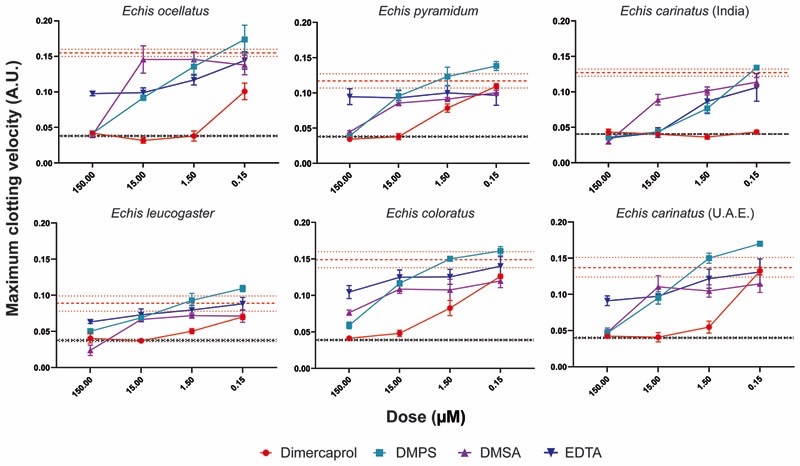
Snakebite envenoming causes 138,000 deaths annually, and ~400,000 victims are left with permanent disabilities. Envenoming by saw-scaled vipers (Viperidae: Echis) leads to systemic hemorrhage and coagulopathy and represents a major cause of snakebite mortality and morbidity in Africa and Asia. The only specific treatment for snakebite, antivenom, has poor specificity and low affordability and must be administered in clinical settings because of its intravenous delivery and high rates of adverse reactions. This requirement results in major treatment delays in resource-poor regions and substantially affects patient outcomes after envenoming. Here, we investigated the value of metal ion chelators as prehospital therapeutics for snakebite. Among the tested chelators, dimercaprol (British anti-Lewisite) and its derivative 2,3-dimercapto-1-propanesulfonic acid (DMPS) were found to potently antagonize the activity of Zn2+-dependent snake venom metalloproteinases in vitro. Moreover, DMPS prolonged or conferred complete survival in murine preclinical models of envenoming against a variety of saw-scaled viper venoms. DMPS also considerably extended survival in a "challenge and treat" model, where drug administration was delayed after venom injection and the oral administration of this chelator provided partial protection against envenoming. Last, the potential clinical scenario of early oral DMPS therapy combined with a delayed, intravenous dose of conventional antivenom provided prolonged protection against the lethal effects of envenoming in vivo. Our findings demonstrate that the safe and affordable repurposed metal chelator DMPS can effectively neutralize saw-scaled viper venoms in vitro and in vivo and highlight the promise of this drug as an early, prehospital, therapeutic intervention for hemotoxic snakebite envenoming.


Conflict of interest statement

Competing interests: The authors declare that they have no competing interests.

Figures

Fig. 1
Fig. 1. The geographical distribution and venom toxin composition of saw-scaled vipers (genus Echis).
Map of saw-scaled viper distribution with the locales of the studied species indicated by red stars. Echis distribution areas and corresponding venom proteomes are highlighted by the following colors: light orange (E. leucogaster), blue (E. ocellatus), green (E. carinatus), pink (E. pyramidum), violet (E. coloratus). Toxin proteome abundances were taken from (11, 58) and generated in this study for E. leucogaster. Toxin family key: SVMP, snake venom metalloproteinase; SVSP, snake venom serine proteinase; PLA2, phospholipase A2; CTL, C-type lectins; LAAO, L-amino acid oxidase; SVMPi, SVMP inhibitors; DIS, disintegrin; CRISP, cysteine-rich secretory protein.
Fig. 2
Fig. 2. Metal chelators inhibit SVMP toxin activity of saw-scaled viper venoms.
The neutralizing capability of four metal chelators against the SVMP activity of six Echis venoms. Data are presented for four drug concentrations, from 150 µM to 150 nM (highest to lowest dose), expressed as percentages of the venom-only sample (100%, dotted red line). The negative control is presented as an interval (dotted black lines) and represents the values recorded in the PBS-only samples (expressed as % of venom activity), where the highest and the lowest values for each set of experiments are depicted. Inhibitors are color-coded (dimercaprol, red; DMPS, blue; DMSA, purple; EDTA, dark blue). The data represent triplicate independent repeats with SEMs, where each repeat represents the average of n≥2 technical replicates.
Fig. 3
Fig. 3. Metal chelators inhibit the procoagulant activity of saw-scaled viper venoms.
The neutralizing capability of four metal chelators against the procoagulant activity of six Echis venoms. Data are presented for four drug concentrations, from 150 µM to 150 nM (highest to lowest dose), expressed as the maximum clotting velocity (middle dotted red line and surrounding interval indicates the maximum clotting velocity of the venom and represents the average ± SEM). The negative control is presented as an interval (dotted black lines, including the average and average ± SEM; this may appear as a single black line if the interval is very narrow and lines overlap) and represents normal plasma clotting. Inhibitors are color-coded (dimercaprol, red; DMPS, blue; DMSA, purple; EDTA, dark blue). The data represent triplicate independent repeats with SEMs, where each repeat represents the average of n≥2 technical replicates.
Fig. 4
Fig. 4. Metal chelators prevent or delay lethality in vivo when preincubated with Echis ocellatus venom.
Kaplan-Meier survival graphs for experimental animals (n=5) receiving venom preincubated (30 min at 37 C) with different metal chelators via the intravenous route. Survival of mice receiving 45 µg of E. ocellatus venom (2.5 × LD50 dose) with and without 60 µg of dimercaprol (A), 60 µg of DMPS (B), or 60 µg of DMSA (C). Drug-only controls are presented as black dotted lines at the top of each graph, and the end of the experiment was at 6 h. None of the drugs exhibited toxicity at the given doses. (D) Quantification of TAT concentrations in envenomed animals. Where the time of death was the same within experimental groups (early deaths or complete survival), TAT concentrations were quantified for n=3, and where times of death varied, n=5. The data are displayed as means of the duplicate technical repeats plus SDs.
Fig. 5
Fig. 5. The efficacy of metal chelators against other medically important Echis venoms.
Kaplan-Meier survival graphs for experimental animals (n=5) receiving Echis pyramidum or E. carinatus (India) venom preincubated (30 min at 37 C) with different metal chelators via the intravenous route. Survival of mice receiving 40 µg of E. pyramidum venom (A), and 47.5 µg of E. carinatus (India) venom (both represent the 2.5 × LD50 dose) (B) with or without 60 µg of dimercaprol or DMPS. Drug-only controls are presented as black dotted lines at the top of each graph. None of the drugs exhibited toxicity at the given doses. The end of the experiment was at 6 h after injection. Quantification of TAT concentrations in envenomed animals is displayed in the right panels. Where the time of death was the same within experimental groups (early deaths or complete survival), TAT concentrations were quantified for n=3, and where times of death varied, n=5. The data are displayed as means of the duplicate technical repeats plus SDs.
Fig. 6
Fig. 6. DMPS delays lethality in vivo in a ‘challenge and treat’ model of envenoming.
Kaplan-Meier survival graphs for experimental animals (n=5) receiving venom (intraperitoneal administration), followed by delayed drug treatment (intraperitoneally 15 min later). (A) Survival of mice receiving 5 x the intravenous LD50 dose of E. ocellatus venom (90 µg) with or without 120 µg of drug (dimercaprol or DMPS) 15 min later. (B) Survival of mice receiving 7 x intravenous LD50 dose of E. pyramidum venom (112 µg) with or without 120 µg of DMPS 15 min later. (C) Survival of mice receiving 5 x intravenous LD50 dose of E. carinatus (India) venom (95 µg) with or without 120 µg of DMPS 15 min later. For (A), (B), and (C), drug-only controls are presented as black dotted lines at the top of each graph (none of the drugs exhibited toxicity at the given doses), and the end of the experiment was at 24 h. (D) Quantification of TAT concentrations in envenomed animals. Where the time of death was the same within experimental groups (early deaths or complete survival), TAT concentrations were quantified for n=3, and where times of death varied, n=5. The data are displayed as means of the duplicate technical repeats plus SDs. EOC, E. ocellatus; EPL, E. pyramidum; ECAR, E. carinatus.
Fig. 7
Fig. 7. Oral DMPS followed by later administration of antivenom protects against in vivo lethality caused by Echis ocellatus venom.
Kaplan-Meier survival graphs for experimental animals (n=5) receiving E. ocellatus venom (90 µg, 5 × intravenous LD50 dose, via intraperitoneal administration), followed by delayed drug treatment (intraperitoneal or oral) and/or antivenom (intraperitoneal or intravenous). (A) Survival of mice receiving E. ocellatus venom (intraperitoneally) followed by 168 µl of the E. ocellatus monospecific antivenom EchiTAbG either intraperitoneally 15 min later or intravenously 1 h later. Corresponding TAT concentrations for the envenomed animals are depicted on the right. Note: For the venom + AV intraperitoneally (15 min) dataset, the mouse that succumbed early to the effects of the venom could not be sampled for TAT, therefore the data displayed only reflect the animals that survived until the end of the experiment. (B) Survival of mice receiving E. ocellatus venom (intraperitoneally) followed by DMPS 15 min later (120 µg, intraperitoneally) and antivenom 1 h after venom administration (168 µl, intravenously). Right panel shows corresponding TAT concentrations. (C) Survival of mice receiving E. ocellatus venom (intraperitoneally), followed immediately by: (i) oral DMPS (600 µg, ~1 min after venom injection), (ii) oral DMPS (600 µg, ~1 min after venom injection) and EchiTAbG antivenom (168 µl, intravenously, 1 h later), and (iii) EchiTAbG antivenom (168 µl, intravenously, 1 h later). Right panel shows corresponding TAT concentrations. For all survival experiments, drug- or antivenom-only controls are presented as black dotted lines at the top of each graph (none exhibited toxicity at the given doses), and the end of the experiment was at 24 h. For quantification of TAT, where the time of death was the same within experimental groups (early deaths or complete survival), TAT concentrations were quantified for n=3, and where times of death varied, n=5. The data are displayed as means of the duplicate technical repeats plus SDs.
Fig. 8
Fig. 8. DMPS effectively neutralizes local hemorrhage caused by E. ocellatus venom.
(A) The resulting murine hemorrhagic lesions observed 2 h after the intradermal injection of (from left to right): (i) 10 µg E. ocellatus venom, (ii) 10 µg E. ocellatus venom preincubated (30 min at 37 C) with 13.3 µg DMPS, (iii) 13.3 µg DMPS, and (iv) PBS control. For all experimental groups, n=3. Black lines represent scale bars equivalent to 2 mm. (B) Quantification of hemorrhagic lesion sizes expressed as the hemorrhagic area (mm2) observed 2 h after injection. The data are expressed as the mean of triplicate measurements ± SEM. A two-tailed unpaired t-test was used to compare the lesion sizes in the venom-only vs. venom+ DMPS groups, and significance is indicated by asterisks (p=0.0007).

Comment in

References

    1. Gutiérrez JM, Calvete JJ, Habib AG, Harrison RA, Williams DJ, Warrell DA. Snakebite envenoming. Nat Rev Dis Prim. 2017;3 17063. - PubMed
    1. Habib AG, Kuznik A, Hamza M, Abdullahi MI, Chedi BA, Chippaux J-P, Warrell DA. Snakebite is Under Appreciated: Appraisal of Burden from West Africa. PLoS Negl Trop Dis. 2015;9:e0004088. - PMC - PubMed
    1. Harrison RA, Hargreaves A, Wagstaff SC, Faragher B, Lalloo DG. Snake envenoming: A disease of poverty. PLoS Negl Trop Dis. 2009;3 doi: 10.1371/journal.pntd.0000569. - DOI - PMC - PubMed
    1. Longbottom J, Shearer FM, Devine M, Alcoba G, Chappuis F, Weiss DJ, Ray SE, Ray N, Warrell DA, Ruiz de Castañeda R, Williams DJ, et al. Vulnerability to snakebite envenoming: a global mapping of hotspots. Lancet (London, England) 2018;392:673–684. - PMC - PubMed
    1. Mohapatra B, Warrell DA, Suraweera W, Bhatia P, Dhingra N, Jotkar RM, Rodriguez PS, Mishra K, Whitaker R, Jha P, for the Collaborators M. D. S Snakebite Mortality in India: A Nationally Representative Mortality Survey. PLoS Negl Trop Dis. 2011;5:e1018. - PMC - PubMed

Publication types