. 2020 Jul;10(7):1038-1057.
doi: 10.1158/2159-8290.CD-19-1242. Epub 2020 May 6.

Somatic Tissue Engineering in Mouse Models Reveals an Actionable Role for WNT Pathway Alterations in Prostate Cancer Metastasis

Affiliations

Somatic Tissue Engineering in Mouse Models Reveals an Actionable Role for WNT Pathway Alterations in Prostate Cancer Metastasis

Josef Leibold et al. Cancer Discov. 2020 Jul.

Abstract

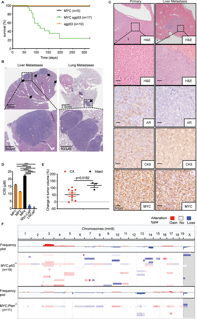
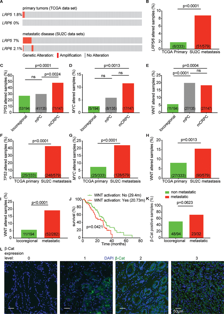
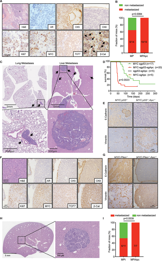
To study genetic factors influencing the progression and therapeutic responses of advanced prostate cancer, we developed a fast and flexible system that introduces genetic alterations relevant to human disease directly into the prostate glands of mice using tissue electroporation. These electroporation-based genetically engineered mouse models (EPO-GEMM) recapitulate features of traditional germline models and, by modeling genetic factors linked to late-stage human disease, can produce tumors that are metastatic and castration-resistant. A subset of tumors with Trp53 alterations acquired spontaneous WNT pathway alterations, which are also associated with metastatic prostate cancer in humans. Using the EPO-GEMM approach and an orthogonal organoid-based model, we show that WNT pathway activation drives metastatic disease that is sensitive to pharmacologic WNT pathway inhibition. Thus, by leveraging EPO-GEMMs, we reveal a functional role for WNT signaling in driving prostate cancer metastasis and validate the WNT pathway as therapeutic target in metastatic prostate cancer. SIGNIFICANCE: Our understanding of the factors driving metastatic prostate cancer is limited by the paucity of models of late-stage disease. Here, we develop EPO-GEMMs of prostate cancer and use them to identify and validate the WNT pathway as an actionable driver of aggressive metastatic disease.This article is highlighted in the In This Issue feature, p. 890.


Conflict of interest statement

CONFLICTS OF INTEREST

W.A. received an honorarium from CARET, and is a consultant for Clovis Oncology, Janssen, MORE Health, and ORIC Pharmaceuticals. C.L.S. serves on the Board of Directors of Novartis, is a co-founder of ORIC Pharmaceuticals and co-inventor of enzalutamide and apalutamide. He is a science advisor to Agios, Beigene, Blueprint, Column Group, Foghorn, Housey Pharma, Nextech, KSQ, Petra, and PMV. S.W.L. is a co-founder and scientific advisory board member of Blueprint Medicines, ORIC Pharmaceuticals, Mirimus, Inc., Faeth Therapeutics, and Geras Bio, and is an advisor to Petra Pharmaceuticals, Constellation Pharmaceuticals, and Boehringer Ingelheim. L.E.D is an advisory board member for Mirimus Inc.

Figures

Figure 1.
Figure 1.. Somatic induction of oncogenic lesions by in vivo electroporation of the prostate gland
(A) Schematic of the electroporation-induced genetically engineered mouse model (EPO-GEMM) of prostate cancer. A MYC transposon vector in combination with a Sleeping Beauty transposase (SB13) and/or a CRISPR/Cas9 vector targeting Pten (sgPten) were delivered into the prostate by direct in vivo electroporation. (B) Kaplan-Meier survival curve of C57BL/6 mice electroporated with a MYC transposon vector and a Sleeping Beauty transposase (MYC; black), a CRISPR/Cas9 vector targeting Pten (sgPten; orange), or the combination of all vectors (MYC sgPten; blue). (C) Representative hematoxylin and eosin (H&E) and immunohistochemical staining of a well differentiated MPt EPO-GEMM (top) or Nkx3.1CreERT2/+;Ptenfl/fl;ARR2/Pbsn-MYC (NPHiMYC) classic GEMM prostate tumor (bottom). (D) Representative H&E and immunohistochemical staining of a poorly differentiated MPt EPO-GEMM (top) or NPHiMYC classic GEMM prostate tumor (bottom).
Figure 2.
Figure 2.. Engineering advanced prostate cancer de novo using EPO-GEMMs
(A) Kaplan-Meier survival curve of mice electroporated with the MYC transposon vector and a Sleeping Beauty transposase (MYC; black), a CRISPR/Cas9 vector targeting p53 (sgp53; orange), or all vectors (MYC sgp53; green). (B) Representative H&E staining of liver and lungs isolated from mice with MYC;sgp53 (MP) EPO-GEMM prostate tumors. Arrows, metastatic nodules. (C) Representative H&E and immunohistochemical staining of a MP EPO-GEMM prostate tumor (left) and a corresponding liver metastasis (right). (D) IC50 values for enzalutamide in indicated murine and human prostate cancer cell lines (n=3; error bars, mean ± s.e.m; **** p < 0.0001; One-way ANOVA). (E) Change in tumor volume of MP EPO-GEMM prostate tumors in intact or castrated (CX) mice one-week post-surgery (n=3–10; error bars, mean ± s.e.m; unpaired two-tailed t test). (F) Frequency plot of copy number variation (CNV) analysis of MP (n=19) and MPt (n=11 (from 6 tumors)) EPO-GEMM prostate tumors.
Figure 3.
Figure 3.. A subset of MYC/p53-driven tumors acquire WNT pathway activation.
(A) Principle component analysis (PCA) of the transcriptional output of MP EPO-GEMM prostate tumors (n=10) compared to wild type (WT) (n=6) murine prostate tissue. MP tumors segregate into two clusters (group 1 and group 2). (B) Gene set enrichment analysis (GSEA) of group 1 and group 2 clusters of MP prostate tumors from (A) reveals an enrichment for β-catenin signaling in one of the populations (hereafter MP WNThi). (C) Heat map of WNT pathway gene expression in MP WNThi and MP WNTlo MP prostate tumors (n=5). (D) Frequency of metastases in the liver in cohorts of mice with either MP WNThi or MP WNTlo prostate tumors (n=5; two-sided Fisher’s exact test). (E) Representative immunohistochemical staining of MP WNThi and MP WNTlo EPO-GEMM prostate tumors. (F) Close up views of clonal CNVs in WNT pathway genes Lrp6 (left) or Wnt2b (right) in individual MP WNThi EPO-GEMM prostate tumors (see arrows). (G) Diagram of the Apc gene and the position of a point mutation found in a MP WNThi EPO-GEMM prostate tumor.
Figure 4.
Figure 4.. WNT pathway alterations are associated with metastatic disease in patients with advanced prostate cancer
(A) Oncoprint displaying the genomic status of LRP5 or LRP6 in prostate cancer patient samples isolated from either primary tumors (TCGA dataset (31)) or from metastatic sites (SU2C datasets (3,4)). (B) Frequency of LRP5 or LRP6 amplifications in the same cohorts of patients as in (A) (two-sided Fisher’s exact test). (C) Frequency of TP53 alterations in patients with locoregional prostate cancer, metastatic but castration sensitive prostate cancer (mPC), or metastatic castration resistant prostate cancer (mCRPC) from datasets in (5) (ns, not significant; two-sided Fisher’s exact test). (D) Frequency of amplifications in MYC in the same cohorts of patients as in (C) (ns, not significant; two-sided Fisher’s exact test). (E) Frequency of activating mutations in the WNT pathway genes APC or CTNNB1 (encoding β-catenin) in the same cohorts of patients as in (C) (ns, not significant; two-sided Fisher’s exact test). (F) Frequency of TP53 alterations in prostate cancer patient samples isolated from either primary tumors (TCGA dataset (31)) or from metastatic sites (SU2C dataset (3,4)) (two-sided Fisher’s exact test). (G) Frequency of amplifications in MYC in the same cohorts of patients as in (F) (two-sided Fisher’s exact test). (H) Frequency of activating mutations in the WNT pathway genes APC or CTNNB1 (encoding β-catenin) in the same cohorts of patients as in (F) (two-sided Fisher’s exact test). (I) Frequency of activating mutations in the WNT pathway genes APC or CTNNB1 in patients with locoregional or metastatic prostate cancer (mPC and mCRPC combined) from the same cohorts of patients as in (C) (two-sided Fisher’s exact test). (J) Kaplan-Meier survival curve of patients with advanced prostate cancer with (red; n=47) or without (green; n=81) activating mutations in the WNT signaling pathway from the SU2C dataset (3) (log-rank test). Median survival in months (m) shown inset. (K) Quantification of immunofluorescence staining for β-catenin in tumor microarrays (TMAs) containing prostate tumor specimens from patients with locoregional or metastatic disease. The percentage of samples that stained positive for β-catenin is shown (two-sided Fisher’s exact test). (L) Representative immunofluorescence staining for β-catenin in TMAs containing prostate tumor specimens from patients with locoregional or metastatic disease. Samples scored as 0 or 1 were considered negative, and those scored as 2 or 3 as positive for β-catenin expression.
Figure 5.
Figure 5.. WNT pathway activation promotes prostate cancer metastasis
(A) Representative H&E and immunohistochemical staining of a primary MPApc prostate tumor. Arrows, nuclear β-catenin localization. (B) Frequency of mice with macrometastatic disease in cohorts with either MP or MPApc prostate EPO-GEMM tumors (one-sided Fisher’s exact test). (C) H&E staining of liver and lung metastases isolated from mice with MPApc EPO-GEMM prostate tumors. Arrows, metastatic nodules. (D) Kaplan-Meier survival curve of mice with indicated EPO-GEMM prostate tumors (log-rank test). (E) Representative immunohistochemical staining of primary MP and MPApc EPO-GEMM prostate tumors. (F) Representative H&E and immunohistochemical staining of a primary MPtApc prostate tumor. (G) Representative immunohistochemical staining of a primary MPtApc EPO-GEMM prostate tumor. (H) H&E staining of a liver metastasis isolated from a mouse with a MPtApc EPO-GEMM prostate tumor. (I) Frequency of mice with macrometastatic disease in cohorts with either MPt or MPtApc prostate EPO-GEMM tumors (one-sided Fisher’s exact test).
Figure 6.
Figure 6.. Apc mutations drive disease and metastatic progression in prostate cancer organoid models
(A) Representative gross bright field (top) and H&E (bottom) images of prostates of NOD-scid IL2Rγnull (NSG) mice 15 weeks after orthotopic transplantation of prostate organoids with indicated genotypes. (B) Representative immunohistochemical staining of prostates of NSG mice 15 weeks after orthotopic transplantation of prostate organoids with indicated genotypes. (C-D) Representative bioluminescence images of NSG mice 4 weeks after tail vein injection of mouse prostate organoids with indicated genotypes (n=3–4).
Figure 7.
Figure 7.. Targeting WNT signaling disrupts prostate cancer metastasis
(A) Growth assay of indicated mouse prostate cancer cell lines or primary murine fibroblasts treated with 1μM of tankyrase inhibitor G007-LK for 72 hours (n=2–3; error bars, mean ± s.e.m; One-way ANOVA). Growth is relative to treatment with vehicle control. (B) Frequency of metastases in prostate tumor-bearing MPApc EPO-GEMM mice after treatment with the tankyrase inhibitor G007-LK (30 mg/kg body weight) or vehicle control (one-sided Fisher’s exact test). (C) Kaplan-Meier survival curve of prostate tumor-bearing MPApc EPO-GEMM mice treated as in (B) (log-rank test). (D) Schematic of in vivo metastasis formation assay. MPApc prostate cancer cell lines were tail vein injected into Nu/Nu (Nude) mice and treatment with G007-LK or vehicle control initiated on the same day. (E) Representative images of H&E stained livers isolated from mice after tail vein injection of MPApc prostate cancer cell lines and treatment with G007-LK (30mg/kg body weight) or vehicle control for 6 weeks (N, normal liver; T, tumor nodules). (F) Frequency of liver metastases in mice after tail vein injection of MPApc prostate cancer cell lines and treatment as in (E) (one-sided Fisher’s exact test). (G) Schematic of orthotopic transplantation assay. MP WNThi prostate cancer cells harboring a WNT reporter construct (7TCF-luciferase) were orthotopically transplanted into C57BL/6 mice. Treatment with G007-LK or vehicle control was initiated upon confirmation of tumor formation by luciferase imaging. (H) Representative images of H&E stained livers isolated from mice after orthotopic injection of MP WNThi prostate cancer cells and treatment with G007-LK (30mg/kg body weight) or vehicle control for 4 weeks. N, normal liver. Arrows, metastatic tumor nodules. (I) Number of metastatic liver nodules in mice after orthotopic injection of MP WNThi prostate cancer cells and treatment as in (H) (n=9–10; error bars, mean ± s.e.m; two-tailed Mann-Whitney test).

References

    1. Siegel RL, Miller KD, Jemal A. Cancer statistics, 2018. CA Cancer J Clin 2018;68(1):7–30 doi 10.3322/caac.21442. - DOI - PubMed
    1. Huggins C Control of cancers of man by endocrinologic methods. Cancer Res 1957;17(5):467–72. - PubMed
    1. Abida W, Cyrta J, Heller G, Prandi D, Armenia J, Coleman I, et al. Genomic correlates of clinical outcome in advanced prostate cancer. Proc Natl Acad Sci U S A 2019;116(23):11428–36 doi 10.1073/pnas.1902651116. - DOI - PMC - PubMed
    1. Robinson D, Van Allen EM, Wu YM, Schultz N, Lonigro RJ, Mosquera JM, et al. Integrative Clinical Genomics of Advanced Prostate Cancer. Cell 2015;162(2):454 doi 10.1016/j.cell.2015.06.053. - DOI - PubMed
    1. Abida W, Armenia J, Gopalan A, Brennan R, Walsh M, Barron D, et al. Prospective Genomic Profiling of Prostate Cancer Across Disease States Reveals Germline and Somatic Alterations That May Affect Clinical Decision Making. JCO Precis Oncol 2017;2017 doi 10.1200/PO.17.00029. - DOI - PMC - PubMed

Publication types