. 2020 May 6;10(1):7655.
doi: 10.1038/s41598-020-64479-2.

Radiographic analysis of adult ankle fractures using combined Danis-Weber and Lauge-Hansen classification systems

Affiliations

Radiographic analysis of adult ankle fractures using combined Danis-Weber and Lauge-Hansen classification systems

Shu-Man Han et al. Sci Rep. .

Abstract

This study was to analyze ankle fractures for determining the epidemiology, types, distribution, possible mechanisms and diagnosis precision. Between January 2013 and December 2017, all Chinese patients older than 16 years of age with ankle fractures excluding old ankle fractures and pathological fractures in a tertiary care hospital were analyzed by using the Danis-Weber and Lauge-Hansen classification systems. Among 3952 patients with ankle fractures, 1225 fractures (31%) were Danis-Weber type A, 1640 (42%) were type B, 751 (19%) were type C, and 336 (9%) were perpendicular compression fracture. There were 1949 fractures on the left side and 2003 on the right with no significant difference (P > 0.05). Male patients between 16 and 50 years of age and women over 50 years had a higher incidence of ankle fractures accounting for 38.4% (1517/3952) and 22.2% (800/3952), respectively. Posterior malleolar fractures, fibular fractures above the inferior tibiofibular joint and Tillaux fractures were easily missed in the diagnosis, with 38 fractures (0.96%) being missed in the diagnosis. In conclusion, young and middle-aged men and older women have a higher incidence of ankle fractures, and use of the Lauge-Hansen and Danis-Weber classification systems can better help assessing the varied and complex ankle fractures, predicting the injuries, increasing diagnostic precision and decreasing misdiagnosis rate.


Conflict of interest statement

The authors declare no competing interests.

Figures
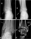
Figure 1
Figure 1
Lateral and medial malleolus fractures. (A,B) The lateral malleolus fracture was shown (arrow) with the fracture line below the inferior tibiofibular joint level. This is Weber type A stage 1 and supination-adduction stage 1 of the Lauge-Hansen classification. (C,D) The lateral malleolus fracture (short arrow) and the medial malleolus fracture (long arrow) were demonstrated. According to the Danis-Weber classification, this is Weber A stage 2. According to the Lauge-Hansen classification system, this is supination-adduction stage 2. The lateral malleolus fracture line is transverse below the inferior tibiofibular joint level. The medial malleolus fracture is higher than the lateral malleolus fracture with the medial malleolus fracture line being vertical or oblique.
Figure 2
Figure 2
Fibular fracture (A,B) is shown at the inferior tibiofibular joint level (arrow). It is stage 2 of Weber Type B fracture or of supination-external rotation of the Lauge-Hansen classification.
Figure 3
Figure 3
Lateral and medial malleolus fractures. (A) Anteroposterior radiograph shows lateral and medial malleolus fractures with inhomogeneous density at the distal tibial end which suggests posterior malleolus fracture. The inferior tibiofibular joint space is increased. B&C. Lateral radiograph (B) reveals the posterior malleolus fracture (B) which is confirmed by computed tomographic scan (C). This is stage 4 of the Weber type B fracture or of the supination-external rotation of the Lauge-Hansen classification.
Figure 4
Figure 4
Medial malleolus fracture and high-location fibular fracture. (A). The medial malleolus fracture (short arrow) and the fibular fracture (long arrow) are shown. (B). Lateral radiograph shows inhomogeneous density at the posterior malleolus, suggesting possible fracture at this site. (C). Computed tomographic scan confirms the posterior malleolus fracture (arrow). This is stage 4 of the pronation-external rotation type of the Lauge-Hansen classification. The medial malleolus fracture line is transverse. The fibular fracture is oblique or spiral and is frequently located above the inferior tibiofibular joint level.
Figure 5
Figure 5
Medial malleolus fracture and fibular fracture above the lower tibiofibular syndesmosis. The medial malleolus fracture (short arrow in A) and the fibular fracture (long arrow in both A,B) are demonstrated. This is stage 3 of the Lauge-Hansen pronation-abduction fracture. The fibular fracture line is transverse above the inferior tibiofibular joint level sometimes with small bone fragments but without accompanying posterior malleolus fracture.
Figure 6
Figure 6
Distribution of ankle factures in the Lauge-Hansen type and stage, sex and age. SAD, supination-adduction; SE, supination-external rotation; PE, pronation-external rotation; PAB, pronation-abduction.
Figure 7
Figure 7
Distribution of ankle fractures in Danis-Weber classification and of compression fractures in age and sex. There were 1225 ankle fractures in Danis-Weber type A, 1640 in type B, 751 in type C, and 336 in compression fractures.

Similar articles

Cited by

References

    1. Barker HB, Beynnon BD, Renstrom PA. Ankle injury risk factors in sports. Sports Med. 1997;23:69–74. doi: 10.2165/00007256-199723020-00001. - DOI - PubMed
    1. Boszczyk A, et al. X-ray features to predict ankle fracture mechanism. Forensic Sci Int. 2018;291:185–192. doi: 10.1016/j.forsciint.2018.08.042. - DOI - PubMed
    1. Boyce SH, Quigley MA. Review of sports injuries presenting to an accident and emergency department. Emerg Med J. 2004;21:704–706. doi: 10.1136/emj.2002.002873. - DOI - PMC - PubMed
    1. Eastgate P, Davidson R, McPhail SM. Radiographic imaging for traumatic ankle injuries: a demand profile and investigation of radiological reporting timeframes from an Australian tertiary facility. J Foot Ankle Res. 2014;7:25. doi: 10.1186/1757-1146-7-25. - DOI - PMC - PubMed
    1. Fonseca LLD, et al. Reproducibility of the Lauge-Hansen, Danis-Weber, and AO classifications for ankle fractures. Rev Bras Ortop. 2018;53:101–106. doi: 10.1016/j.rboe.2017.11.013. - DOI - PMC - PubMed