. 2020 May;581(7806):71-76.
doi: 10.1038/s41586-020-2247-3. Epub 2020 Apr 29.

APOE4 leads to blood-brain barrier dysfunction predicting cognitive decline

Affiliations

APOE4 leads to blood-brain barrier dysfunction predicting cognitive decline

Axel Montagne et al. Nature. 2020 May.

Abstract

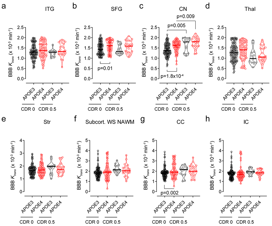
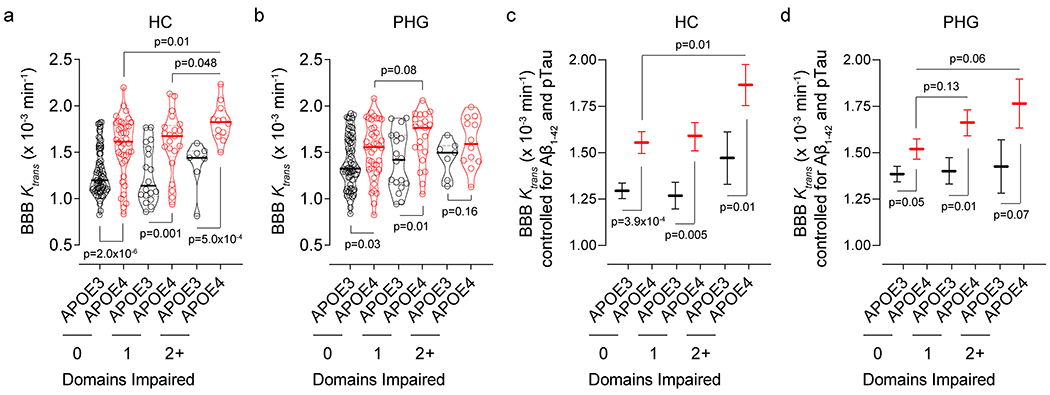
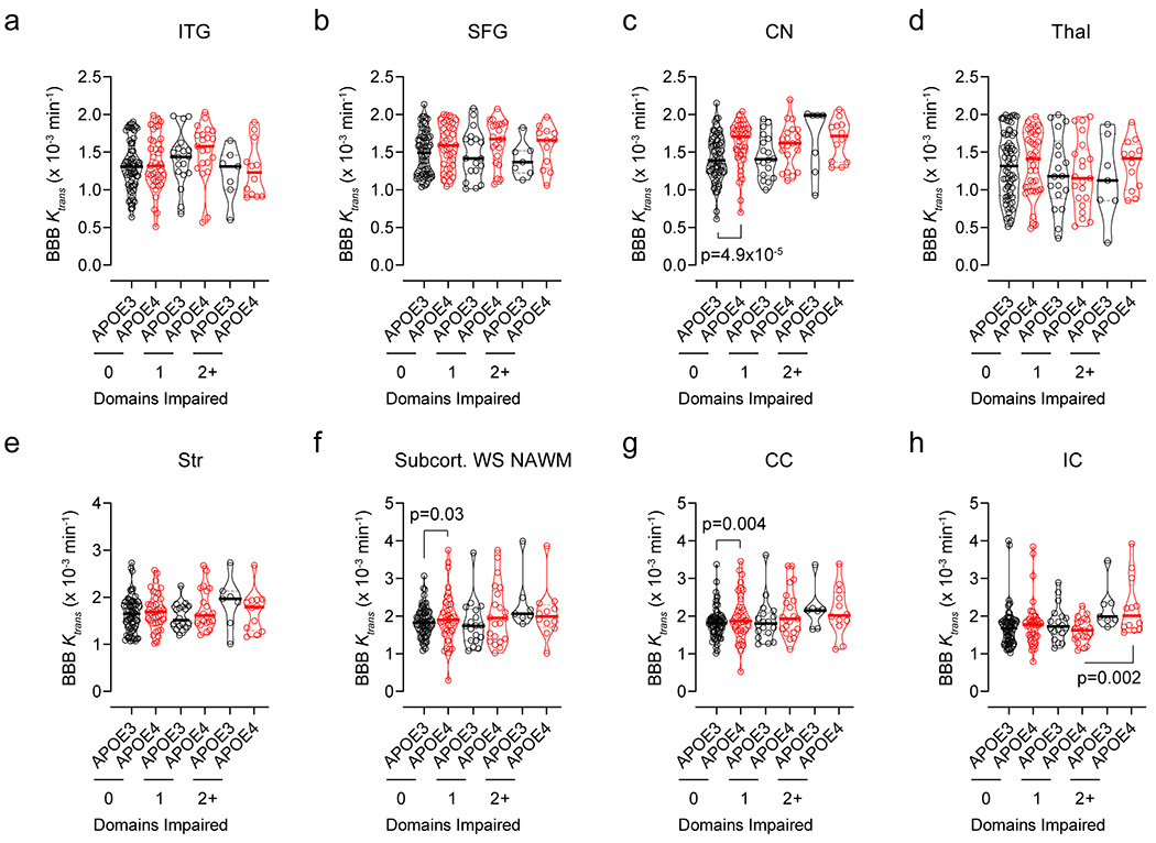
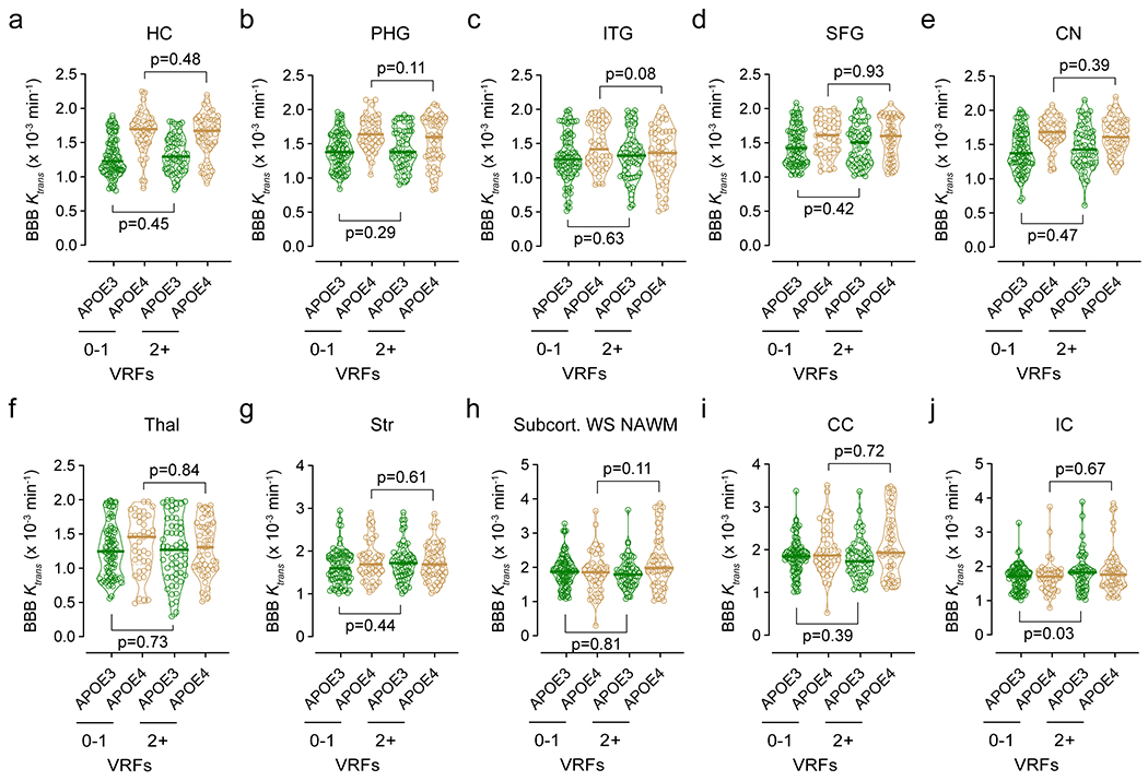
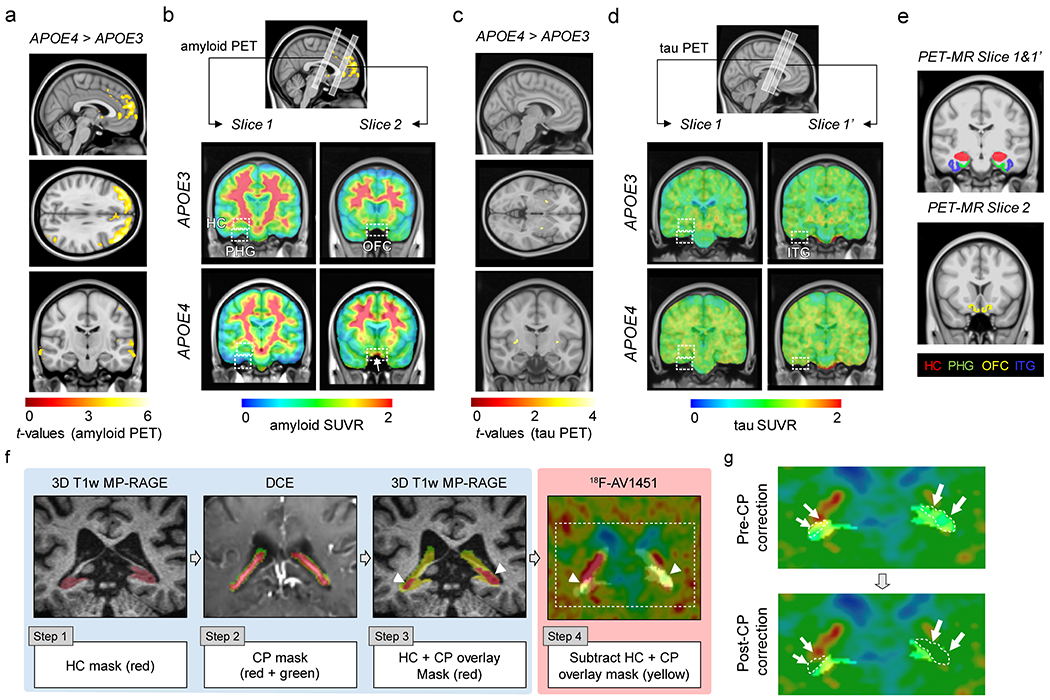
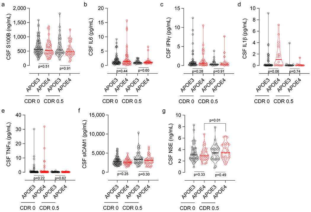
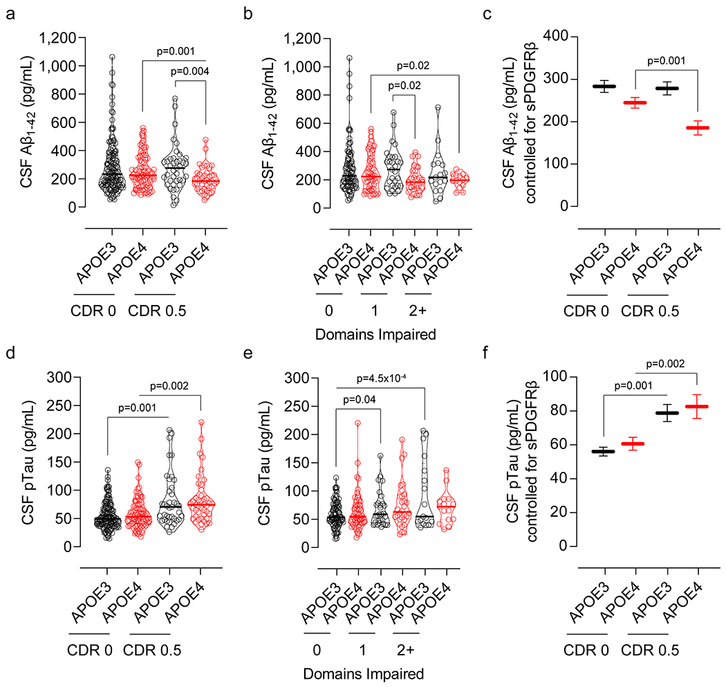
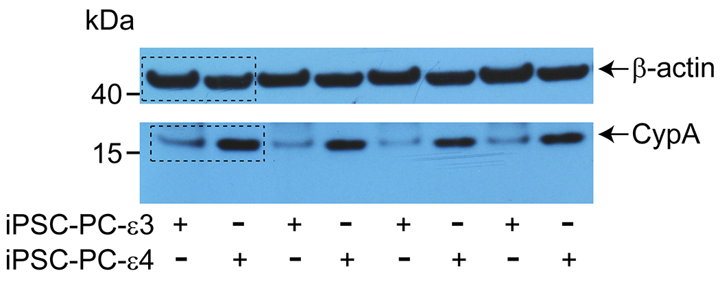
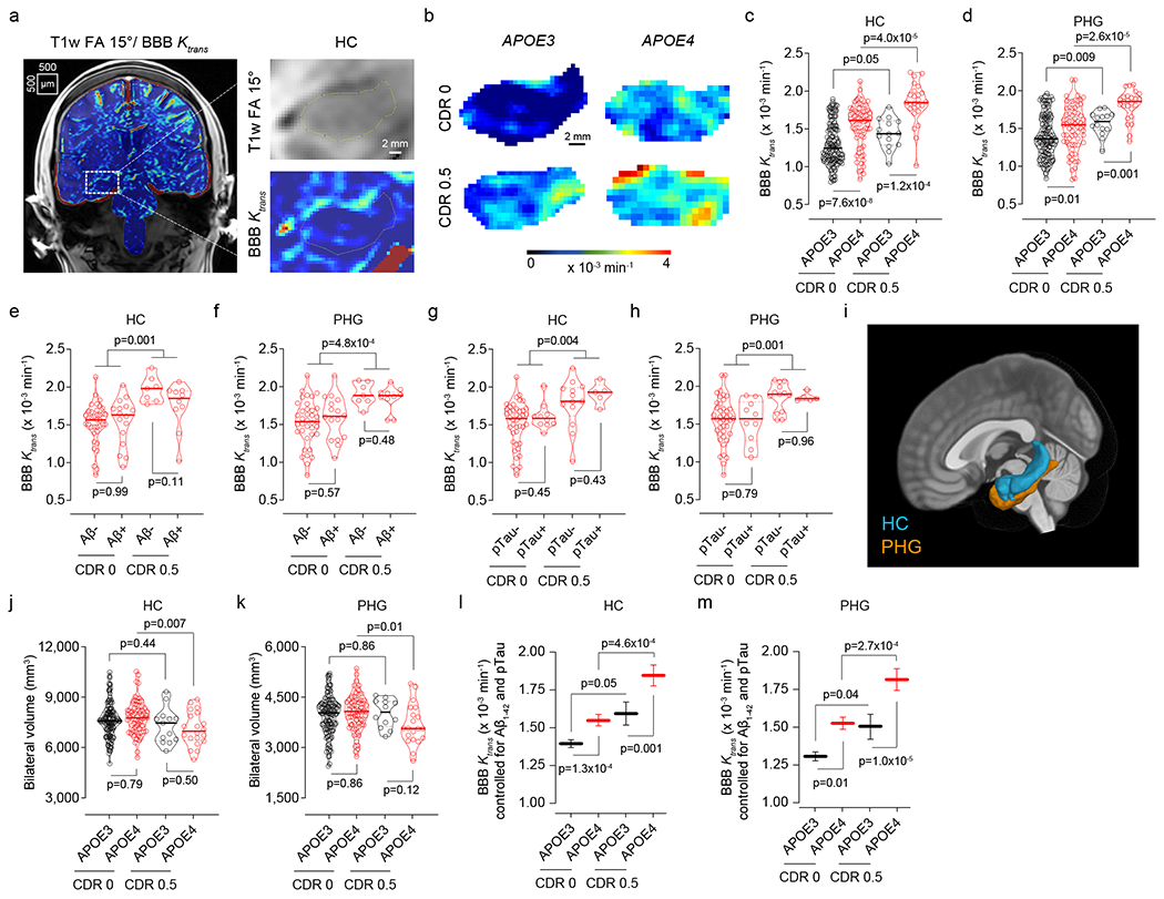
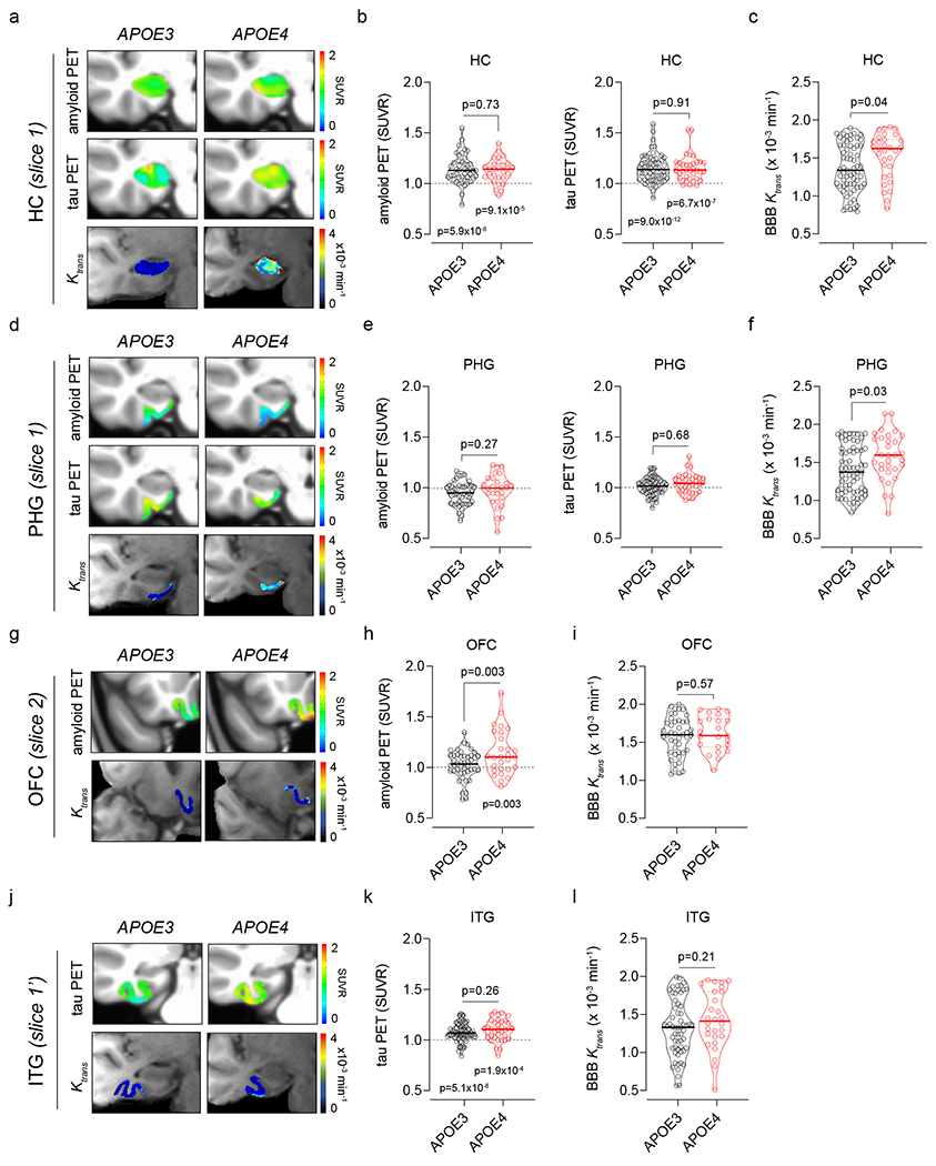
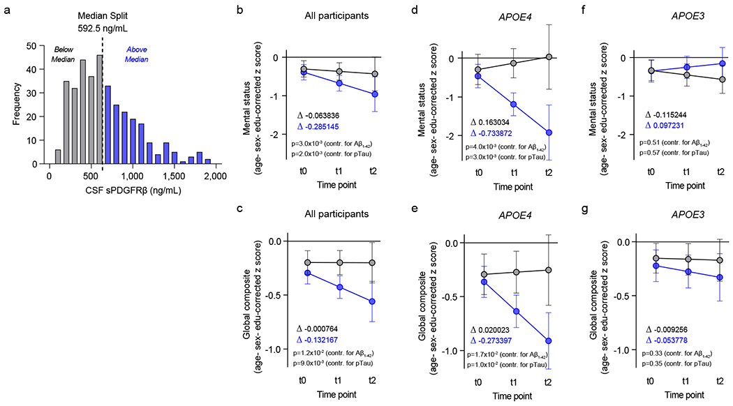
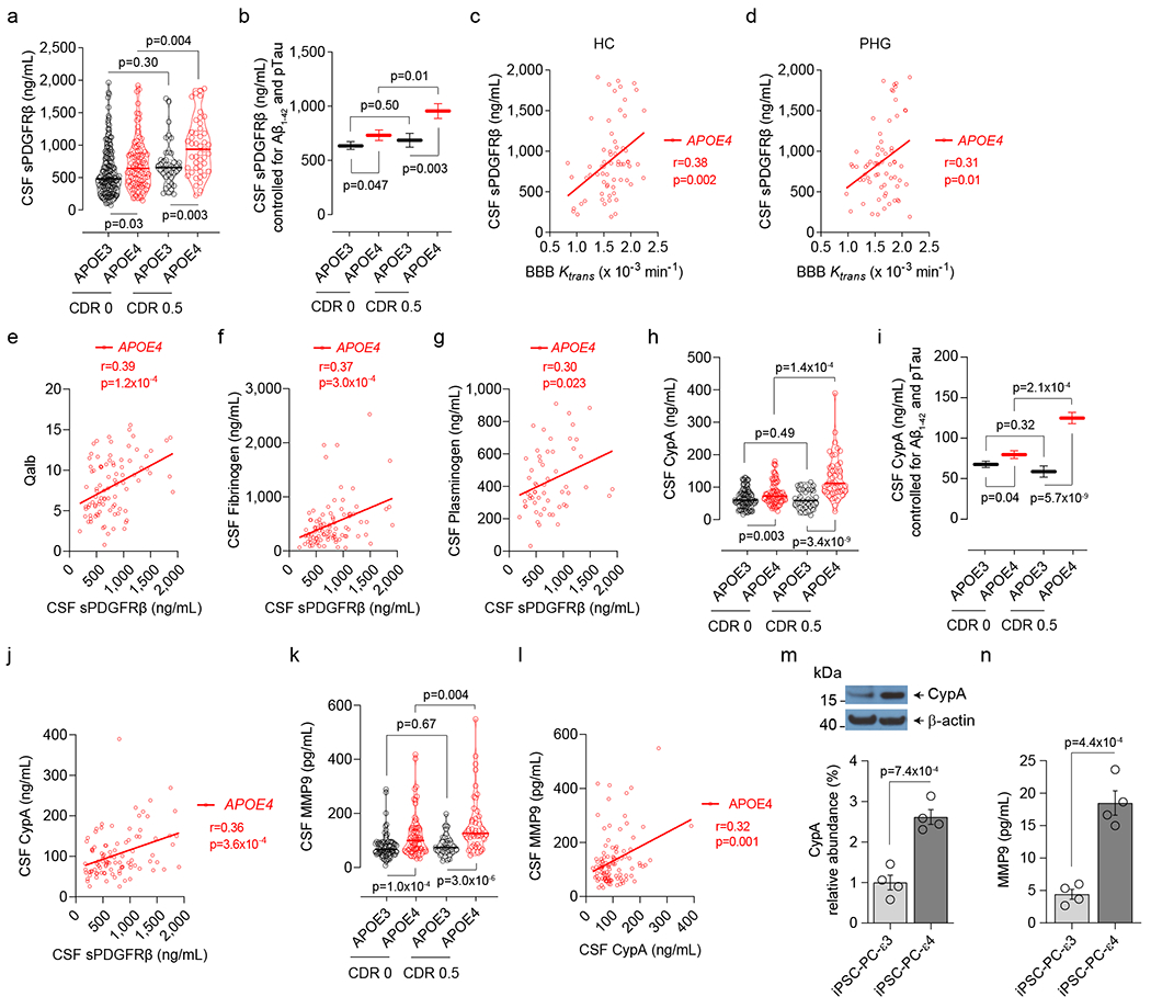
Vascular contributions to dementia and Alzheimer's disease are increasingly recognized1-6. Recent studies have suggested that breakdown of the blood-brain barrier (BBB) is an early biomarker of human cognitive dysfunction7, including the early clinical stages of Alzheimer's disease5,8-10. The E4 variant of apolipoprotein E (APOE4), the main susceptibility gene for Alzheimer's disease11-14, leads to accelerated breakdown of the BBB and degeneration of brain capillary pericytes15-19, which maintain BBB integrity20-22. It is unclear, however, whether the cerebrovascular effects of APOE4 contribute to cognitive impairment. Here we show that individuals bearing APOE4 (with the ε3/ε4 or ε4/ε4 alleles) are distinguished from those without APOE4 (ε3/ε3) by breakdown of the BBB in the hippocampus and medial temporal lobe. This finding is apparent in cognitively unimpaired APOE4 carriers and more severe in those with cognitive impairment, but is not related to amyloid-β or tau pathology measured in cerebrospinal fluid or by positron emission tomography23. High baseline levels of the BBB pericyte injury biomarker soluble PDGFRβ7,8 in the cerebrospinal fluid predicted future cognitive decline in APOE4 carriers but not in non-carriers, even after controlling for amyloid-β and tau status, and were correlated with increased activity of the BBB-degrading cyclophilin A-matrix metalloproteinase-9 pathway19 in cerebrospinal fluid. Our findings suggest that breakdown of the BBB contributes to APOE4-associated cognitive decline independently of Alzheimer's disease pathology, and might be a therapeutic target in APOE4 carriers.


Conflict of interest statement

Competing Interests

The authors declare no competing interests.

Figures

Extended Data Figure 1.
Extended Data Figure 1.. Regional BBB Ktrans constant in eight additional brain regions in APOE4 carriers and non-carriers (APOE3) with CDR status 0 and 0.5.
DCE-MRI BBB permeability, Ktrans constant, in the inferior temporal gyrus (ITG, a), superior frontal gyrus (SFG, b), caudate nucleus (CN, c), thalamus (Thal, d), striatum (Str, e), subcortical watershed normal-appearing white matter (Subcort. WS NAWM, f), corpus callosum (CC, g), and internal capsule (IC, h) in CDR 0 APOE3 (black, n=128) and APOE4 (red, n=68) carriers, CDR 0.5 APOE3 (black, n=14) and APOE4 (red, n=25) carriers. Violin plot continuous lines indicate median values and dotted lines indicate interquartile range. Significance by ANCOVAs for main effects and post-hoc comparisons controlling for age, sex, and education.
Extended Data Figure 2.
Extended Data Figure 2.. BBB breakdown in the hippocampus and parahippocampal gyrus in APOE4 carriers increases with cognitive domain impairment.
(a,b) DCE-MRI BBB permeability, Ktrans constant, in the hippocampus (HC, a) and parahippocampal gyrus (PHG, b) in individuals with 0 cognitive domain impaired that are APOE3 (black, n=70) and APOE4 (red, n=40) carriers, 1 cognitive domain impaired that are APOE3 (black, n=18) and APOE4 (red, n=21) carriers, and 2+ cognitive domains impaired that are APOE3 (black, n=7) and APOE4 (red, n=12) carriers. (c,d) Ktrans (estimated marginal means ± SEM from ANCOVA models corrected for age, sex, education, CSF Aβ1-42 and pTau status, and HC and PHG volumes) in the HC (c) and PHG (d) in individuals with 0 cognitive domain impaired that are APOE3 (black, n=70) and APOE4 (red, n=40) carriers, 1 cognitive domain impaired that are APOE3 (black, n=18) and APOE4 (red, n=21) carriers, and 2+ cognitive domains impaired that are APOE3 (black, n=7) and APOE4 (red, n=12) carriers. Panels a and b: Violin plot continuous lines indicate median values and dotted lines indicate interquartile range. Significance by ANCOVA for main effects and post-hoc comparisons controlling for age, sex, and education. All ANCOVA omnibus tests remained significant at false discovery rate threshold of 0.05.
Extended Data Figure 3.
Extended Data Figure 3.. Regional BBB Ktrans constant in eight additional brain regions in APOE4 carriers and non-carriers (APOE3) with different degree of cognitive domain impairment.
DCE-MRI BBB permeability, Ktrans constant, in the inferior temporal gyrus (ITG, a), superior frontal gyrus (SFG, b), caudate nucleus (CN, c), thalamus (Thal, d), striatum (Str, e), subcortical watershed normal-appearing white matter (Subcort. WS NAWM, f), corpus callosum (CC, g), and internal capsule (IC, h) in individuals with 0 cognitive domain impaired that are APOE3 (black, n=70) and APOE4 (red, n=40) carriers, 1 cognitive domain impaired that are APOE3 (black, n=18) and APOE4 (red, n=21) carriers, and 2+ cognitive domains impaired that are APOE3 (black, n=7) and APOE4 (red, n=12) carriers. Violin plot continuous lines indicate median values and dotted lines indicate interquartile range. Significance tests from ANCOVAs for main effects and post-hoc comparisons controlling for age, sex, and education.
Extended Data Figure 4.
Extended Data Figure 4.. Regional BBB Ktrans constant in all studied brain regions in APOE4 carriers and non-carriers (APOE3) in relation to vascular risk factors.
DCE-MRI BBB permeability, Ktrans constant, in the hippocampus (HC, a), parahippocampal gyrus (PHG, b), inferior temporal gyrus (ITG, c), superior frontal gyrus (SFG, d), caudate nucleus (CN, e), thalamus (Thal, f), striatum (Str, g), subcortical watershed normal-appearing white matter (Subcort. WS NAWM, h), corpus callosum (CC, i), and internal capsule (IC, j) in APOE3 (black, n=80) and APOE4 (red, n=42) carriers with 0-1 vascular risk factors (VRFs), and APOE3 (black, n=58) and APOE4 (red, n=51) carriers with 2+ VRFs. Violin plot continuous lines indicate median values and dotted lines indicate interquartile range. Significance by ANCOVAs for main effects and post-hoc comparisons controlling for age, sex, and education (ns=non-significant).
Extended Data Figure 5.
Extended Data Figure 5.. Amyloid and tau PET analysis in APOE4 carriers and correction of 18F-AV1451 off-target binding in the choroid plexus.
All studies were performed in individuals with clinical dementia rating score 0. Amyloid and tau PET studies were conducted using 18F-Florbetaben (FBB) or 18F-Florbetapir (FBP), and 18F-Flortaucipir (AV1451), respectively. For amyloid PET data analysis, FBP and FBB datasets were combined. (a) Uptake of amyloid tracers by the orbital frontal cortex (OFC) in APOE4 (n=29) relative to APOE3 (n=45) carriers (voxel-wise 2-sample one-tailed t-tests). (b) Representative amyloid PET Standardized Uptake Value Ratios (SUVR) maps from APOE3 homozygote (APOE3) (upper) and APOE4 carrier (APOE4) (lower). Slices 1 and 2, regions-of-interest (ROIs) for amyloid PET and BBB DCE-MRI scans (see e). Arrow, amyloid tracer uptake by OFC. The APOE3 and APOE4 representative images used FBP. (c) Uptake of tau tracer shows undetectable tau accumulation in APOE3 (n=60) or APOE4 (n=37) carriers (voxel-wise 2-sample one-tailed t-tests). (d) Representative tau PET SUVR maps from APOE3 (upper) and APOE4 (lower) carriers. Slice 1 and slice 1’, ROIs for tau PET and BBB DCE-MRI scans (see e). (e) Coronal 3D scans of regions studied in main Figure 2: hippocampus (HC; red), parahippocampal gyrus (PHG; green), medial orbital frontal cortex (OFC; yellow), and inferior temporal gyrus (ITG; blue). (f) Correction of 18F-AV1451 off-target binding in the choroid plexus: Step 1) Hippocampus (HC) masks were generated from the 3D T1-weighted MP-RAGE; Step 2) Choroid plexus (CP) masks were generated from the T1-weighted VIBE image post-GBCA (flip angle = 15°) image; Step 3) HC and CP masks were overlaid (arrowheads, red); and Step 4) CP overlap with HC masks (arrowheads, yellow) were subtracted to obtain CP-corrected HC tau PET signal after adding 6-mm voxel size on top of CP mask generated from DCE data. (g) Representative images of HC tau PET signal before (left) and after (right) applying the CP correction (arrows and white dotted lines = overlap between HC and CP).
Extended Data Figure 6.
Extended Data Figure 6.. CSF biomarkers of glia and inflammatory response and endothelial and neuronal cell injury in APOE4 carriers and non-carriers (APOE3).
(a) CSF astrocytic S100 calcium-binding protein B (S100B) levels in CDR 0 APOE3 (black, n=77) and APOE4 (red, n=41) carriers, and CDR 0.5 APOE3 (black, n=39) and APOE4 (red, n=32) carriers. (b) CSF interleukin 6 (IL6) levels in CDR 0 APOE3 (black, n=71) and APOE4 (red, n=47) carriers, and CDR 0.5 APOE3 (black, n=34) and APOE4 (red, n=32) carriers. (c) CSF interferon gamma (IFNγ) levels in CDR 0 APOE3 (black, n=54) and APOE4 (red, n=29) carriers, and CDR 0.5 APOE3 (black, n=25) and APOE4 (red, n=17) carriers. (d) CSF interleukin 1β (IL1β) levels in CDR 0 APOE3 (black, n=43) and APOE4 (red, n=18) carriers, and CDR 0.5 APOE3 (black, n=17) and APOE4 (red, n=13) carriers. (e) CSF tumor necrosis factor α (TNFα) levels in CDR 0 APOE3 (black, n=70) and APOE4 (red, n=46) carriers, and CDR 0.5 APOE3 (black, n=34) and APOE4 (red, n=32) carriers. (f) CSF soluble intercellular adhesion molecule 1 (sICAM1) levels in CDR 0 APOE3 (black, n=77) and APOE4 (red, n=40) carriers, and CDR 0.5 APOE3 (black, n=39) and APOE4 (red, n=33) carriers. (g) CSF Neuron-Specific Enolase (NSE) levels in CDR 0 APOE3 (black, n=47) and APOE4 (red, n=32) carriers, and CDR 0.5 APOE3 (black, n=29) and APOE4 (red, n=29) carriers. Violin plot continuous lines indicate median values and dotted lines indicate interquartile range. Panels a and b had one outlier each, which were removed prior to statistical analysis using methods described in Statistical Analyses section. Significance by ANCOVAs for main effects and post-hoc comparisons controlling for age, sex, and education (ns=non-significant).
Extended Data Figure 7.
Extended Data Figure 7.. Lower CSF Aβ1-42 and increased pTau levels in APOE4 carriers with cognitive impairment.
(a) CSF Aβ1-42 levels in CDR 0 APOE3 (black, n=141) and APOE4 (red, n=83) and CDR 0.5 APOE3 (black, n=39) and APOE4 (red, n=41) carriers. (b) CSF Aβ1-42 levels in APOE3 (black, n=89) and APOE4 (red, n=55) carriers with 0 cognitive domain impaired, APOE3 (black, n=29) and APOE4 (red, n=31) carriers with 1 cognitive domain impaired, and APOE3 (black, n=17) and APOE4 (red, n=14) carriers with 2+ cognitive domains impaired. (c) CSF Aβ1-42 levels (estimated marginal means ± SEM from ANCOVA models corrected for age, sex, education, and CSF sPDGFRβ levels) in CDR 0 APOE3 (black, n=141) and APOE4 (red, n=83) and CDR 0.5 APOE3 (black, n=39) and APOE4 (red, n=41) carriers. (d) CSF pTau levels in CDR 0 APOE3 (black, n=141) and APOE4 (red, n=82) and CDR 0.5 APOE3 (black, n=39) and APOE4 (red, n=43) carriers. (e) CSF pTau levels in APOE3 (black, n=89) and APOE4 (red, n=56) carriers with 0 cognitive domain impaired, APOE3 (black, n=29) and APOE4 (red, n=30) carriers with 1 cognitive domain impaired, and APOE3 (black, n=17) and APOE4 (red, n=15) carriers with 2+ cognitive domains impaired. (f) CSF pTau levels (estimated marginal means ± SEM from ANCOVA models corrected for age, sex, education, and CSF sPDGFRβ levels) in CDR 0 APOE3 (black, n=141) and APOE4 (red, n=82) and CDR 0.5 APOE3 (black, n=39) and APOE4 (red, n=43) carriers. Violin plot continuous lines indicate median values and dotted lines indicate interquartile range. CSF Aβ1-42 and pTau values were log10-transformed due to non-normal distribution prior to statistical analysis. Significance tests from ANCOVAs for main effects and post-hoc comparisons controlling for age, sex, and education.
Extended Data Figure 8.
Extended Data Figure 8.. Full scans of western blots.
Full scans of western blots for CypA shown in Figure 4 panel m (top).
Figure 1.
Figure 1.. Blood-brain barrier breakdown in the hippocampus and parahippocampal gyrus in APOE4 carriers increases with cognitive impairment independently of CSF Aβ and tau status.
(a,b) Blood-brain barrier (BBB) permeability Ktrans maps generated by dynamic contrast-enhanced magnetic resonance imaging in the hippocampus (HC) of APOE3 homozygotes (APOE3) and APOE4 carriers (APOE4) with clinical dementia rating (CDR) score 0 or 0.5. (c,d) BBB permeability, Ktrans constant, in the HC (c) and parahippocampal gyrus (PHG, d) in CDR 0 APOE3 (black, n=128) and APOE4 (red, n=68) and CDR 0.5 APOE3 (black, n=14) and APOE4 (red, n=25) carriers. (e,f) Ktrans values in the HC (e) and PHG (f) in APOE4 carriers CDR 0 Aβ1-42 negative (Aβ-; n=37) or positive (Aβ+; n=16), and CDR 0.5 Aβ- (n=7) or Aβ+ (n=10). (g,h) Ktrans values in the HC (g) and PHG (h) in APOE4 carriers CDR 0 pTau negative (pTau−; n=42) or positive (pTau+; n=10), and CDR 0.5 pTau− (n=13) or pTau+ (n=5). (i) HC (blue) and PHG (orange) overlaid on a 3D template. (j,k) HC (j) and PHG (k) volumes in CDR 0 APOE3 (n=124) and APOE4 (n=75) and CDR 0.5 APOE3 (n=13) and APOE4 (n=20) carriers. (l,m) Ktrans (estimated marginal means ± SEM from ANCOVA models corrected for age, sex, education, CSF Aβ1-42 and pTau status, and HC and PHG volumes) in the HC (l) and PHG (m) in CDR 0 APOE3 (black, HC n=125; PHG n=128) and APOE4 (red, HC and PHG n=68) and CDR 0.5 APOE3 (black, HC n=12; PHG n=14) and APOE4 (red, HC n=20; PHG n=25) carriers. In c-h, j, and k, continuous line indicates median value; dotted line indicates interquartile range. Significance by ANCOVA for main effects and post-hoc comparisons controlling for age, sex, and education. All ANCOVA omnibus tests remained significant at false discovery rate threshold of 0.05.
Figure 2.
Figure 2.. Blood-brain barrier breakdown in APOE4 carriers is independent of amyloid and tau brain accumulation.
All studies were performed in individuals with clinical dementia rating score 0. (a) Representative superimposed left-hippocampus (HC) amyloid PET (top), tau PET (middle), and BBB Ktrans maps (bottom) from APOE3 (left) and APOE4 (right) carriers. (b,c) HC: amyloid and tau tracer uptake (b) and BBB Ktrans constant (c) in APOE3 (n=45, 60, and 65) and APOE4 (n=29, 37, and 31) carriers. (d) Representative superimposed left-parahippocampal gyrus (PHG) amyloid PET (top), tau PET (middle), and BBB Ktrans maps (bottom) from APOE3 (left) and APOE4 (right) carriers. (e,f) PHG: amyloid and tau tracer uptake (e) and BBB Ktrans constant (f) in APOE3 (n=45, 60, and 65) and APOE4 (n=29, 37, and 31) carriers. (g) Representative superimposed left-medial orbital frontal cortex (OFC) amyloid PET (top) and BBB Ktrans maps (bottom) from APOE3 (left) and APOE4 (right) carriers. (h,i) OFC: amyloid tracer uptake (h) and BBB Ktrans constant (i) in APOE3 (n=45 and 44) and APOE4 (n=29 and 23) carriers. (j) Representative superimposed left-inferior temporal gyrus (ITG) tau PET (top) and BBB Ktrans maps (bottom) from APOE3 (left) and APOE4 (right) carriers. (k,l) ITG: tau tracer uptake (l) and BBB Ktrans constant (l) in APOE3 (n=60 and 59) and APOE4 (n=37 and 28) carriers. In b, c, e, f, h, i, k, and l, continuous lines indicate median values and dotted lines indicate interquartile range. The BBB Ktrans constant was determined in all participants (see Extended Data Tables 2a,b) who received either both amyloid and tau tracers (n=58), only amyloid tracer (n=9) or only tau tracer (n= 29). Significance by ANCOVA for group comparisons controlling for age, sex, and education, and one-tailed t-tests for comparison of PET values to SUVR = 1.
Figure 3.
Figure 3.. Elevated baseline CSF levels of pericyte injury biomarker sPDGFRβ predict cognitive decline in APOE4 carriers.
(a) Histogram frequency distribution of CSF sPDGFRβ values using median split to divide participants into two groups with high (blue, above median 600-2,000 ng/ml) and low (grey, below median; 0-600 ng/ml) baseline CSF sPDGFRβ levels. All longitudinal analyses used baseline CSF sPDGFRβ as a continuous predictor of future cognitive decline. (b-c) Linear mixed model analysis of all studied participants (n=146) followed over 2-year intervals for up to 4.5 years after baseline lumbar puncture (LP) shows that higher baseline CSF sPDGFRβ (blue) predicts greater decline in demographically-corrected mental status exam scores over time (p=0.01), which remains significant after controlling for CSF Aβ (p=0.002) and pTau (p=0.002) status (b), and in global cognitive composite scores (p=0.01), which also remains significant after controlling for CSF Aβ (p=0.017) and pTau (p=0.01) status (c). (d-e) Higher CSF sPDGFRβ (blue) in APOE4 carriers (n=58) predicts future decline in mental status exam scores (p=0.005) after controlling for CSF Aβ (p=0.004) and pTau (p=0.003) status (d), and in global cognitive composite scores (p=0.02) after controlling for CSF Aβ (p=0.02) and pTau (p=0.01) status (e). (f-g) Baseline CSF sPDGFRβ does not predict decline in APOE3 homozygotes (n=88) on either mental status (f) or global composite (g) scores regardless of CSF Aβ or pTau status. Panels b-g, for graphical depiction, separate lines indicate median split of baseline CSF sPDGFRβ (grey, below median; blue, above median). ∆ slopes provided for median split of baseline CSF sPDGFRβ groups (grey, below median; blue, above median). Time was modeled as t0 = −1 to 0.5 years post-LP, t1 = 0.5 to 2.5 years post-LP, and t2 = 2.5 to 4.5 years post-LP. The error bars are SE of the estimate. In panels b-g, significance by linear mixed model analysis; no multiple comparison correction applied. See Supplementary Information Tables 2–4 for detailed statistics.
Figure 4.
Figure 4.. Elevated CSF sPDGFRβ, cyclophilin A and matrix metalloproteinase-9 in APOE4 carriers.
(a) CSF sPDGFRβ in CDR 0 APOE3 (black, n=152) and APOE4 (red, n=95) and CDR 0.5 APOE3 (black, n=42) and APOE4 (red, n=45) carriers. (b) CSF sPDGFRβ (estimated marginal means ± SEM from ANCOVA models corrected for age, sex, education, CSF Aβ1-42 and pTau status) in CDR 0 APOE3 (black, n=152) and APOE4 (red, n=95) and CDR 0.5 APOE3 (black, n=42) and APOE4 (red, n=45) carriers. (c,d) CSF sPDGFRβ and BBB Ktrans correlation in the hippocampus (HC, n=65; c) and parahippocampal gyrus (PHG, n=65; d) in APOE4 carriers. (e-g) Correlations between CSF sPDGFRβ and albumin quotient (Qalb, n=92; e), fibrinogen (n=93; f), and plasminogen (n=57; g) in APOE4 carriers. (h) CSF cyclophilin A (CypA) in CDR 0 APOE3 (black, n=75) and APOE4 (red, n=62) and CDR 0.5 APOE3 (black, n=33) and APOE4 (red, n=45) carriers. (i) CSF CypA (estimated marginal means ± SEM from ANCOVA models corrected for age, sex, education, CSF Aβ1-42 and pTau status) in CDR 0 APOE3 (black, n=75) and APOE4 (red, n=62) and CDR 0.5 APOE3 (black, n=33) and APOE4 (red, n=45) carriers. (j) CSF CypA and sPDGFRβ correlation in APOE4 carriers (n=96). (k) CSF matrix metalloproteinase-9 (MMP9) in CDR 0 APOE3 (black, n=72) and APOE4 (red, n=68) and CDR 0.5 APOE3 (black, n=33) and APOE4 (red, n=45) carriers. (l) CSF MMP9 and CypA correlation in APOE4 carriers (n=104). (m,n) CypA (m; see Extended Data Fig. 8), and secreted MMP9 in the culture medium (n), in human iPSC-derived APOE3 (ε3/ε3) and APOE4 (ε4/ε4) pericytes. Mean + SEM from four independent culture replicates. In a, h and k, continuous lines indicate median values and dotted lines interquartile range. Significance by ANCOVA for main effects and post-hoc comparisons controlling for age, sex, and education. Panels c-g, j, and l: two-tailed simple linear regression; Pearson correlation coefficient (r). Panels m and n, unpaired one-tailed Student t-test.

Comment in

Similar articles

Cited by

References

    1. Wardlaw JM et al. Neuroimaging standards for research into small vessel disease and its contribution to ageing and neurodegeneration. Lancet Neurol. 12, 822–838 (2013). - PMC - PubMed
    1. Kapasi A, DeCarli C & Schneider JA Impact of multiple pathologies on the threshold for clinically overt dementia. Acta Neuropathol. (Berl.) 134, 171–186 (2017). - PMC - PubMed
    1. Iadecola C The Neurovascular Unit Coming of Age: A Journey through Neurovascular Coupling in Health and Disease. Neuron 96, 17–42 (2017). - PMC - PubMed
    1. Iturria-Medina Y et al. Early role of vascular dysregulation on late-onset Alzheimer’s disease based on multifactorial data-driven analysis. Nat. Commun 7, 11934 (2016). - PMC - PubMed
    1. Sweeney MD, Sagare AP & Zlokovic BV Blood-brain barrier breakdown in Alzheimer disease and other neurodegenerative disorders. Nat. Rev. Neurol 14, 133–150 (2018). - PMC - PubMed

MeSH terms