. 2020 May 6;10(1):7614.
doi: 10.1038/s41598-020-64492-5.

Intraoperative Laser Speckle Contrast Imaging For Real-Time Visualization of Cerebral Blood Flow in Cerebrovascular Surgery: Results From Pre-Clinical Studies

Affiliations

Intraoperative Laser Speckle Contrast Imaging For Real-Time Visualization of Cerebral Blood Flow in Cerebrovascular Surgery: Results From Pre-Clinical Studies

Antonella Mangraviti et al. Sci Rep. .

Abstract

Cerebrovascular surgery can benefit from an intraoperative system that conducts continuous monitoring of cerebral blood flow (CBF). Such a system must be handy, non-invasive, and directly integrated into the surgical workflow. None of the currently available techniques, considered alone, meets all these criteria. Here, we introduce the SurgeON™ system: a newly developed non-invasive modular tool which transmits high-resolution Laser Speckle Contrast Imaging (LSCI) directly onto the eyepiece of the surgical microscope. In preclinical rodent and rabbit models, we show that this system enabled the detection of acute perfusion changes as well as the recording of temporal response patterns and degrees of flow changes in various microvascular settings, such as middle cerebral artery occlusion, femoral artery clipping, and complete or incomplete cortical vessel cautery. During these procedures, a real-time visualization of vasculature and CBF was available in high spatial resolution through the eyepiece as a direct overlay on the live morphological view of the surgical field. Upon comparison with indocyanine green angiography videoangiography (ICG-VA) imaging, also operable via SurgeON, we found that direct-LSCI can produce greater information than ICG-VA and that continuous display of data is advantageous for performing immediate LSCI-guided adjustments in real time.


Conflict of interest statement

Authors A.R., J.C., K.R., S.I.C. and M.J.B. were employed by Vasoptic Medical, Inc. when the research was carried out. A.R., K.R., M.J.B have ownership interests in Vasoptic Medical, Inc. One or more patents pertaining to the system and methods described in this manuscript are assigned to Vasoptic Medical, Inc. and list A.R. and M.J.B. as inventors.

Figures

Figure 1
Figure 1
SurgeON System Schematic and Specifications. (A) As shown in the schematic, the SurgeON System comprises a surgical microscope modified to include the following key components: a near infrared (NIR) laser source (green) irradiates the target ROI images which are then captured by the NIR camera (blue). This camera is connected to a PC via MATLAB environment where these acquired laser speckle data are processed in real-time and a video-feed of the resulting blood flow information is transferred to the LSCI projector (yellow) to be seen by the operator through the microscope eyepiece. (B) The SurgeON System has imaging specifications that are suitable for neurosurgery.
Figure 2
Figure 2
SurgeON shows in vivo blood flow changes in real-time. (A) Frames of the recorded video during real-time visualization of the femoral artery and vein through the microscope’s eyepiece using morphologic, LSCI, LSCI overlay on morphological images and ICG-VA. (B) Frame of the real-time LSCI through the eyepiece microscope during the femoral artery clipping procedure. The bar graphs show the Mean + /- SE of the relative blood flow velocity indices (1/tauc values) acquired in 5 sets per each condition in 5 different rats in separate experiments.
Figure 3
Figure 3
SurgeON tracks CBF changes during MCA cautery. Frames of the real-time MCA images through the microscope’s eyepiece: morphologic view with overlaid LSCI, LSCI and ICG-VA images. Bar graphs showing significant (p ≤ 0.0001) decrease in blood flow velocity indices estimated using laser speckle contrast imaging after vessel cautery in each rat.
Figure 4
Figure 4
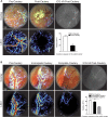
SurgeON provides more information than ICG-VA on CBF variation following MCAO. (A) Frames of real-time visualization of the cortical vessels through the microscope’s eyepiece: morphologic, LSCI, overlay of morphologic and LSCI and ICG-VA for comparison. (B) Representative real-time morphologic images with overlaid LSCI and LSCI only during pre-cautery, 2 hours post-cautery, 24 hours post-cautery and ICG-VA at 24 hours showing CBF decrease at 2 hours compared to the basal CBF and a partial restored blood flow at 24 hours. Bar graph showing mean + /−SE of blood flow velocity indices measured and aggregated across three vessels in each rat brain, in a total of five rats that underwent this procedure as part of three separate experiments. Note that ICG-VA reveals lingering fluorescence even 24 hours post cautery, possibly exacerbated by lack of clearance in a flow-arrested vessel whereas the noninvasive, quantitative LSCI technique clearly demonstrates a CBF reduction after MCAO.
Figure 5
Figure 5
Cortical CBF changes following MCAO resulted in proportional infarcted brain tissue. (A) Schematic representation of the MCAO procedure and brain area analyzed by TCC. (a) Illustration and pictures of the brain analyzed, the brains were harvested from wild type control rats and from the rats that underwent MCAO 24 hours post-surgery, and all were treated with TCC staining. (b) The brains were sliced and the infarcted area (white area) was measured via ImageJ. (c) Bar graphs show for each rat the mean + /- SE of the total and ischemic areas of the all sectioned slices per brain (white and black, respectively). (B) The table shows that the CBF (1/tauc) values in the cortical vessel of the analyzed area 24 hours post MCAO correlate with the extent of the infarcted area, which tends to be bigger in the groups of rats with a lower CBF at 24 hours.
Figure 6
Figure 6
SurgeON detects in real time incomplete vessel occlusion and guides immediate adjustments during a vascular procedure. (A) Frames of the recorded real-time imaging of the rabbit cortical vessels through the microscope’s eyepiece: morphologic, LSCI, overlay of morphologic and LSCI and ICG-VA. (B) Real-time overlay and LSCI of the surgical field of view after the first unsuccessful vessel’s cautery attempt, and then after the second when upon LSCI guidance we performed a complete vessel cautery. The bar graph shows the significant CBF reduction obtained only after the complete vessel cautery. ICG-VA imaging confirms the absence of flow in the brain trunk of the target vessel however gives no information on the reduced CBF in the vessel branches.

Similar articles

Cited by

References

    1. Gruber A, Dorfer C, Standhardt H, Bavinzski G, Knosp E. Prospective Comparison of Intraoperative Vascular Monitoring Technologies During Cerebral Aneurysm Surgery. Neurosurgery. 2011;68:657–673. doi: 10.1227/NEU.0b013e31820777ee. - DOI - PubMed
    1. Zaidi, H. A. et al. Indocyanine green angiography in the surgical management of cerebral arteriovenous malformations: lessons learned in 130 consecutive cases. Neurosurgery10 Suppl 2, 246–251; discussion 251, 10.1227/neu.0000000000000318 (2014). - PubMed
    1. Ma CY, et al. Intraoperative indocyanine green angiography in intracranial aneurysm surgery: Microsurgical clipping and revascularization. Clinical neurology and neurosurgery. 2009;111:840–846. doi: 10.1016/j.clineuro.2009.08.017. - DOI - PubMed
    1. Dashti, R., Laakso, A., Niemela, M., Porras, M. & Hernesniemi, J. Microscope-integrated near-infrared indocyanine green videoangiography during surgery of intracranial aneurysms: the Helsinki experience. Surgical neurology71, 543–550; discussion 550, 10.1016/j.surneu.2009.01.027 (2009). - PubMed
    1. Raabe A, et al. Prospective evaluation of surgical microscope-integrated intraoperative near-infrared indocyanine green videoangiography during aneurysm surgery. Journal of neurosurgery. 2005;103:982–989. doi: 10.3171/jns.2005.103.6.0982. - DOI - PubMed

Publication types