A Case Report: Neonatal Torsional Ovarian Cyst
- PMID: 32377121
- PMCID: PMC7192291
- DOI: 10.14744/SEMB.2018.48154
A Case Report: Neonatal Torsional Ovarian Cyst
Abstract
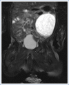
The majority of abdominal masses detected in the neonatal period are benign (85%) and usually originate in the urinary tract (50%), genital system (15%), gastrointestinal system (15%), or the hepatobiliary tract (5%). Ovarian cysts comprise one-third of the masses with a genital origin. Presently described is a case of an ovarian cyst that developed during the antenatal period and transformed into a hemorrhagic cystic mass as a result of torsion. A female infant born at 37 weeks of gestation with the prediagnosis of nephroma was admitted to the neonatal intensive care unit. Abdominal ultrasonography revealed a smooth cystic mass approximately 50x45x35 mm in size in the left upper quadrant that was not associated with the kidney. Magnetic resonance imaging revealed a 55x44x49-mm cystic mass in the left adnexal region containing multiple septations that were not enhanced with contrast material, and the mass was then interpreted as a hemorrhagic fetal ovarian cyst. The left ovary, compromised by 2 full torsions, was removed during a laparoscopy performed on the postnatal seventh day. The infant was subsequently discharged without complications. It should be kept in mind that cystic masses detected in the prenatal period may be of ovarian origin. An appropriate follow-up and treatment should be planned according to the size of the ovarian cyst and the clinical findings.
Keywords: Complication; newborn; ovarian cyst.
Copyright: © 2019 by The Medical Bulletin of Sisli Etfal Hospital.
Conflict of interest statement
Conflict of interest: None declared.
Figures
References
-
- Meizner I, Levy A, Katz M, Maresh AJ, Glezerman M. Fetal ovarian cysts:prenatal ultrasonographic detection and postnatal evaluation and treatment. Am J Obstet Gynecol. 1991;164:874–8. - PubMed
-
- Bryant AE, Laufer MR. Fetal ovarian cysts:incidence, diagnosis and management. J Reprod Med. 2004;49:329–37. - PubMed
-
- Özdilek B, Nalbantoğlu B, Donma MM, Çelik C, Paketçi C, Karasu E, et al. Yenidoğanda over kisti. Çocuk Dergisi. 2013;13:36–9.
-
- Sakala EP, Leon ZA, Rouse GA. Management of antenatally diagnosed fetal ovarian cysts. Obstet Gynecol Surv. 1991;46:407–14. - PubMed
-
- Turgal M, Ozyuncu O, Yazicioglu A. Outcome of sonographically suspected fetal ovarian cysts. J Matern Fetal Neonatal Med. 2013;26:1728–32. - PubMed
Publication types
LinkOut - more resources
Full Text Sources


