. 2020 Jul;22(1):454-464.
doi: 10.3892/mmr.2020.11111. Epub 2020 May 4.

Upregulation of microRNA‑1 inhibits proliferation and metastasis of breast cancer

Affiliations

Upregulation of microRNA‑1 inhibits proliferation and metastasis of breast cancer

Jing Peng et al. Mol Med Rep. 2020 Jul.

Abstract

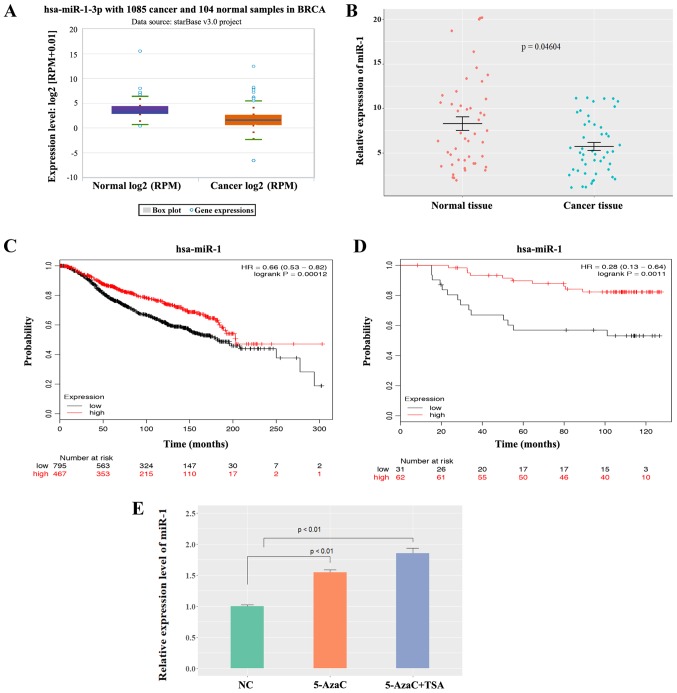
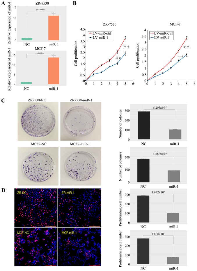
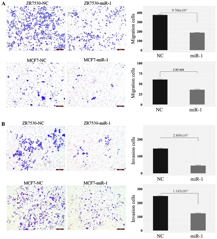
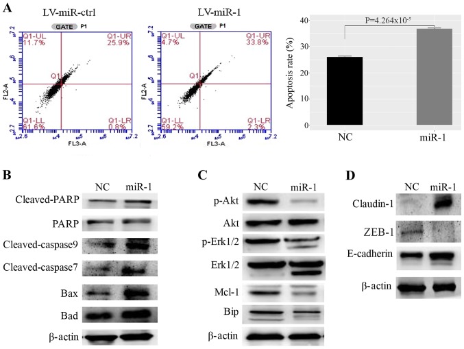
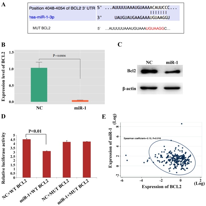
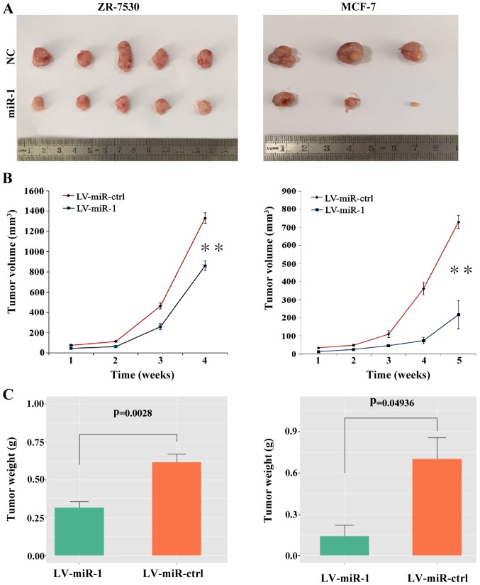
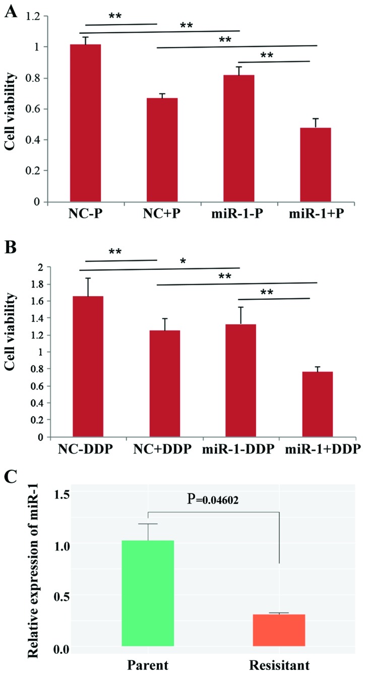
Recent studies have shown that microRNAs (miRs) play a key role in the regulation of cancer development. In the present study, reverse transcription‑quantitative PCR was used to detect the expression of miR‑1 in breast cancer and adjacent tissues, and survival analysis was performed to compare the low‑expression groups with the Kaplan-Meier method. Overexpression of miR‑1 was used to observe the effects on the proliferation, migration and invasion of breast cancer cells in vitro and in vivo. Moreover, Bcl‑2 expression was measured by western blotting and luciferase assays after the overexpression of miR‑1. The present study reported that miR‑1 is expressed at low levels in breast cancer and that cell proliferation, migration and invasion are inhibited in miR‑1‑overexpressing cells. Enhanced miR‑1 expression can also increase cell apoptosis. The present study also demonstrated that Bcl‑2 is a potential target of miR‑1. In vivo studies indicate that overexpression of miR‑1 decreases tumor volume and weight in nude mice. The data from the present study demonstrated for the first time that overexpression of miR‑1 increases the sensitivity of breast cancer cells to paclitaxel and cisplatin. The present study provided new evidence for the important role of miR‑1 in the tumorigenesis and drug sensitivity of breast cancer.

Keywords: breast cancer; microrna-1; Bcl-2; proliferation; metastasis.


Figures

Figure 1.
Figure 1.
Expression level of miR-1 in BC. (A) miR-1 was expressed at low levels in BC tissues analyzed by the StarBase3 database. (B) miR-1 was expressed at low levels in BC tissues compared with normal breast tissues (n=47, P=0.04604). (C) A high level of miR-1 was correlated with a good overall survival in BC patients according to data in METABRIC, P=0.00012. (D) A low level of miR-1 was correlated with poor overall survival in BC patients according to Dataset GSE19783, P=0.0011. (E) The miR-1 level was increased in MCF-7 human BC cells following 5-AzaC and TSA treatment (n=3, P<0.01). miR, microRNA; BC, breast cancer; 5-AzaC, 5-Azacytidine; TSA, trichostatin A; RPM, reads per million.
Figure 2.
Figure 2.
miR-1 inhibits BC cell growth. (A) Validation of miR-1 overexpression in BC cells (n=3, P<0.01). (B) A high level of miR-1 inhibited BC cell proliferation, as shown by CCK-8 assay (1-5: 12, 24, 48, 72 and 96 h), n=6, **P<0.01. (C) A high level of miR-1 inhibited BC cell clone formation, n=3, P<0.01. (D) A high level of miR-1 inhibited BC cell proliferation, as demonstrated by EdU essay, n=3, P<0.01. miR, microRNA; BC, breast cancer; NC, normal control; ctrl, control; LV, lentiviral.
Figure 3.
Figure 3.
Overexpression of miR-1 inhibits cell migration and invasion. (A) A high level of miR-1 inhibits the migration ability of BC cells, n=3, P<0.01). (B) A high level of miR-1 inhibits the invasion ability of BC cells, n=3, P<0.01, (scale bar=200 µm). miR, microRNA; NC, normal control.
Figure 4.
Figure 4.
Upregulation of miR-1 promotes MCF-7 cell apoptosis. (A) miR-1 promotes apoptosis of MCF7 cells, as demonstrated by fluorescence activated cell sorting, n=3, P<0.01. (B) Proteins that promote apoptosis were upregulated in miR-1-overexpressing cells. (C) Proteins that inhibit apoptosis and induce proliferation were decreased in miR-1- overexpressing cells. (D) The cell adhesion protein claudin-1 was upregulated and the EMT-related proteins ZEB1 and E-cadherin were upregulated in miR-1-overexpressing cells. miR, microRNA; ctrl, control; NC, normal control; PARP, poly ADP ribose polymerase; Mcl-1, myeloid cell leukemia 1; Bip, binding immunoglobulin protein; EMT, epithelial-mesenchymal transition; ZEB1, zinc finger E-box binding homeobox 1.; LV, lentiviral.
Figure 5.
Figure 5.
Bcl-2 is a direct target of miR-1. (A) The Bcl-2 5′UTR contains binding sites for miR-1 and the mutated sequence of the Bcl-2 3′UTR. (B) Bcl2 mRNA was decreased in miR-1- overexpressing MCF-7 cells, n=3, P<0.01. (C) Bcl2 protein was also decreased in miR-1-overexpressing MCF-7 cells. (D) Luciferase reporter assays in MCF7 cells showed that miR-1 can directly bind to the 3′UTR of WT Bcl2 and decreased the luciferase activity, n=3, P<0.01. (E) The expression levels of miR-1 and Bcl-2 in BC tissues were negatively correlated according to data downloaded from the TCGA, P=0.016. miR, microRNA; BC, breast cancer; NC, normal control; MUT, mutant; WT, wild type.
Figure 6.
Figure 6.
Upregulation of miR-1 leads to tumor suppression in vivo. (A) Tumors were resected 4 weeks or 5 weeks after initial cell inoculation in the ZR-7530 group and the MCF-7 group, respectively (n=5 for the ZR-7530 group; n=3 for the MCF-7 group). (B) Upregulation of miR-1 decreased the tumor volume compared with the control. (C) Upregulation of miR-1 decreased the tumor weight compared with the control. **P<0.01. miR, microRNA; NC, normal control; ctrl, control; LV, lentiviral; ctrl, control.
Figure 7.
Figure 7.
miR-1 enhances paclitaxel and cisplatin sensitivities in breast cancer. (A) Paclitaxel (P) sensitivity was increased in miR-1-overexpressing MCF-7 cells (miR-1+P), and cell viability was decreased compared with the control group (NC+P). (B) Cisplatin (DDP) sensitivity was increased in miR-1-overexpressing MCF-7 cells (miR-1+P), and cell viability was decreased compared with the control group (NC+P). (C) miR-1 was downregulated in cisplatin-resistant BC cells (P=0.046) (n=3), *P<0.05 and **P<0.01. miR, microRNA; BC, breast cancer.

References

    1. Siegel RL, Miller KD, Jemal A. Cancer statistics. CA Cancer J Clin. 2018;68:7–30. doi: 10.3322/caac.21442. - DOI - PubMed
    1. Minemura H, Takagi K, Miki Y, Shibahara Y, Nakagawa S, Ebata A, Watanabe M, Ishida T, Sasano H, Suzuki T. Abnormal expression of miR-1 in breast carcinoma as a potent prognostic factor. Cancer Sci. 2015;106:1642–1650. doi: 10.1111/cas.12808. - DOI - PMC - PubMed
    1. Gerratana L, Fanotto V, Bonotto M, Bolzonello S, Minisini AM, Fasola G, Puglisi F. Pattern of metastasis and outcome in patients with breast cancer. Clin Exp Metastasis. 2015;32:125–133. doi: 10.1007/s10585-015-9697-2. - DOI - PubMed
    1. Higgins MJ, Baselga J. Targeted therapies for breast cancer. J Clin Invest. 2011;121:3797–3803. doi: 10.1172/JCI57152. - DOI - PMC - PubMed
    1. Ali S, Coombes RC. Endocrine-responsive breast cancer and strategies for combating resistance. Nat Rev Cancer. 2002;2:101–112. doi: 10.1038/nrc721. - DOI - PubMed