. 2020 Sep;69(9):1905-1916.
doi: 10.1007/s00262-020-02594-9. Epub 2020 May 6.

Prognostic role of PD-L1 and immune-related gene expression profiles in giant cell tumors of bone

Affiliations

Prognostic role of PD-L1 and immune-related gene expression profiles in giant cell tumors of bone

Jasna Metovic et al. Cancer Immunol Immunother. 2020 Sep.

Abstract

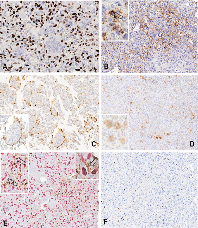
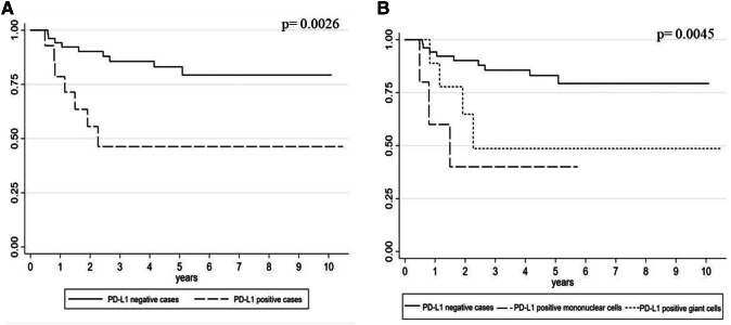
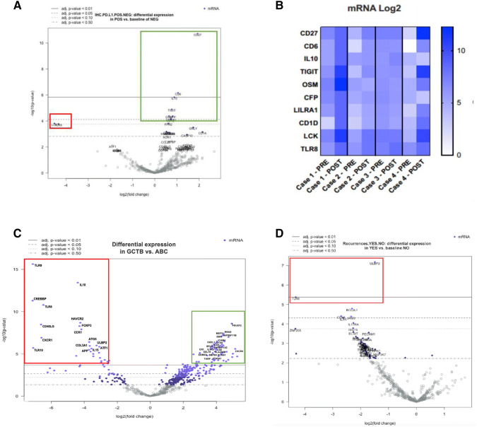
Giant cell tumor of bone (GCTB) is a locally aggressive and rarely metastatic tumor, with a relatively unpredictable clinical course. A retrospective series of 46 GCTB and a control group of 24 aneurysmal bone cysts (ABC) were selected with the aim of investigating the PD-L1 expression levels and immune-related gene expression profile, in correlation with clinicopathological features. PD-L1 and Ki67 were immunohistochemically tested in each case. Furthermore, comprehensive molecular analyses were carried out using NanoString technology and nCounter PanCancer Immune Profiling Panel, and the gene expression results were correlated with clinicopathological characteristics. PD-L1 expression was observed in 13/46 (28.3%) GCTB (and in 1/24, 4.2%, control ABC, only) and associated with a shorter disease free interval according to univariate analysis. Moreover, in PD-L1-positive lesions, three genes (CD27, CD6 and IL10) were significantly upregulated (p < 0.01), while two were downregulated (LCK and TLR8, showing borderline significance, p = 0.06). Interestingly, these genes can be related to maturation and immune tolerance of bone tissue microenvironment, suggesting a more immature/anergic phenotype of giant cell tumors. Our findings suggest that PD-L1 immunoreactivity may help to select GCTB patients with a higher risk of recurrence who could potentially benefit from immune checkpoint blockade.

Keywords: Giant cell tumor of bone; Immune-related genes; NanoString technology; PD-L1; Prognosis.


Conflict of interest statement

The authors declare that they have no conflict of interest.

Figures

Fig. 1
Fig. 1
a In a giant cell tumor of bone, strong immunoreactivity of neoplastic mononuclear stromal cells for H3.3 G35W mutation-specific antibody is present (400x). b* Strong PD-L1 immunoreactivity at the cell membrane of mononuclear stromal cells (200x, insert 400x). c, d PD-L1 expression by giant cells is mainly confined at the membrane level of the same giant cell (400x, insert 500x) and only occasionally occurs in the cytoplasm (80x, insert 200x). e* Double PD-L1 and H3F3A immunostaining in a controversial case of giant cell tumor of bone helps to distinguish neoplastic mononuclear stromal cells from adjacent immune cells (200x, inserts 400x and 500x). f FOXP3-positive tumor infiltrating lymphocytes in a case of giant cell tumor of bone (200x). *The arrows in images B and E indicate mononuclear tumor cells reactive for PD-L1
Fig. 2
Fig. 2
a Kaplan–Meier curve of DFI according to PD-L1 expression in giant cell tumor of bone (log-rank test p = 0.0026). b Kaplan–Meier curve of DFI according to PD-L1 expression in different cell types of giant cell tumor of bone (log-rank test p = 0.0045)
Fig. 3
Fig. 3
a Different gene expression profiles according to PD-L1 immunoreactivity. Upregulated genes CD27, CD6, IL10 (p < 0.01) are marked by the green rectangle. Red rectangle highlights downregulated genes LCK and TLR8 although not reaching statistical significance (p = 0.06). b Different gene expressions in cases pre- and post-treatment with Denosumab. Particularly, cases 1 and 4 show higher levels of CD27, CD6, TIGIT, OSM, LCK and TLR8 genes in the post-treatment samples compared to those of initial biopsies. c Giant cell tumors of bone, compared to aneurysmal bone cysts, show significantly higher expression of MEFV, TNFRSF11B, EBI3, CXCL1, ICOS, CD6, CCL19, CR1, EOMES, KLRG1 and CXCR4 genes (p < 0.01) indicated by the green rectangle. Downregulated genes TLR9, IL10, TLR8, FOXP3, CXCR1, ATG5, ATF1 and IL15 (p < 0.01) are marked by the red rectangle. dTLR8 and ULBP2 genes were found significantly downregulated in giant cell tumors of bone that developed locoregional recurrences

References

    1. Cowan RW, Singh G. Giant cell tumor of bone: a basic science perspective. Bone. 2013;52:238–246. doi: 10.1016/j.bone.2012.10.002. - DOI - PubMed
    1. Athanasou NA, Bansal M, Forsyth R, Reid RP, Sapi Z. Giant cell tumour of bone. In: Fletcher CDM, Bridge JA, Hogendoorn PCW, Mertens F, editors. WHO classification of tumours of soft tissue and bone. 4. Lyon: IARC Press; 2013. pp. 321–324.
    1. Errani C, Ruggieri P, Asenzio MA, Toscano A, Colangeli S, Rimondi E, et al. Giant cell tumor of the extremity: a review of 349 cases from a single institution. Cancer Treat Rev. 2010;36:1–7. doi: 10.1016/j.ctrv.2009.09.002. - DOI - PubMed
    1. Werner M. Giant cell tumour of bone: morphological, biological and histogenetical aspects. Int Orthop. 2006;30:484–489. doi: 10.1007/s00264-006-0215-7. - DOI - PMC - PubMed
    1. Luo G, Li F, Li X, Wang ZG, Zhang B. TNF-α and RANKL promote osteoclastogenesis by upregulating RANK via the NF-κB pathway. Mol Med Rep. 2018;17:6605–6611. doi: 10.3892/mmr.2018.8698. - DOI - PMC - PubMed

MeSH terms