Review
. 2020 Jun 30;48(3):1089-1099.
doi: 10.1042/BST20191084.

Biomolecular complex viewed by dynamic nuclear polarization solid-state NMR spectroscopy

Affiliations
Review

Biomolecular complex viewed by dynamic nuclear polarization solid-state NMR spectroscopy

Arnab Chakraborty et al. Biochem Soc Trans. .

Abstract

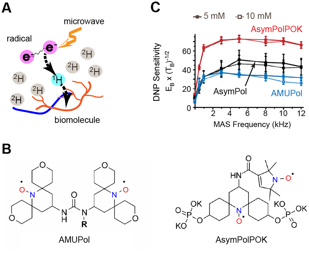
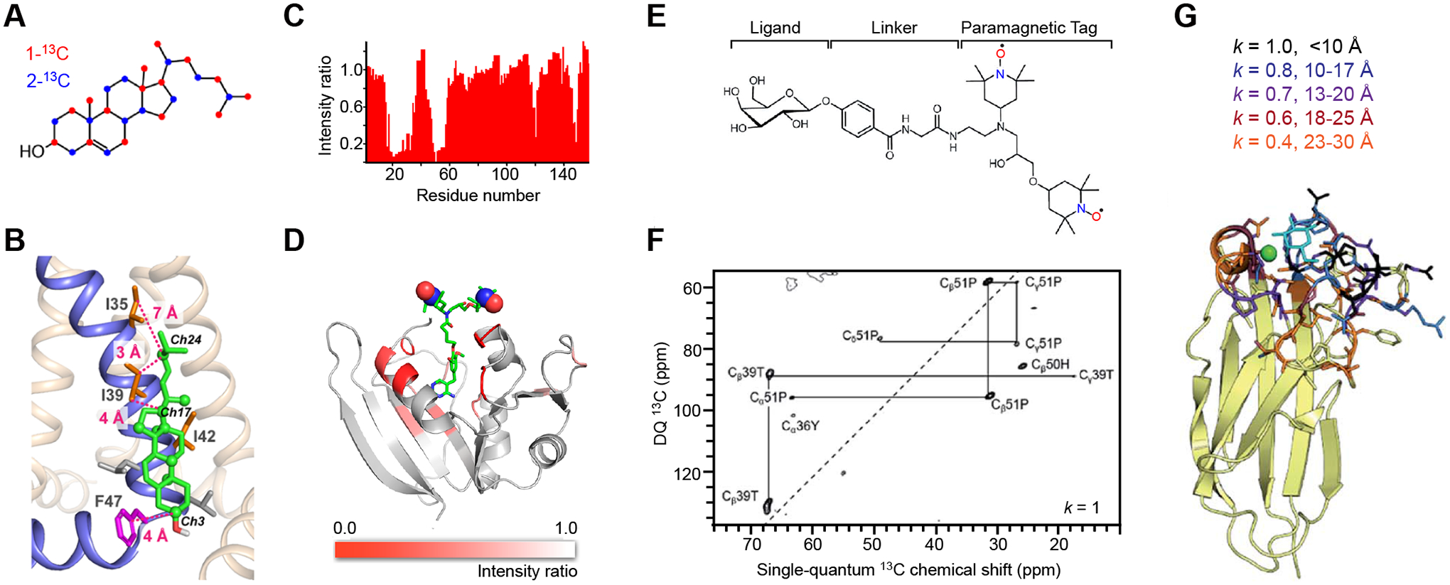
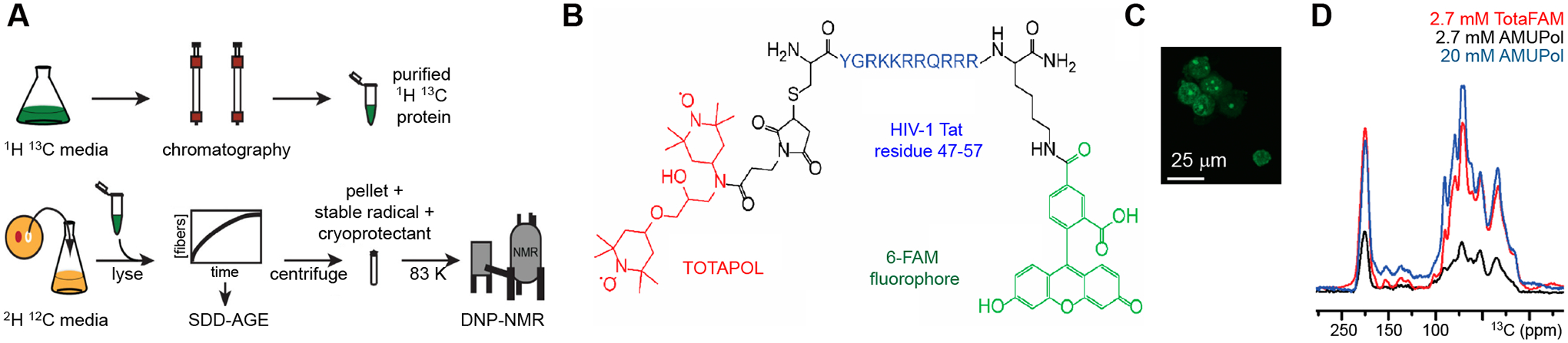
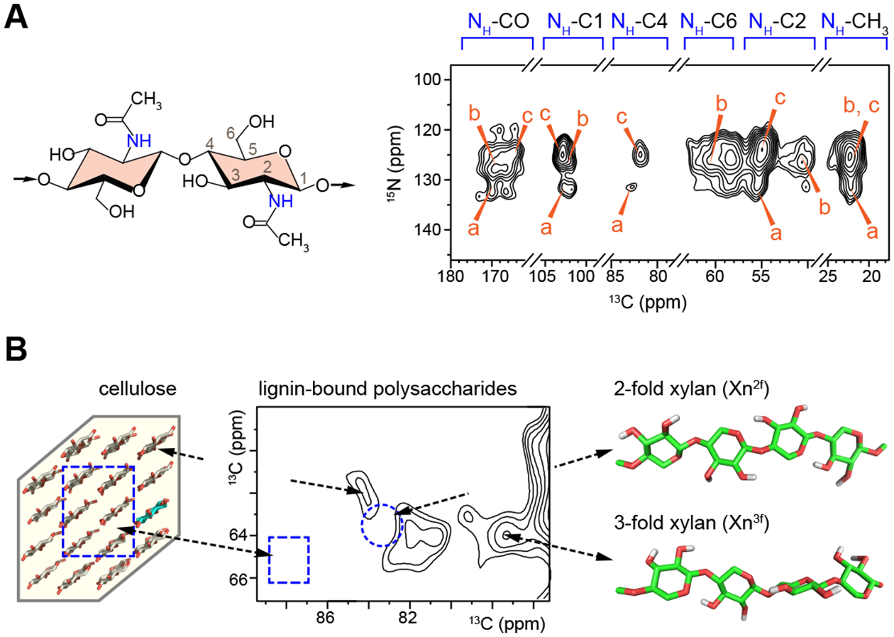
Solid-state nuclear magnetic resonance (ssNMR) is an indispensable tool for elucidating the structure and dynamics of insoluble and non-crystalline biomolecules. The recent advances in the sensitivity-enhancing technique magic-angle spinning dynamic nuclear polarization (MAS-DNP) have substantially expanded the territory of ssNMR investigations and enabled the detection of polymer interfaces in a cellular environment. This article highlights the emerging MAS-DNP approaches and their applications to the analysis of biomolecular composites and intact cells to determine the folding pathway and ligand binding of proteins, the structural polymorphism of low-populated biopolymers, as well as the physical interactions between carbohydrates, proteins, and lignin. These structural features provide an atomic-level understanding of many cellular processes, promoting the development of better biomaterials and inhibitors. It is anticipated that the capabilities of MAS-DNP in biomolecular and biomaterial research will be further enlarged by the rapid development of instrumentation and methodology.

Keywords: cell wall; dynamic nuclear polarization; membrane proteins; pathogenic fungi; polysaccharides; solid-state NMR.


Figures

Figure 1.
Figure 1.. MAS-DNP technique boosts NMR sensitivity.
(A) Illustration of the MAS-DNP mechanism. (B) Representative structure of two bi-radicals, AMUPol and AsymPolPOK. (C) Enhancement of the sensitivity of spectra collected on 13C-urea at a variety of magic-angle spinning frequencies at 9.4 T and 105 K using three different bi-radicals, including AMUPol, AsymPolPOK, and AsymPol at two different concentrations (5 and 10 mM). The MAS-DNP sensitivity is quantified as the signal intensity per unit square root of time. Figures 1B and 1C are adapted from reference [26] with copyright permission and reference [27] (an open-access article).
Figure 2.
Figure 2.. MAS-DNP methods for probing protein-ligand binding.
(A) Yeast-based 13C labeling of cholesterol using site-specifically 13C-labeled glucose. The labeled carbon sites on cholesterol are in red and blue for cholesterols produced from 1-13C and 2-13C glucose molecules, respectively. (B) A structural model of a cholesterol molecule bound to the influenza M2 proteins. The key Ile and Phe residues, as well as their distances to cholesterol carbons, are shown. (C) Signal bleaching quantified in solution 1H-15N HSQC spectra due to the binding of radicals to dihydrofolate reductase. (D) A model of E. coli dihydrofolate reductase with DNP bleaching information represented by the intensity ratios of 13C-13C DARR spectra collected on two samples containing either bound radicals or exogenous radicals. (E) Scheme for incorporating a carbohydrate ligand to a paramagnetic tag for selective DNP. (F) Selective DNP 13C–13C INADEQUATE difference spectrum of LecA obtained using k=1: only the tightly bound residues are observed. (G) Sideview of LecA. Residues observed using selective DNP are highlighted, with the corresponding k values given. Figures 2A-2D are adapted from references [38, 51] with copyright permission. Figures 2E-2G are adapted from reference [31], an open-access article.
Figure 3.
Figure 3.. Cellular MAS-DNP of protein structure.
(A) Preparation of proteins at endogenous levels for MAS-DNP in biological environments. (B) Chemical structure of the trimodal polarizing agent TotaFAM. (C) A fluorescent image confirms the cellular uptake of TotaFAM. (D) 1D 13C spectra of HEK293F cells at <6 K using different radicals. Figures are adapted from references [13, 14] with copyright permission.
Figure 4.
Figure 4.. Polysaccharide structure and polymer binding in plant and fungal cell walls.
(A) Representative structure and MAS-DNP spectra of chitin in cell walls of intact A. fumigatus. Three major types of chitin signals have been resolved (Types a-c). (B) The aromatic-edited spectrum of maize stems shows the signals of lignin-bound carbohydrates. Arrows and black dotted lines connect the spectral regions with polysaccharide structures. The dashline circle and rectangle on the spectrum highlight the missing signals of the carbohydrate components that are far from lignin. Figures are adapted from references [10, 11], which are open-access publications.

References

    1. McDermott A (2009) Structure and Dynamics of Membrane Proteins by Magic Angle Spinning Solid-State NMR. Annu. Rev. Biophys 38, 385–403 - PubMed
    1. Marchanka A, Simon B, Althoff-Ospelt G and Carlomagno T (2015) RNA structure determination by solid-state NMR spectroscopy. Nat. Commun 6, 7024. - PMC - PubMed
    1. Meier BH, Riek R and Bockmann A (2017) Emerging Structural Understanding of Amyloid Fibrils by Solid-State NMR. Trends Biochem. Sci 42, 777–787 - PubMed
    1. Comellas G and Rienstra CM (2013) Protein Structure Determination by Magic-Angle Spinning Solid-State NMR, and Insights into the Formation, Structure, and Stability of Amyloid Fibrils. Annu. Rev. Biophys 42, 515–536 - PubMed
    1. Mandala VS, Williams JK and Hong M (2018) Structure and Dynamics of Membrane Proteins from Solid-State NMR. Annu. Rev. Biophys 47, 201–222 - PMC - PubMed

Publication types