. 2020 Jun 3;11(11):1555-1562.
doi: 10.1021/acschemneuro.0c00210. Epub 2020 May 19.

Expression of the SARS-CoV-2 Entry Proteins, ACE2 and TMPRSS2, in Cells of the Olfactory Epithelium: Identification of Cell Types and Trends with Age

Affiliations

Expression of the SARS-CoV-2 Entry Proteins, ACE2 and TMPRSS2, in Cells of the Olfactory Epithelium: Identification of Cell Types and Trends with Age

Katarzyna Bilinska et al. ACS Chem Neurosci. .

Abstract

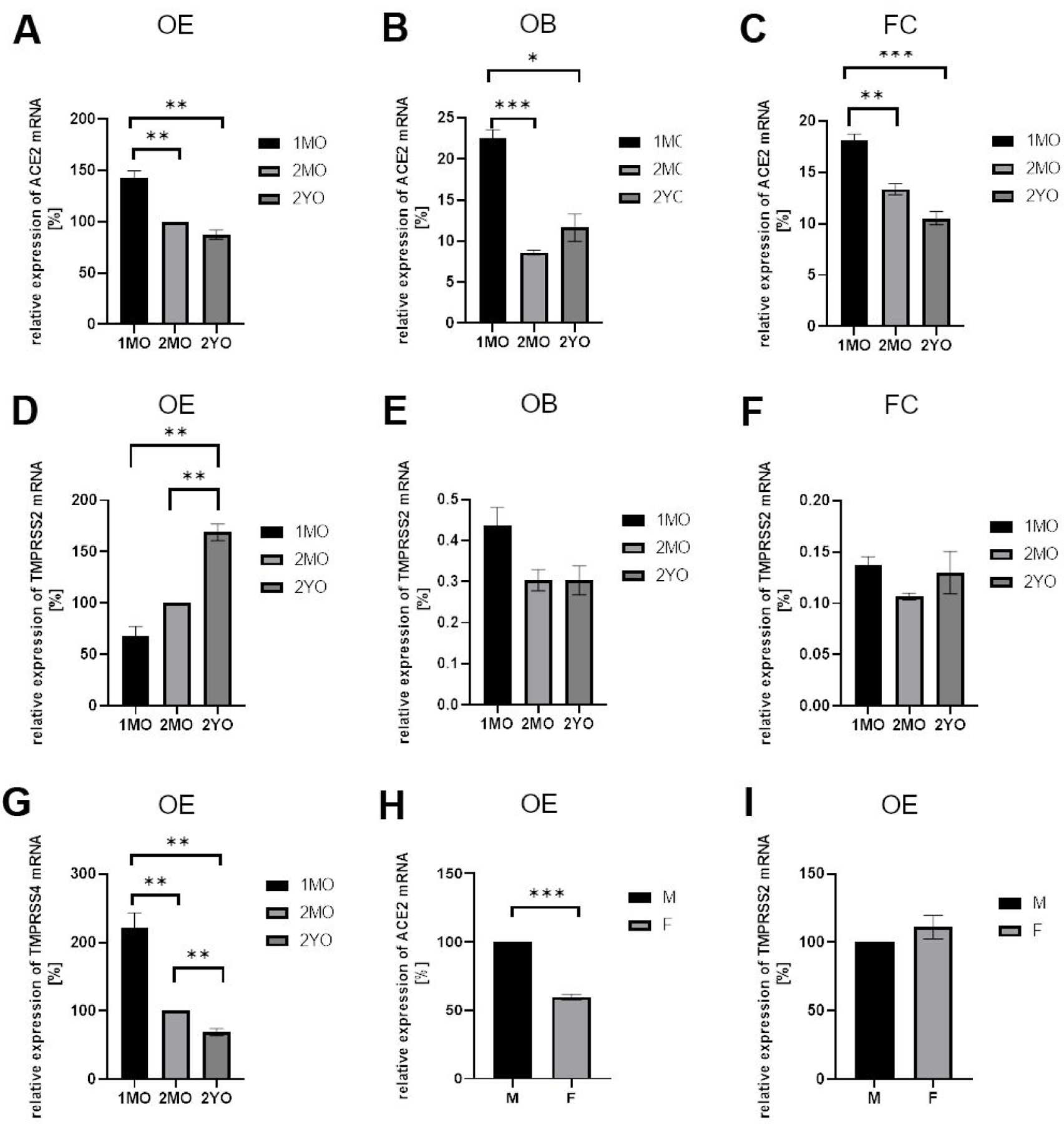
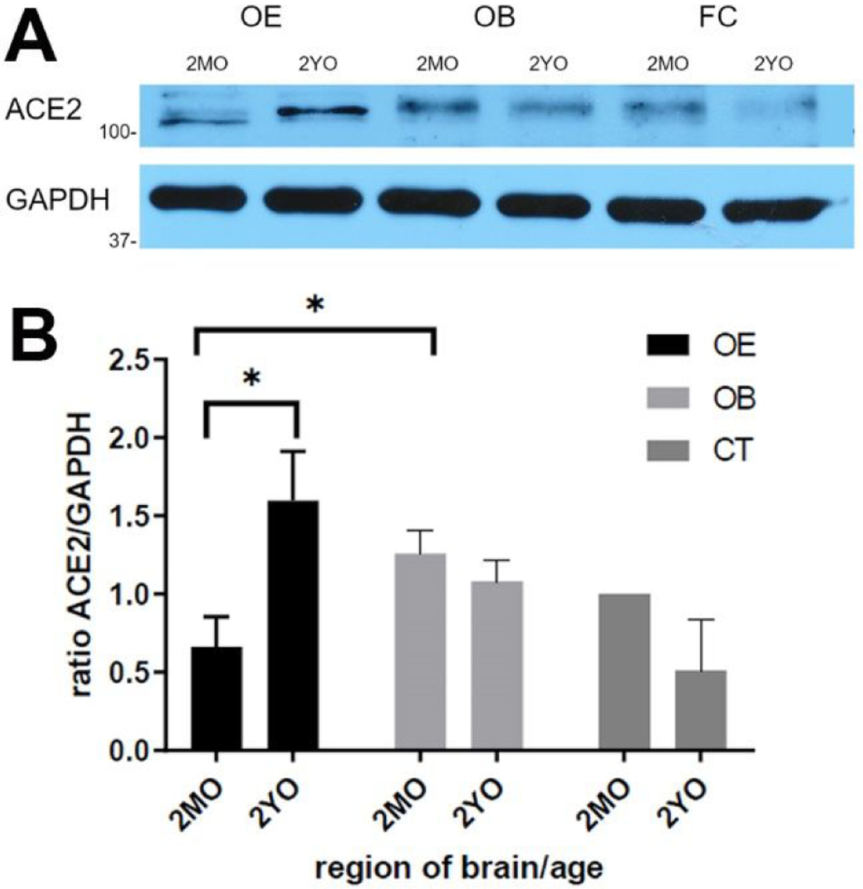
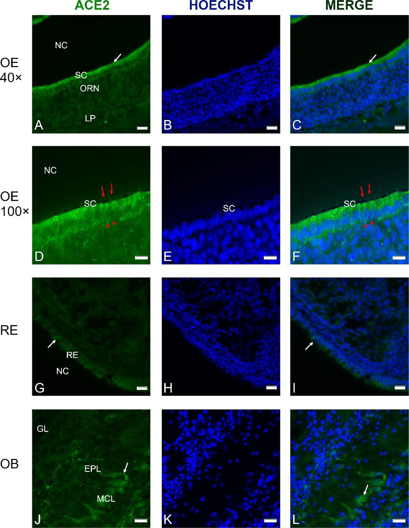
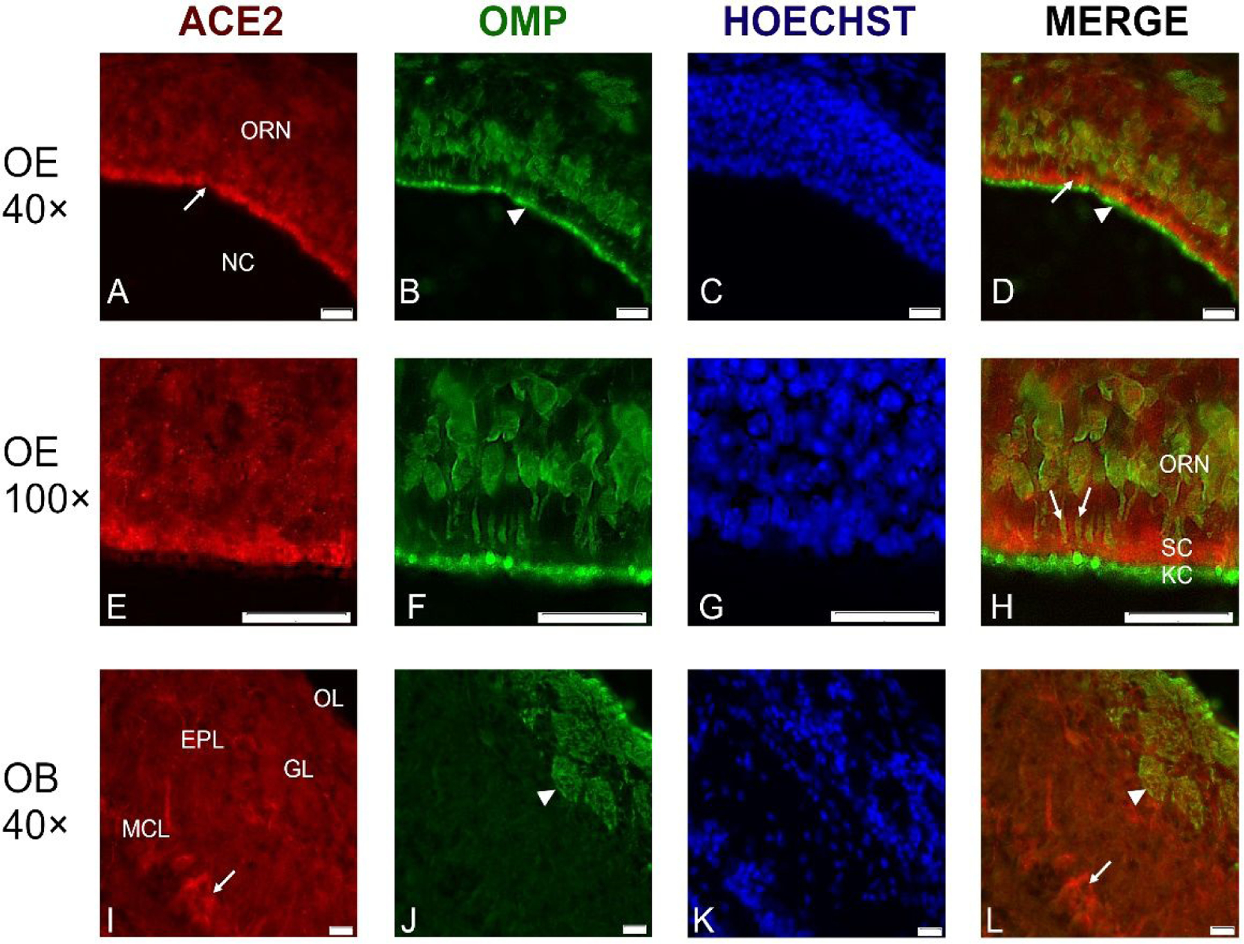
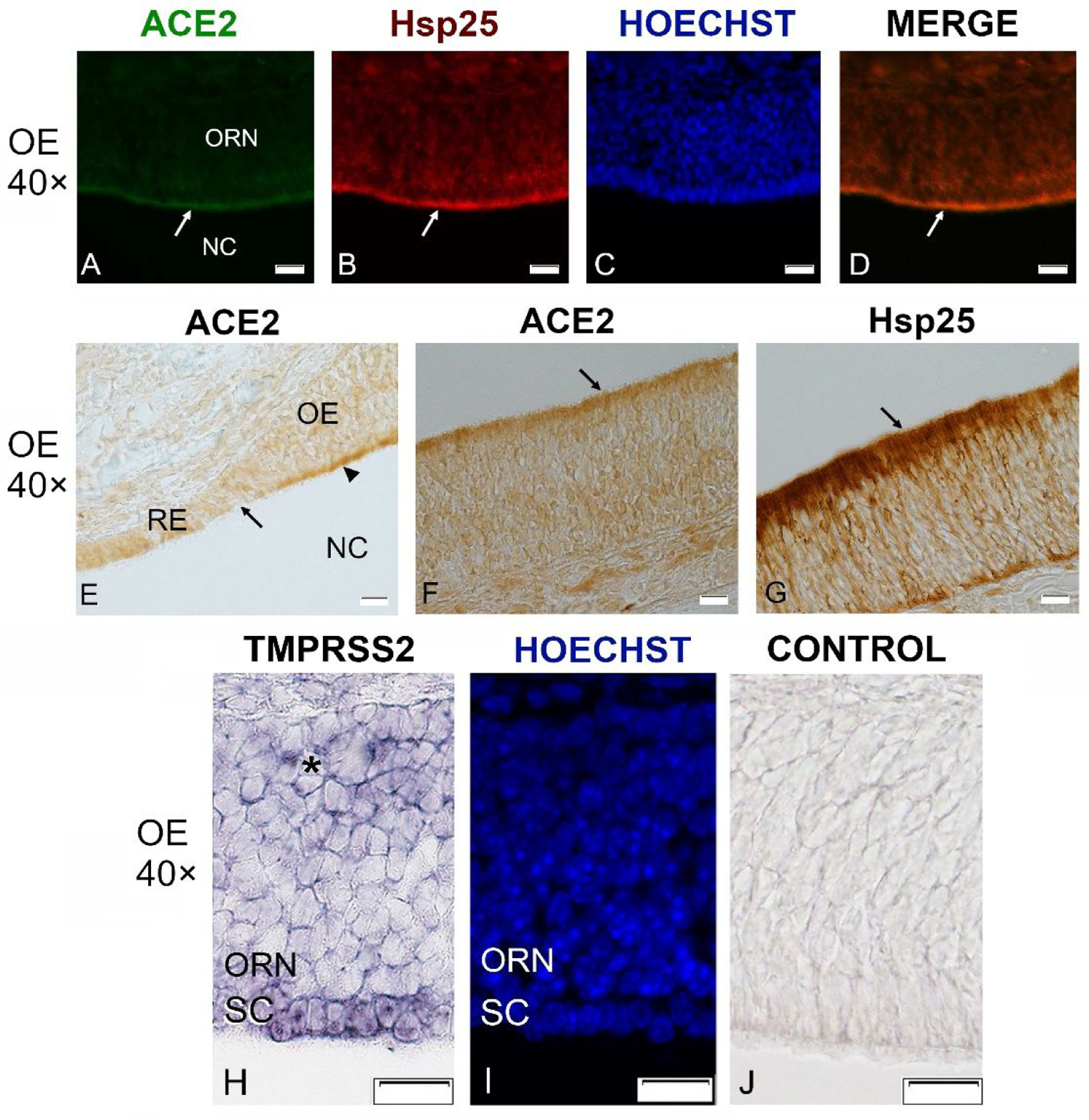
The COVID-19 pandemic revealed that there is a loss of smell in many patients, including in infected but otherwise asymptomatic individuals. The underlying mechanisms for the olfactory symptoms are unclear. Using a mouse model, we determined whether cells in the olfactory epithelium express the obligatory receptors for entry of the SARS-CoV-2 virus by using RNAseq, RT-PCR, in situ hybridization, Western blot, and immunocytochemistry. We show that the cell surface protein ACE2 and the protease TMPRSS2 are expressed in sustentacular cells of the olfactory epithelium but not, or much less, in most olfactory receptor neurons. These data suggest that sustentacular cells are involved in SARS-CoV-2 virus entry and impairment of the sense of smell in COVID-19 patients. We also show that expression of the entry proteins increases in animals of old age. This may explain, if true also in humans, why individuals of older age are more susceptible to the SARS-CoV-2 infection.

Keywords: ACE2 expression; COVID-19; SARS-CoV-2; aging; anosmia; olfactory epithelium.


Figures

Fig. 1
Fig. 1
Gene expression analysis by RT-PCR for ACE2 and TMPRSS2 in murine olfactory epithelium (OE), olfactory bulb (OB), and frontal cortex (FC) at different ages. Relative expression levels were normalized to expression in 2-month old OE (100%) using GAPDH as housekeeping gene. Similar results were obtained with beta actin as the housekeeping gene. A. Relative expression of ACE2 in OE decreases from juvenile 1-month old (1MO) to 2-month old (2MO), but it is stable in 2-year old (2YO). B, C. ACE2 levels observed in OB and FC are lower as compared to OE and stable during aging. Note that OE has higher expression level than OB and FC at each stage analyzed. D. For TMPRSS2, much higher expression was detected in OE as compared to OB (E) and FC (F). Note that with aging the level of TMPRSS2 increases significantly in OE but not in OB or FC. G. Expression of another protease, TMPRSS4, in OE significantly decreases during aging. H. Lower expression of ACE2 was noted in OE of female as compared to male 2-month old mice. I. TMPRSS2 expression in OE did not show gender-specific differences. All graphs give the mean values, and error bars represent ± SEM. * P ≤ 0.01, ** P ≤ 0.005, *** P ≤ 0.001.
Fig. 2.
Fig. 2.
Detection of ACE2 protein in tissue lysate by Western blot. A. One or more bands just above 100 kDa is visible which is more intense in aged olfactory epithelium (OE) (2YO) than in 2-month old (2MO). In contrast, in olfactory bulb (OB) and frontal cortex (FC), ACE2 bands are weaker and decrease with age. GAPDH verifies similar loading per lane. B. Quantification of the relative band intensity shows significant increase of ACE2 in aged OE but not in aged OB and FC. Intensity of ACE2 band for 2MO FC was set at 100%. Graphs give the mean values, and error bars represent ± SEM. * P ≤ 0.01.
Fig. 3
Fig. 3
Cross-sections through the olfactory epithelium (OE) (A-F), respiratory epithelium (RE) (G-I) and olfactory bulb (OB) (J-L) probed with an anti-ACE2 antibody; nuclei were stained with Hoechst 33258 (B, E, H, K). A thin layer of labeling is visible at the luminal surface of the OE (A, D). Higher magnification shows labeling just above the layer of sustentacular cell nuclei (D-F), while the olfactory knobs and cilia (red arrows) do not contain ACE2, and neither do the olfactory receptor neurons (ORN, asterisks in D and F). C, D. Merging of the images from A and B as well as D and E reveals that the cell bodies of the sustentacular cells (SC) contain most of the ACE2 label. ACE2 label was weaker in the RE of the nasal cavity (G-I). In the OB, ACE2 antibodies labeled some mitral cells, including their proximal dendrites (J-L, arrows). Control sections probed without primary antibodies or with control rabbit IgG had no detectable signal (not shown). Other abbreviations: NC- nasal cavity; MCL- mitral cell layer; EPL- external plexiform layer; GL- glomerular layer. Scale bars: 20 μm (A-C, G-I), 10 μm (D-F), and 25 μm (J-L).
Fig. 4
Fig. 4
Representative cross-sections through the olfactory epithelium (OE) in the center of the nasal cavity (NC) probed with antibodies against ACE2 (red channel) and against olfactory marker protein (OMP) (green channel). ACE2 immunoreactivity was located just below the luminal surface (A, D; arrows), between the OMP-labeled olfactory receptor knobs and cilia (arrowhead) and the layer of the olfactory receptor neurons (ORN) (A-D). At higher magnification, it is apparent that OMP-labeled ORN dendrites (arrows in H) penetrate the layer of ACE2-labeled sustentacular cells (SC), while the knobs and cilia (KC) of the ORNs at the luminal surface only contain OMP, but not ACE2 (E-H). The border between the KC and the SC layers is indicated in the merged image (H, arrowhead). In the olfactory bulb (OB), ACE2 label is mainly in some mitral neurons (arrow), while glomeruli (GL) and olfactory nerve layer at the top of OB are not labeled (I, L). Nuclei were stained with Hoechst 33258. ORN- olfactory receptor neurons, MCL- mitral cell layer; EPL- external plexiform layer; GL- glomerular layer; OL- olfactory nerve layer. Scale bars: 20 μm.
Fig. 5
Fig. 5
Sections through the olfactory epithelium (OE) probed with antibodies against ACE2 (21115-1-AP, green channel) and Hsp25 (red channel), and with in situ hybridization for TMPRSS2 in a P21 mouse. ACE2 immunoreactivity is visible mainly at the luminal surface of the OE (A, arrow) where Hsp25 antibodies label sustentacular cells (B, arrow). Nuclei were stained with Hoechst 33258 (C, I). Merging of ACE2 and Hsp25 signal shows that the two fluorescence signals largely overlap (D, arrow). Section stained at the border between the respiratory epithelium (RE) and olfactory epithelium (OE) reveals much stronger labeling of the OE (arrowhead) as compared to the RE (E). ACE2 labeling using chromogenic DAB-based visualization shows the same localization at the OE surface (F). Hsp25 shows the same localization but much stronger label (G). In situ hybridization for TMPRSS2 produces label in the sustentacular cells (SC), but very little or no label in the mature olfactory receptor neurons (ORN), and label in only a few cells of the deeper layers of OE where immature olfactory neurons are located (asterisk, H). The same section is stained with Hoechst to show the nuclei (I), and an adjacent section is the sense control (J). NC- nasal cavity. All scale bars: 20 μm.

Comment in

References

    1. Lechien JR, Chiesa-Estomba CM, De Siati DR, Horoi M, Le Bon SD, Rodriguez A, Dequanter D, Blecic S, El Afia F, Distinguin L, Chekkoury-Idrissi Y, Hans S, Delgado IL, Calvo-Henriquez C, Lavigne P, Falanga C, Barillari MR, Cammaroto G, Khalife M, Leich P, Souchay C, Rossi C, Journe F, Hsieh J, Edjlali M, Carlier R, Ris L, Lovato A, De Filippis C, Coppee F, Fakhry N, Ayad T, Saussez S (2020) Olfactory and gustatory dysfunctions as a clinical presentation of mild-to-moderate forms of the coronavirus disease (COVID-19): a multicenter European study [Apr 6]. Eur Arch Otorhinolaryngol 2020;10.1007/s00405-020-05965-1. doi:10.1007/s00405-020-05965-1 [Epub ahead of print] - DOI - PMC - PubMed
    1. Moein ST, Hashemian SMR, Mansourafshar B, Khorram-Tousi A, Tabarsi P, Doty RL (2020) Smell dysfunction: a biomarker for COVID-19. Int Forum Allergy Rhinol 2020 Apr 17. doi:10.1002/alr.22587. [Epub ahead of print] - DOI - PMC - PubMed
    1. Netland J, Meyerholz DK, Moore S, Cassell M, Perlman S (2008) Severe acute respiratory syndrome coronavirus infection causes neuronal death in the absence of encephalitis in mice transgenic for human ACE2. J. Virol 82(5):7264–75. - PMC - PubMed
    1. Baig AM, Khaleeq A, Ali U, Syeda H (2020) Evidence of the COVID-19 virus targeting the CNS: host-virus interactions and proposed neurotropic mechanisms. ACC Chem. Neurosci 11(7):995–998. doi: 10.1021/acschemneuro.0c00122 - DOI - PubMed
    1. Mao L, Jin H, Wang M, Yu H, Chen S, He Q, Chang J, Hong C, Zhou Y, Wang D, Miao X, Li Y, Hu B (2020) Neurological manifestations of hospitalized patients with coronavirus disease 2019 in Wuhan, China. JAMA Neurol doi:10.1001/jamaneurol.2020.1127. (ahead of print) - DOI - PMC - PubMed

MeSH terms