. 2020 May 7;15(5):e0232575.
doi: 10.1371/journal.pone.0232575. eCollection 2020.

Prevalence of pectus excavatum in an adult population-based cohort estimated from radiographic indices of chest wall shape

Affiliations

Prevalence of pectus excavatum in an adult population-based cohort estimated from radiographic indices of chest wall shape

Mikaela Biavati et al. PLoS One. .

Abstract

Background: Pectus excavatum is the most common chest wall skeletal deformity. Although commonly evaluated in adolescence, its prevalence in adults is unknown.

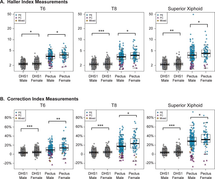
Methods and findings: Radiographic indices of chest wall shape were analyzed for participants of the first (n = 2687) and second (n = 1780) phases of the population-based Dallas Heart Study and compared to clinical cases of pectus (n = 297). Thoracic computed tomography imaging studies were examined to calculate the Haller index, a measure of thoracic axial shape, and the Correction index, which quantitates the posterior displacement of the sternum relative to the ribs. At the level of the superior xiphoid, 0.5%, 5% and 0.4% of adult Dallas Heart Study subjects have evidence of pectus excavatum using thresholds of Haller index >3.25, Correction index >10%, or both, respectively. Radiographic measures of pectus are more common in females than males and there is a greater prevalence of pectus in women than men. In the general population, the Haller and Correction indices are associated with height and weight, independent of age, gender, and ethnicity. Repeat imaging of a subset of subjects (n = 992) demonstrated decreases in the mean Haller and Correction indices over seven years, suggesting change to a more circular axial thorax, with less sternal depression, over time.

Conclusions: To our knowledge, this is the first study estimating the prevalence of pectus in an unselected adult population. Despite the higher reported prevalence of pectus cases in adolescent boys, this study demonstrates a higher prevalence of radiographic indices of pectus in adult females.


Conflict of interest statement

The authors have declared that no competing interests exist.

Figures

Fig 1
Fig 1. Calculation of the haller index and correction index from axial images of chest computed tomography scans of pectus cases.
Anatomic schematic of the lateral view of the thorax (A) demonstrating the position of the T6 and T8 vertebral bodies relative to the body of the sternum and the superior xiphoid process. Measurements of chest wall dimensions (red lines) used to calculate the Haller Index (B) and Correction Index (C) from a patient with pectus excavatum (left) and a patient with pectus carinatum (right). Measurements in B and C are shown at the level of the superior xiphoid. Haller (D) and Correction (E) Index measurements of patients with pectus excavatum (PE, n = 274, blue circles), pectus carinatum (PC, n = 19, red triangles) and those with a mixed pectus excavatum and carinatum defect (Mixed, n = 4, orange diamonds) at the level of T6, T8, Superior Xiphoid, as well as the point of maximal (max.) sternal depression (n = 278) or protrusion (n = 23). Patients with a mixed defect are included in both the maximal sternal depression and protrusion data sets. Box and whisker plots (black) are superimposed.
Fig 2
Fig 2. Comparison of haller and correction index measurements for men and women from the Dallas Heart Study (DHS) and pectus cohorts.
Haller (A) and Correction (B) indices of the DHS1 cohort (n = 2687) and pectus cases (n = 297) at T6, T8 and superior xiphoid axial levels. Individuals from DHS1 (gray circles) and subjects with pectus excavatum (PE, n = 274, blue circles) pectus carinatum (PC, n = 19, red triangles) or a mixed pectus excavatum and carinatum defect (Mixed, n = 4, orange diamonds) are individually plotted with superimposed box and whisker plots (black). *, ** and *** indicative of P-value <0.05, <0.001 and <0.0001, respectively.
Fig 3
Fig 3. Correlation of the correction index with the haller index for pectus and Dallas Heart Study (DHS) cohorts.
The Correction and Haller index at the level of the superior xiphoid is plotted for each of the pectus excavatum (PE, n = 274, blue circles), pectus carinatum (PC, n = 19, red triangles) and mixed pectus excavatum and carinatum (Mixed, n = 4, orange diamonds) patients (A, B) and for each of the DHS1 subjects (C) (n = 2687). Indices are measured at the point of maximal sternal deformity, that is, at the point of maximal sternal depression for the PE cases and the point of maximal sternal protrusion for the PC cases (A), or at the superior xiphoid (B, C). Dashed lines indicate a Haller Index of 3.25 or a Correction Index of 10%. Note the different axes.
Fig 4
Fig 4. Haller and correction indices of the Dallas Heart Study subjects.
Haller index (A) and Correction Index (B) measured at the superior xiphoid axial level is plotted against the age in years, height in cm, weight in kg and BMI in kg/m2 for DHS1 (n = 2687) subjects. Individual subjects are represented as gray circles; the trend line is superimposed. Correlation (C) and box and whisker plots (D) of Haller and Correction indices for DHS Subjects (n = 992) with available imaging from DHS1 and DHS2. A DHS subject (E) demonstrating an increase in the Haller index in the repeat imaging study at the level of the superior xiphoid. The magnitude of Haller index increase is within the top 1%. Imaging shows the development of a more circular, and less oval, axial chest wall shape.

References

    1. Chung CS, Myrianthopoulos NC. Factors affecting risks of congenital malformations. I. Analysis of epidemiologic factors in congenital malformations. Report from the Collaborative Perinatal Project. Birth defects original article series. 1975;11(10):1–22. . - PubMed
    1. Rajabi-Mashhadi MT, Ebrahimi M, Mobarhan MG, Moohebati M, Boskabady MH, Kazemi-Bajestani SM. Prevalence of chest wall deformities in a large sample of Iranian children aged 7–14 years. Iran J Pediatr. 2010;20(2):221–4. Epub 2010/06/01. - PMC - PubMed
    1. Coskun ZK, Turgut HB, Demirsoy S, Cansu A. The prevalence and effects of Pectus Excavatum and Pectus Carinatum on the respiratory function in children between 7–14 years old. Indian J Pediatr. 2010;77(9):1017–9. Epub 2010/09/04. 10.1007/s12098-010-0155-5 . - DOI - PubMed
    1. Westphal FL, Lima LC, Lima Neto JC, Chaves AR, Santos Junior VL, Ferreira BL. Prevalence of pectus carinatum and pectus excavatum in students in the city of Manaus, Brazil. J Bras Pneumol. 2009;35(3):221–6. Epub 2009/04/25. 10.1590/s1806-37132009000300005 . - DOI - PubMed
    1. Yucesan S, Dindar H, Olcay I, Okur H, Kilicaslan S, Ergoren Y, et al. Prevalence of congenital abnormalities in Turkish school children. Eur J Epidemiol. 1993;9(4):373–80. Epub 1993/07/01. 10.1007/bf00157393 . - DOI - PubMed

Publication types

MeSH terms