. 2020 Jun 7;20(11):2009-2019.
doi: 10.1039/d0lc00142b. Epub 2020 May 7.

Evaluation of intercellular communication between breast cancer cells and adipose-derived stem cells via passive diffusion in a two-layer microfluidic device

Affiliations

Evaluation of intercellular communication between breast cancer cells and adipose-derived stem cells via passive diffusion in a two-layer microfluidic device

Sharif M Rahman et al. Lab Chip. .

Abstract

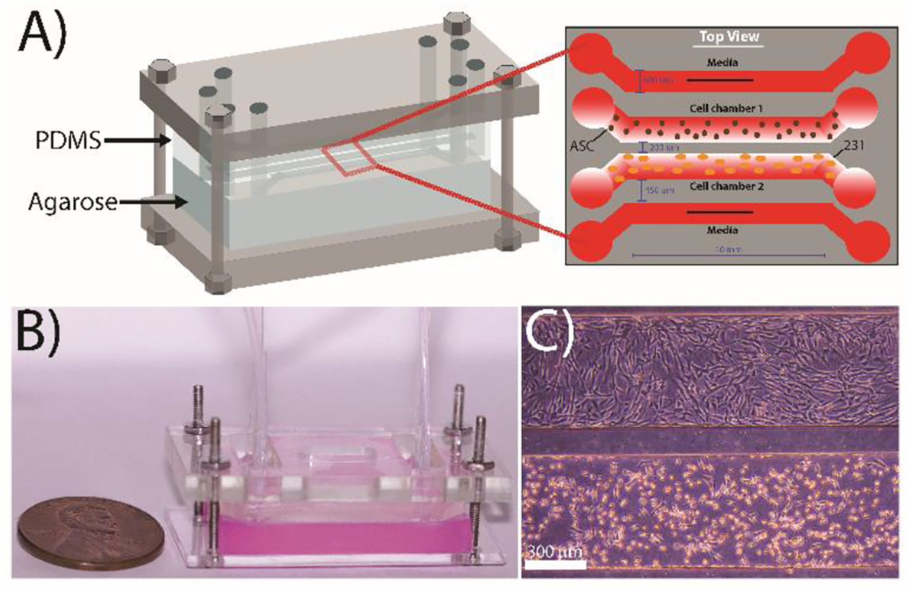
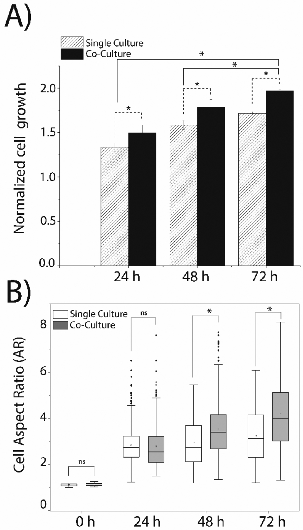
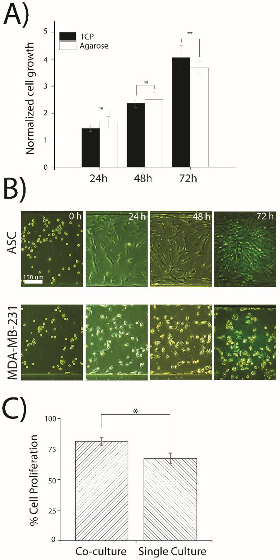
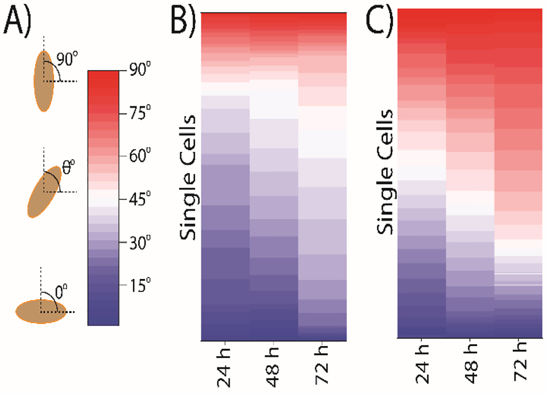
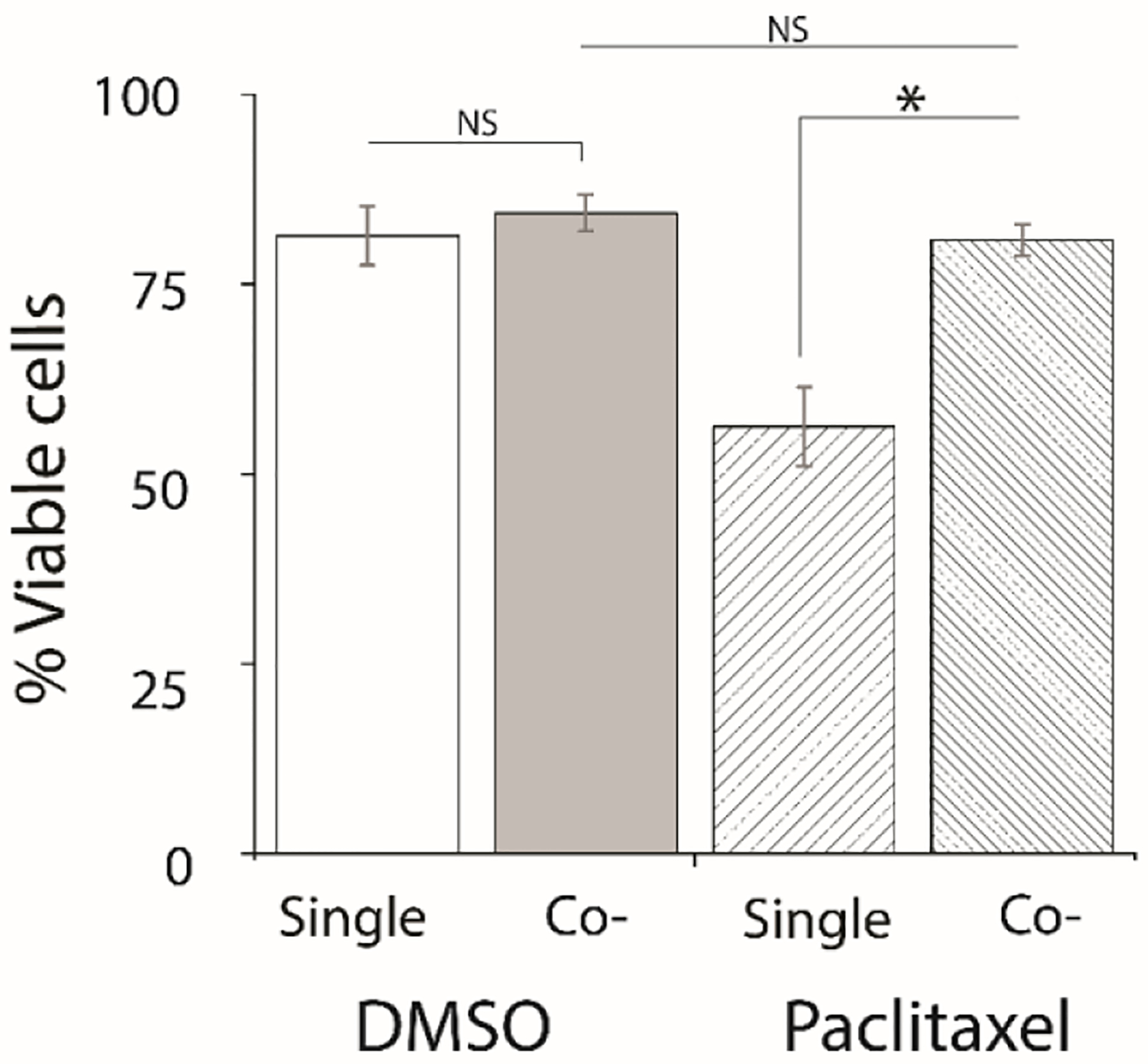
Breast cancer tumorigenesis and response to therapy is regulated by cancer cell interactions with the tumor microenvironment (TME). Breast cancer signaling to the surrounding TME results in a heterogeneous and diverse tumor microenvironment, which includes the production of cancer-associated fibroblasts, macrophages, adipocytes, and stem cells. The secretory profile of these cancer-associated cell types results in elevated chemokines and growth factors that promote cell survival and proliferation within the tumor. Current co-culture approaches mostly rely on transwell chambers to study intercellular signaling between adipose-derived stem cells (ASCs) and cancer cells; however, these methods are limited to endpoint measurements and lack dynamic control. In this study, a 4-channel, "flow-free" microfluidic device was developed to co-culture triple-negative MDA-MB-231 breast cancer cells and ASCs to study intercellular communication between two distinct cell types found in the TME. The device consists of two layers: a top PDMS layer with four imprinted channels coupled with a bottom agarose slab enclosed in a Plexiglas chamber. For dynamic co-culture, the device geometry contained two centered, flow-free channels, which were supplied with media from two outer flow channels via orthogonal diffusion through the agarose. Continuous fresh media was provided to the cell culture channel via passive diffusion without creating any shearing effect on the cells. The device geometry also allowed for the passive diffusion of cytokines and growth factors between the two cell types cultured in parallel channels to initiate cell-to-cell crosstalk. The device was used to show that MDA-MB-231 cells co-cultured with ASCs exhibited enhanced growth, a more aggressive morphology, and polarization toward the ASCs. The MDA-MB-231 cells were found to exhibit a greater degree of resistance to the drug paclitaxel when co-cultured with ASCs when compared to single culture studies. This microfluidic device is an ideal platform to study intercellular communication for many types of cells during co-culture experiments and allows for new investigations into stromal cell-mediated drug resistance in the tumor microenvironment.


Conflict of interest statement

Conflicts of interest

“There are no conflicts to declare”.

Figures

Figure 1:
Figure 1:
Design of the microfluidic co-culture device. (A) The flow-free microfluidic device consisted of four fluidic channels imprinted into a PDMS slab placed on top of 3 wt% agarose. The device contains four parallel channels: the two outermost ‘flow’ channels are constantly supplied with media while the two innermost “flow-free” channels are used to culture the MDA-MB-231 cells and adipose stem cells. All channels are 600 μm wide with a contact length of 10 mm and a height of 150 μm. The spacing between the media channels and the culture channels is 450 μm, while the spacing between the two cell culture channels is 200 μm to facilitate cellular crosstalk. (B) Image of a completely assembled device with agarose stained red for enhanced visualization. (C) Representative image of MDA-MB-231 cells (bottom) and ASCs (top) cultured in the device after 72 h.
Figure 2:
Figure 2:
COMSOL simulation of mass transfer in “flow-free” cell culture channels. (A) Diffusion of a model biomolecule from the outer flow channel into the center culture channels. (Inset image) Visual representation of the COMSOL simulations showing diffusion across the device. (B) Simulation of biomolecules between the two flow-free culture channels. (Inset image) Visual representation of the COMSOL simulation. In both (A) and (B) biomolecules were approximated at 140 nM SDF-1α in the source channels(C) Simulation of the oxygen diffusion from outside of the device, through PDMS to cell culture channel. External oxygen was approximated to be ≈21%.
Figure 3:
Figure 3:
Simultaneous co-culture of MDA-MB-231 cells and ASCs alters breast cancer cell growth and morphology. (A) The growth of the MDA-MB-231 cells was observed in single and co-culture using an initial cell density of 0.5×10⁵ cells/mL. (B) Morphological changes in MDA-MB-231 cells in single culture and co-culture was determined by calculating the aspect ratio: measuring the ratio of the major cell axis length to minor cell axis length of individual cells. A minimum of 500 MDA-MB-231 cells was analyzed for each time point. Single culture and co-culture data were compared by student’s t-test with a statistically significant value set at p<0.05.
Figure 4:
Figure 4:
Cell growth and proliferation cultured off-chip and on microfluidic device. (A) Normalized growth of MDA-MB-231 cells cultured off-chip on either TCP or agarose. Cells were cultured in a 100 mm petri dish for 72 hours with an initial total number of 700,000 cells. Cell growth was normalized by dividing the number of cells of each time point by the 0-hour cell numbers. The statistical significance test between cell growth on TCP and agarose with ** representing p<0.05 and ns indicating no significance. (B) Transmitted light (brightfield) images of ASCs (top culture channel) and MDA-MB-231 cells (bottom culture channel) in the device during a 72 h experiment. Images are representative of cell cultures across the entire channel and of triplicate experimentation. (C) Cell proliferation was quantified in the microfluidic device for MDA-MB-231 cells using anti Ki-67 antibodies was compared among cells in co-culture and single culture experiments. Experiments were conducted in duplicate and the statistical differences between two groups were determined by the student’s t-test with a statistical confidence interval value set at p<0.05.
Figure 5:
Figure 5:
MDA-MB-231 cells orient themselves towards the ASC population during simultaneous co-culture. (A) The angle was calculated with the cell major axis line with respect to the horizontal line. The angles were taken within 0° to 90° where 0° refer no directionality of cells in the parallel position of the channel, and 90° indicates 100% directionality of cells in the perpendicular position of channels. All angles were measured (B) when MDA-MB-231 cells were cultured in single culture. (C) when MDA-MB-231 cells were cultured in Co-culture with ASCs for 72 h. The warm color represents angle in the range of 45° to 90°. A minimum 500 cells were analyzed for angle measurement.
Figure 6:
Figure 6:
Co-cultured cancer cells show drug resistance to 20 nM Paclitaxel. The viability of MDA-MB-231 cells treated with either a DMSO control or Paclitaxel was measured on-chip after 3 days of Paclitaxel treatment in single cultured and co-cultured experiments. MDA-MB-231 cells were incubated with Calcein AM (2.5 μM) and Ethidium homodimer-1 (4 μM) after terminating the experiment. Viability experiments were conducted triplicate and the statistical differences between two groups were determined by the student’s t-test with a statistically confidence interval value set at p<0.05.

References

    1. Wirtz D, Konstantopoulos K and Searson PC, Nat. Rev. Cancer, 2011, 11 VN-r, 512–522. - PMC - PubMed
    1. Hanahan D and Weinberg RA, Cell, 2011, 144, 646–674. - PubMed
    1. Rhodes LV, Antoon JW, Muir SE, Elliott S, Beckman BS and Burow ME, Mol. Cancer, 2010, 9, 295. - PMC - PubMed
    1. Sabol RA, Beighley A, Giacomelli P, Wise RM, Harrison MAA, O’donnnell BA, Sullivan BN, Lampenfeld JD, Matossian MD, Bratton MR, Wang G, Collins-Burow BM, Burow ME and Bunnell BA, Int. J. Mol. Sci, 2019, 20, 1–13. - PMC - PubMed
    1. Strong AL, Ohlstein JF, Biagas BA, Rhodes LV, Pei DT, Tucker HA, Llamas C, Bowles AC, Dutreil MF, Zhang S, Gimble JM, Burow ME and Bunnell BA, Breast Cancer Res, 2015, DOI:10.1186/s13058-015-0622-z. - DOI - PMC - PubMed

Publication types