. 2020 Jul;88(1):170-182.
doi: 10.1002/ana.25762. Epub 2020 May 28.

Hippocampal Shape Is Associated with Memory Deficits in Temporal Lobe Epilepsy

Affiliations

Hippocampal Shape Is Associated with Memory Deficits in Temporal Lobe Epilepsy

Tjardo S Postma et al. Ann Neurol. 2020 Jul.

Abstract

Objective: Cognitive problems, especially disturbances in episodic memory, and hippocampal sclerosis are common in temporal lobe epilepsy (TLE), but little is known about the relationship of hippocampal morphology with memory. We aimed to relate hippocampal surface-shape patterns to verbal and visual learning.

Methods: We analyzed hippocampal surface shapes on high-resolution magnetic resonance images and the Adult Memory and Information Processing Battery in 145 unilateral refractory TLE patients undergoing epilepsy surgery, a validation set of 55 unilateral refractory TLE patients, and 39 age- and sex-matched healthy volunteers.

Results: Both left TLE (LTLE) and right TLE (RTLE) patients had lower verbal (LTLE 44 ± 11; RTLE 45 ± 10) and visual learning (LTLE 34 ± 8, RTLE 30 ± 8) scores than healthy controls (verbal 58 ± 8, visual 39 ± 6; p < 0.001). Verbal learning was more impaired the greater the atrophy of the left superolateral hippocampal head. In contrast, visual memory was worse with greater bilateral inferomedial hippocampal atrophy. Postsurgical verbal memory decline was more common in LTLE than in RTLE (reliable change index in LTLE 27% vs RTLE 7%, p = 0.006), whereas there were no differences in postsurgical visual memory decline between those groups. Preoperative atrophy of the left hippocampal tail predicted postsurgical verbal memory decline.

Interpretation: Memory deficits in TLE are associated with specific morphological alterations of the hippocampus, which could help stratify TLE patients into those at high versus low risk of presurgical or postsurgical memory deficits. This knowledge could improve planning and prognosis of selective epilepsy surgery and neuropsychological counseling in TLE. ANN NEUROL 2020 ANN NEUROL 2020;88:170-182.


Conflict of interest statement

Nothing to report.

Figures
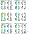
FIGURE 1
FIGURE 1
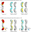
Association of hippocampal shape alterations with presurgical verbal and visual memory in temporal lobe epilepsy (TLE). Upper panels show findings common to both left and right TLE, correcting for epilepsy lateralization (A, verbal memory; B, visual memory). Also displayed are separate analyses of left (C, verbal memory; D, visual memory) and right (E, verbal memory; F, visual memory) TLE subgroups. Each panel shows T‐value maps on the left, representing deformations related to memory scores (blue colors: more atrophy related to worse memory). Significant p values are displayed on the right thresholded to p < 0.05 corrected for multiple comparisons using random field theory. The right and left hippocampi are visualized from a superior and an inferior perspective. An approximation of major hippocampal subregional boundaries is overlaid on hippocampal surfaces, description in A. CA = cornu ammonis; DG = dentate gyrus; FWE = familywise error; Sub = subiculum.
FIGURE 2
FIGURE 2
Association of hippocampal shape alterations with presurgical verbal and visual delayed recall in temporal lobe epilepsy (TLE). Panels show findings common to both left and right TLE, correcting for epilepsy lateralization (A, verbal delayed recall; B, visual delayed recall). Each panel shows T‐value maps on the left, representing deformations related to memory scores (blue colors: more atrophy related to worse memory). Significant p values are displayed on the right thresholded to p < 0.05 corrected for multiple comparisons using random field theory. The right and left hippocampi are visualized from a superior and an inferior perspective. An approximation of major hippocampal subregional boundaries is overlaid on hippocampal surfaces, description in A. CA = cornu ammonis; DG = dentate gyrus; FWE = familywise error; Sub = subiculum.
FIGURE 3
FIGURE 3
Association of presurgical hippocampal shape alterations with verbal and visual memory decline after anterior temporal lobe resection (ATLR). Displayed are results after left (A) and right (B) ATLRs. The mean extent of hippocampal resection projected on the hippocampal surface is shown on the left. T‐value maps represent deformations related to decline in memory scores (blue colors: more presurgical atrophy related to postsurgical worsening of memory). Significant p values are displayed on the right thresholded to p < 0.05 corrected for multiple comparisons using random field theory. The right and left hippocampi are visualized from a superior and an inferior perspective. An approximation of major hippocampal subregional boundaries is overlaid on hippocampal surfaces, description in A. CA = cornu ammonis; DG = dentate gyrus; FWE = familywise error; Sub = subiculum.
FIGURE 4
FIGURE 4
Three‐dimensional (3D) reconstructions of hippocampi in example cases. Displayed are 3D reconstructions of the hippocampal surface. The z scores of inward (blue, ie, atrophy) or outward (yellow/red, ie, hypertrophy) displacements from a mean normal hippocampal template are projected onto these surfaces. (A) Hippocampi of a 41‐year‐old female with left temporal lobe epilepsy with poor presurgical verbal memory (25 points) and slightly impaired visual memory (30 points) but no verbal memory decline after surgery (35 points). She had left hippocampal atrophy mainly affecting the left hippocampal head. (B) Hippocampi of a 63‐year‐old female with left temporal lobe epilepsy with slightly impaired presurgical verbal (41 points) and visual (26 points) memory and verbal memory decline following surgery (24 points). She had left hippocampal atrophy affecting the left hippocampal head and tail. (C) Hippocampi of a 65‐year‐old female with right temporal lobe epilepsy with normal presurgical verbal memory (44 points) and poor visual memory (12 points) but no verbal or visual memory decline after surgery (49 and 10 points, respectively). Despite normal hippocampal volumes, there was localized atrophy that affected the inferomedial hippocampal surface mainly on the right. (D) Hippocampi of a 30‐year‐old healthy male with good verbal and visual memory performance (73 and 43 points, respectively).

Similar articles

Cited by

References

    1. Helmstaedter C, Kockelmann E. Cognitive outcomes in patients with chronic temporal lobe epilepsy. Epilepsia 2006;47:96–98. - PubMed
    1. Frisk V, Milner B. The role of the left hippocampal region in the acquisition and retention of story content. Neuropsychologia 1990;28:349–359. - PubMed
    1. SML, Milner B. The role of the right hippocampus in the recall of spatial location. Neuropsychologia 1981;19:781–793. - PubMed
    1. Saling MM. Verbal memory in mesial temporal lobe epilepsy: beyond material specificity. Brain 2009;132:570–582. - PubMed
    1. Baxendale S, Thompson P. Defining meaningful postoperative change in epilepsy surgery patients: measuring the unmeasurable? Epilepsy Behav 2005;6:207–211. - PubMed

Publication types