Review
. 2020 May 5;12(5):297.
doi: 10.3390/toxins12050297.

Potential Roles and Functions of Listerial Virulence Factors during Brain Entry

Affiliations
Review

Potential Roles and Functions of Listerial Virulence Factors during Brain Entry

Franjo Banović et al. Toxins (Basel). .

Abstract

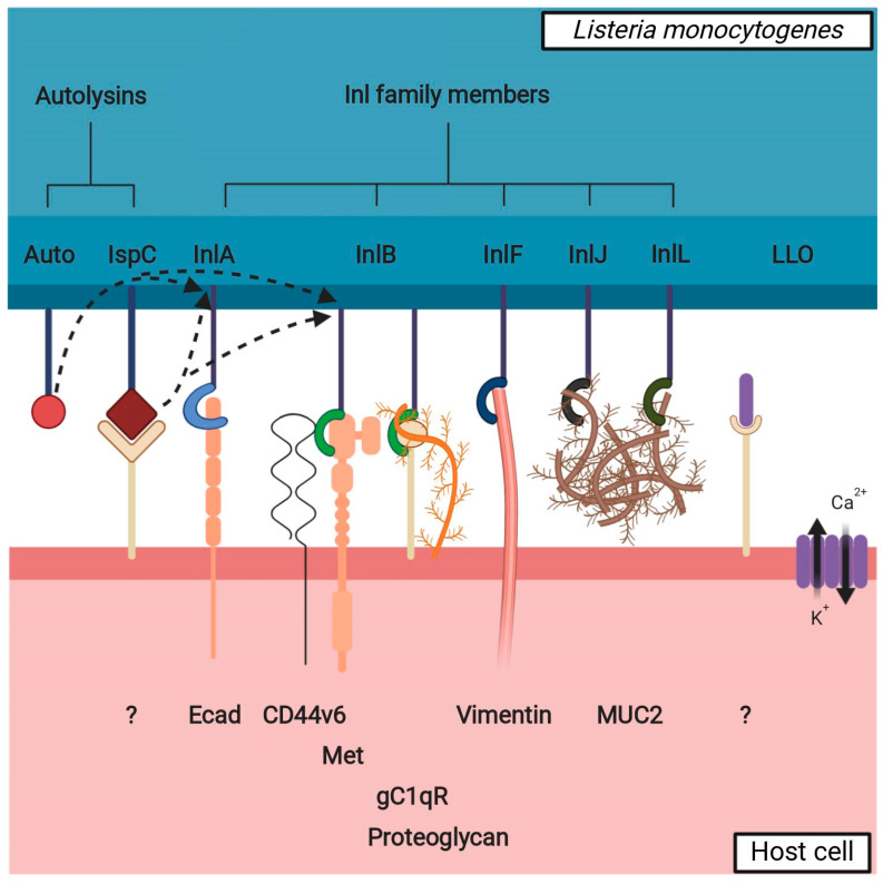
Although it rarely induces disease in humans, Listeria monocytogenes (Lm) is important due to the frequency of serious pathological conditions-such as sepsis and meningitis-it causes in those few people that do get infected. Virulence factors (VF) of Lm-especially those involved in the passage through multiple cellular barriers of the body, including internalin (Inl) family members and listeriolysin O (LLO)-have been investigated both in vitro and in vivo, but the majority of work was focused on the mechanisms utilized during penetration of the gut and fetoplacental barriers. The role of listerial VF during entry into other organs remain as only partially solved puzzles. Here, we review the current knowledge on the entry of Lm into one of its more significant destinations, the brain, with a specific focus on the role of various VF in cellular adhesion and invasion.

Keywords: Listeria monocytogenes; autolysin; brain invasion; internalin; listeriolysin; virulence factors.


Conflict of interest statement

The authors declare no conflict of interest.

Figures

Figure 1
Figure 1
Various proposed routes for listerial entry into the brain. (a) Entry via the blood-brain barrier (BBB). The BBB is comprised of the microvascular endothelial cells connected by tight junctions and supported by astrocytes and pericytes. Passage through the microvascular endothelium proceeds in a transcellular manner, with the bacteria either inducing endocytosis into the cells by interacting with the receptors on the cellular surface or being carried across it in infected monocytes. 1—brain tissue; 2—microvascular capillary lumen; 3—microvascular endothelium; 4—astrocyte; 5—pericyte; 6—macrophage. (b) Entry via the blood-cerebrospinal fluid barrier (BCSFB). The BCSFB is comprised of the choroid plexus epithelial cells connected by tight junctions—the endothelium of the choroid plexus is fenestrated and presents no obstacles to the bacteria. Passage through the choroid plexus epithelium is transcellular, either direct—by induction of endocytosis into the cells—or indirect—within infected monocytes, similar to passage through the BBB. 1—cerebrospinal fluid (CSF); 2—choroid plexus capillary lumen; 3—choroid plexus endothelium; 4—choroid plexus epithelium; 5—connective tissue; 6—macrophage. (c) Entry via the trigeminal nerve. Damage to the stratified squamous epithelium that forms the top layer of oral mucosa opens a path for bacteria from contaminated food to enter the submucosa, where they can be phagocytized by either resident macrophages or recruited phagocytes. Lm is able to survive within these cells and to spread further to the nerve endings of the trigeminal nerve, along which it can travel to the brain stem. 1—trigeminal nerve ending; 2—oral cavity; 3—stratified squamous epithelium of the oral mucosa; 4—submucosa; 5—Lm-contaminated food; 6—wound in the oral mucosa; 7—resident macrophage; 8—recruited phagocyte. (d) Entry via the olfactory epithelium. The ciliated endings of olfactory sensory neurons are located at the surface of olfactory epithelium, supported by epithelial cells and covered in mucus. The axons of olfactory sensory neurons form bundles that pass through the ethmoid bone of the skull and end in the olfactory bulb. Lm can access these neurons when liquids contaminated with Lm (such as vaginal secretions ingested during childbirth) are ingested into the nasal cavity, and traveling along them provides a direct route to the brain. 1—olfactory bulb; 2—nasal cavity; 3—ethmoid bone; 4—supporting epithelium; 5—olfactory neuron; 6—olfactory nerve fiber.
Figure 2
Figure 2
Interaction between listerial VF potentially involved in brain invasion and a non-phagocytic host cell. Inl family members bind to cellular surface receptors to assist Lm in adhesion to and/or invasion into the cell. The interaction partners of presented Inl family members are readily expressed in the brain with the exception of MUC2, which is mainly expressed in the intestinal mucosa—it is possible that InlJ and InlL have other interaction partners in the brain. Autolysins (Auto, IspC) participate in the remodeling of the peptidoglycan layer, affecting the presence of the Inl family members on the bacterial surface (shown by dotted arrows); IspC also has a role in adhesion to the cellular surface. LLO oligomerizes to form pores on the cell membrane and thus destabilizes it, with a twofold effect: influx of Ca2+: a) causes the disruption of tight junctions between the cells (paracellular path) and b) begins a signaling cascade resulting in pre-endocytosis actin remodeling (transcellular path); LLO also possibly interacts with a cellular surface receptor to initiate a downstream cellular signal. Unknown or hypothetical interaction partners on cellular surface are labeled with a question mark.

References

    1. Radoshevich L., Cossart P. Listeria monocytogenes: Towards a complete picture of its physiology and pathogenesis. Nat. Rev. Microbiol. 2018;16:32–46. doi: 10.1038/nrmicro.2017.126. - DOI - PubMed
    1. Maury M.M., Tsai Y.H., Charlier C., Touchon M., Chenal-Francisque V., Leclercq A., Criscuolo A., Gautier C., Roussel S., Brisabois A., et al. Uncovering Listeria monocytogenes hypervirulence by harnessing its biodiversity. Nat. Genet. 2016;48:308–313. doi: 10.1038/ng.3501. - DOI - PMC - PubMed
    1. Maiden M.C., Bygraves J.A., Feil E., Morelli G., Russell J.E., Urwin R., Zhang Q., Zhou J., Zurth K., Caugant D.A., et al. Multilocus sequence typing: A portable approach to the identification of clones within populations of pathogenic microorganisms. Proc. Natl. Acad Sci. USA. 1998;95:3140–3145. doi: 10.1073/pnas.95.6.3140. - DOI - PMC - PubMed
    1. Ragon M., Wirth T., Hollandt F., Lavenir R., Lecuit M., Le Monnier A., Brisse S. A new perspective on Listeria monocytogenes evolution. PLoS Pathog. 2008;4:e1000146. doi: 10.1371/journal.ppat.1000146. - DOI - PMC - PubMed
    1. Bierne H., Milohanic E., Kortebi M. To be cytosolic or vacuolar: The double life of listeria monocytogenes. Front. Cell Infect. Microbiol. 2018;8:136. doi: 10.3389/fcimb.2018.00136. - DOI - PMC - PubMed

Publication types

MeSH terms