The Impact of Intracortical Lesions on Volumes of Subcortical Structures in Multiple Sclerosis
- PMID: 32381540
- PMCID: PMC7228180
- DOI: 10.3174/ajnr.A6513
The Impact of Intracortical Lesions on Volumes of Subcortical Structures in Multiple Sclerosis
Abstract
Background and purpose: Recent studies showed thalamic atrophy in the early stages of MS. We investigated the impact of intracortical lesions on the volumes of subcortical structures (especially the thalamus) compared with other lesions in MS.
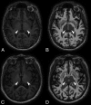
Materials and methods: Seventy-one patients with MS were included. The volumes of intracortical lesions and white matter lesions were identified on double inversion recovery and FLAIR, respectively, by using 3D Slicer. Volumes of white matter T1 hypointensities and subcortical gray matter, thalamus, caudate, putamen, and pallidum volumes were calculated using FreeSurfer. Age, MS duration, and the Expanded Disability Status Scale score were assessed.
Results: Patients with intracortical lesions were older (P = .003), had longer disease duration (P < .001), and higher Expanded Disability Status Scale scores (P = .02). The presence of intracortical lesions was associated with a significant decrease of subcortical gray matter volume (P = .02). In our multiple regression model, intracortical lesion volume was the only predictor of thalamic volume (R 2 = 0.4, b* = -0.28, P = .03) independent of white matter lesion volume and T1 hypointensity volume. White matter lesion volume showed an impact on subcortical gray matter volume in patients with relapsing-remitting MS (P = .04) and those with disease duration of <5 years (P = .04) and on thalamic volume in patients with Expanded Disability Status Scale scores of <4.0 (P = .01). By contrast, intracortical lesion volume showed an impact on subcortical gray matter and thalamic volumes in the secondary-progressive MS subgroup (P = .02 and P < .001) in patients with a long-standing disease course (P < .001 and P = .001) and more profound disability (P < .001 and P < .001).
Conclusions: Thalamic atrophy was explained better by intracortical lesions than by white matter lesion and T1 hypointensity volumes, especially in patients with more profound disability.
© 2020 by American Journal of Neuroradiology.
Figures


Comment in
-
The Development of Subcortical Gray Matter Atrophy in Multiple Sclerosis: One Size Does Not Fit All.AJNR Am J Neuroradiol. 2020 Sep;41(9):E80-E81. doi: 10.3174/ajnr.A6698. Epub 2020 Jul 9. AJNR Am J Neuroradiol. 2020. PMID: 32646951 Free PMC article. No abstract available.
References
Publication types
MeSH terms
LinkOut - more resources
Full Text Sources
Medical
