. 2020 May 7;11(5):328.
doi: 10.1038/s41419-020-2519-8.

The USP10-HDAC6 axis confers cisplatin resistance in non-small cell lung cancer lacking wild-type p53

Affiliations

The USP10-HDAC6 axis confers cisplatin resistance in non-small cell lung cancer lacking wild-type p53

Chen Hu et al. Cell Death Dis. .

Abstract

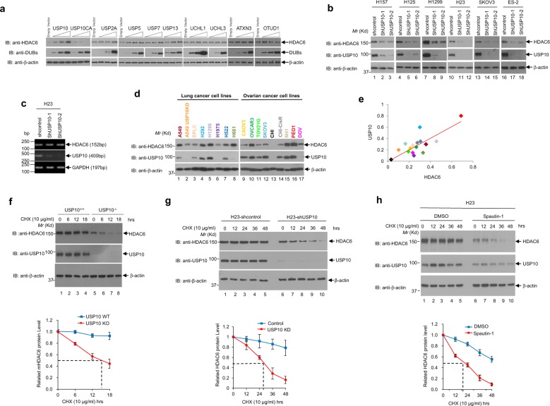
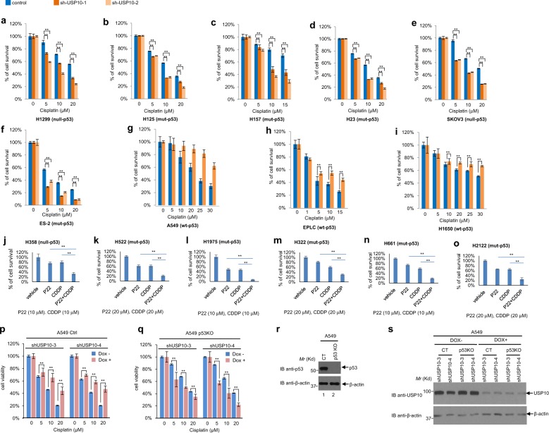
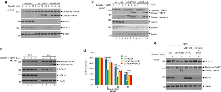
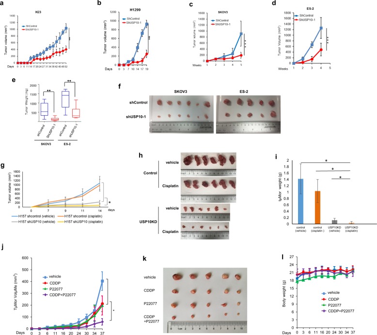
Ubiquitin-specific peptidase 10 (USP10) stabilizes both tumor suppressors and oncogenes in a context-dependent manner. However, the nature of USP10's role in non-small cell lung cancer (NSCLC) remains unclear. By analyzing The Cancer Genome Atlas (TCGA) database, we have shown that high levels of USP10 are associated with poor overall survival in NSCLC with mutant p53, but not with wild-type p53. Consistently, genetic depletion or pharmacological inhibition of USP10 dramatically reduces the growth of lung cancer xenografts lacking wild-type p53 and sensitizes them to cisplatin. Mechanistically, USP10 interacts with, deubiquitinates, and stabilizes oncogenic protein histone deacetylase 6 (HDAC6). Furthermore, reintroducing either USP10 or HDAC6 into a USP10-knockdown NSCLC H1299 cell line with null-p53 renders cisplatin resistance. This result suggests the existence of a "USP10-HDAC6-cisplatin resistance" axis. Clinically, we have found a positive correlation between USP10 and HDAC6 expression in a cohort of NSCLC patient samples. Moreover, we have shown that high levels of USP10 mRNA correlate with poor overall survival in a cohort of advanced NSCLC patients who received platinum-based chemotherapy. Overall, our studies suggest that USP10 could be a potential biomarker for predicting patient response to platinum, and that targeting USP10 could sensitize lung cancer patients lacking wild-type p53 to platinum-based therapy.


Conflict of interest statement

The authors declare that they have no conflict of interest.

Figures

Fig. 1
Fig. 1. USP10 interacts with HDAC6.
a, b Endogenous USP10 and HDAC6 interact with each other in H23 cells. a The anti-USP10 antibody or anti-IgG was used to immunoprecipitate USP10 in H23 cells followed by anti-HDAC6 Western blotting analysis (upper panel). The blot was stripped and reprobed with the anti-USP10 antibody (lower panel). b The reciprocal immunoprecipitation of a was performed. c USP10 physically binds to HDAC6. Bacterially-purified GST or GST-USP10 was incubated with His-HDAC6 isolated from E.coli, and the GST pull-down assays were performed followed by anti-HDAC6 Western blotting analysis (upper panel). His-HDAC6, GST, and GST-USP10 proteins were visualized via Coomassie blue staining (middle and lower panels). d, e Both the DAC1 domain and DAC2 domain of HDAC6 interact with USP10. d The schematic diagrams of HDAC6 full-length (FL) and deletion mutants. e The indicated vector and HDAC6 plasmids were co-transfected with HA-USP10 in 293T cells, followed by anti-Flag immunoprecipitation and subsequent anti-HA Western blotting analysis (upper panel). The blot was then stripped and reprobed with anti-Flag antibodies (middle panel). The input of HA-USP10 is shown in the lower panel. f, g The very C-terminus of USP10 binds to HDAC6. f The schematic diagrams of USP10 full-length (FL) and deletion mutants. g The indicated vector and USP10 plasmids were co-transfected with HA-HDAC6 in 293T cells, and anti-Flag immunoprecipitation was performed followed by anti-HA Western blotting analysis (upper panel). The blot was then reprobed with anti-Flag antibodies (middle panel). The input of HA-HDAC6 is shown in the lower panel.
Fig. 2
Fig. 2. USP10 stabilizes HDAC6.
a 293T cells were transfected with empty vector, wild-type USP10 (Flag-USP10), catalytically-dead mutant of USP10 (Flag-USP10CA) or other DUBs as indicated. Thirty-six hours post-transfection, cells were harvested. Anti-HDAC6, anti-USP10, anti-DUBs, and anti-β-actin Western blotting analyses were performed. b Depletion of USP10 reduces the level of HDAC6. Six lung and ovarian cancer cell lines, H157, H125, H1299, H23, SKOV3, and ES-2, were infected with lentivirus encoding two shRNAs against USP10. The anti-USP10, anti-HDAC6, and anti-β-actin Western blotting analyses were performed as indicated. c Depletion of USP10 does not affect the mRNA level of HDAC6 in H23 cells. Total RNAs extracted from shcontrol, shUSP10-1 or shUSP10-2 lentivirus stably infected H23 cells were subjected to RT-PCR for HDAC6, USP10, and GAPDH, as indicated. d HDAC6 protein levels are positively correlated with USP10 in a panel of lung and ovarian cancer cell lines. HDAC6, USP10, and β-actin protein levels were obtained by Western blotting analyses of 17 cell lines (A549, A549-USP10KD, EPLC, H292, H1299, H1975, H522, H661, CAOV3, OVCAR3, TOV21G, SKOV3, CHI, CHI-CisR, M41, PEO1, and DOV). e HDAC6 and USP10 protein levels were normalized by the level of β-actin and quantified with Image Pro Plus 6.0 software. The correlation analysis was performed by Excel CORREL function and the results were shown as a scatter plot indicating a strong positive correlation coefficient of USP10 and HDAC6 (r = 0.78, p < 0.01). f The half-life of HDAC6 is shortened in USP10KO MEFs. Cycloheximide (CHX) was added to USP10 WT and USP10 KO MEFs at the indicated concentration and time intervals. The anti-HDAC6, anti-USP10, and anti-β-actin Western blotting analyses were performed (top panel). The experiments were repeated three times. The HDAC6 expression was quantified and a plot showing half-life of HDAC6 in USP10 WT and USP10 KO MEFs was drawn (lower panel). g The half-life of HDAC6 is shortened in USP10KD H23 cells. Half-lives of HDAC6 in H23-shcontrol and H23-shUSP10 cells were performed as f. h USP10 inhibitor Spautin-1 shortens the half-life of HDAC6. Spautin-1 was added to the H23 cells at the indicated concentrations and time intervals. The anti-HDAC6, anti-USP10, and anti-β-actin Western blotting analyses were performed. Half-life of HDAC6 in DMSO-treated or Spautin-1-treated H23 cells was performed as f. For graphs in fh, the mean band intensities from three independent experiments as measured by Image-Pro plus 6.0 shows the approximate half-lives in the presence of CHX. The error bars represent the standard deviation.
Fig. 3
Fig. 3. USP10 deubiquitinates HDAC6.
a USP10 regulates the protein level of HDAC6 via the ubiquitin-proteasome pathway. H23 control and H23 USP10 stable knockdown (USP10KD) cells were either left untreated or treated with MG132 for 10 h, then were lysed and subjected to Western blotting analyses as indicated. b Wild-type, but not the catalytically-dead mutant of USP10, deubiquitinates HDAC6 in vivo. 293T cells were transfected with the indicated plasmids. The anti-Flag denatured immunoprecipitation was performed followed by anti-HA Western blotting analysis (upper panel). The blot was stripped and reprobed with anti-Flag antibody (middle panel). The anti-GFP Western blotting analysis was performed to show the input of GFP-USP10WT and GFP-USP10CA. c Wild-type, but not the catalytically-dead mutant of USP10, deubiquitinates HDAC6 in vitro. Ubiquitinated HA-HDAC6 proteins isolated from 293T cells were pulled down by anti-HA agarose beads, followed by incubation with bacterial purified GST, GST-USP10, or GST-USP10CA proteins as described in the Methods. HDAC6 ubiquitination levels were determined by Western blotting with anti-HA (top panel), and the amount of GST, GST-USP10, and GST-USP10CA proteins were confirmed by coomassie blue staining (bottom two panels). d Knockdown of USP10 increases the K48-linked poly-ubiquitination of HDAC6. H1299 cells stably expressing shControl or shUSP10 shRNAs were treated with MG132 (5 µM) overnight. The anti-HDAC6 antibody was used to immunoprecipitate HDAC6 in control and USP10KD cells. Half of the samples were subject to anti-K48 poly-Ub Western blotting analysis; the other half of the samples were subject to anti-HDAC6 Western blotting analysis as indicated. The anti-USP10 and anti-β-actin Western blotting analyses were also performed using total cell lysates. e–g Representative MS2 spectra showing putative ubiquitin binding sites Lysines 51, 116, and 849 within HDAC6. Recombinant HDAC6 was immunoprecipitated, separated by SDS-PAGE and digested in-gel with trypsin. Peptides were analyzed by LC-MS/MS. Ubiquitination commonly occurs as the last amino acid of ubiquitin is covalently linked to a lysine residue on the substrate. Since the last three ubiquitin residues are Arg/Gly/Gly, tryptic cleavage of ubiquitinated histidine residues can by identified by Gly/Gly modification (+114). Inset: Fragmentation patterns of b and y ions show sequence information and localization of the Gly/Gly histidine modification. Also shown are the modified amino acid residue number for HDAC6, m/z and charge state. h Lysines 51, 116, 849 are targeted for ubiquitination of HDAC6. Upper panel: The diagram of HDAC6 showing HDAC6 domains and the three ubiquitination sites. Lower panel: HA-Ub was cotransfected with either Flag-HDAC6 wild-type or Flag-HDAC6 Ub site mutants as indicated into 293T cells. Anti-Flag-M2 agarose beads were used to IP Flag-HDAC6. Anti-HA Western blotting analysis was performed to detect the ubiquitination level of HDAC6. i Mutation of the three ubiquitination sites (K51, K116, and K849) in HDAC6 prolongs HDAC6’s half-life. USP10 stable knockdown 293T cells were transfected with either Flag-HDAC6 wild-type (WT) or Flag-HDAC6 K51/116/849R (3KR) followed by CHX treatment at indicated time intervals. Anti-Flag and anti-β-actin Western blotting analyses were performed (upper panel). A graph of the mean band intensities from three independent experiments as measured by Image-Pro plus 6.0 shows the approximate half-lives of HDAC6 wild type and the triple site mutant in the presence of CHX. The error bars represent the standard deviation (low panel).
Fig. 4
Fig. 4. Knockdown of USP10 sensitizes lung and ovarian cancer cells harboring mutant- or null- p53, but not wild-type p53, to cisplatin by MTT assays.
a–i Indicated control cell line and two USP10 stable knockdown counterparts were subjected to a 3-day MTT assay. The dosage of cisplatin used is indicated. j–o Indicated cell lines were treated with vehicle, P22077 (P22), cisplatin (CDDP), or P22 + CDDP and were subjected to a 2-day MTT assay, except l, which was subject to a 3-day MTT assay. p Doxycycline (Dox)-induced two USP10 knockdowns in A549-control (Ctrl or CT) cell lines: A549-shUSP10-3 and A549-shUSP10-4, were pretreated with 1 μg/ml Doxycycline for 3 days, then were co-treated with cisplatin as indicated concentrations for another 3 days. MTT assays were performed to measure cell viability after treatment. q Dox-induced two USP10 knockdowns in A549-p53KO cell lines: A549-p53KO-shUSP10-3, A549-p53KO-shUSP10-4 were pretreated with 1 μg/ml Doxycycline for 3 days, then were co-treated with cisplatin as indicated concentrations for another 3 days. MTT assays were performed to measure cell viability after treatment. r Anti-p53 and anti−β-actin Western blotting analyses were performed with A549-(CT) and A549-(p53KO) cells. s A549-(CT) and A549-(p53KO) cells were treated with either vehicle or 1 μg/ml Doxycycline for 3 days and whole cell lysate were subjected to Western blot analysis using indicated antibodies. For a–q, the error bars represent the standard deviation. Double asterisk indicates p < 0.01.
Fig. 5
Fig. 5. Knockdown of USP10 sensitizes lung and ovarian cancer cells harboring mutant- or null-p53 to cisplatin by colony formation assays.
a, b Knockdown of USP10 sensitizes three lung and two ovarian cancer cell lines to cisplatin by colony formation assays. Control and USP10 stable knockdown H157, H23, H1299, SKOV3, and ES-2 cells were treated with cisplatin at the various concentrations (H157, 2 μM; H23, 0.5 μM; H1299, 1 μM; SKOV3 and ES-2, 0.3 μM) for 7–14 days. Colony formation assay was performed as described in the Methods. Colonies were visualized by crystal violet staining (a). Colonies numbers were quantified with OpenCFU software. The percentage of colony formation of indicated cells are shown in (b). c, d Inducible knockdown USP10 sensitizes H23 cells to cisplatin by colony formation assays. H23 control and stable inducible USP10 knockdown pool cells were plated at 100 cells/well into six-well dishes and cultured with or without Dox and then treated with cisplatin as indicated for ~14 days. Colonies were fixed, stained with methanol/crystal violet dye, and images captured (c). Colony numbers were counted by OpenCFU software (d). e, f Inhibition of USP10 by spautin 1 sensitizes H1299, H157 and H23 cells to cisplatin in colony formation assays. H1299, H157, and H23 cells were treated with various concentrations of cisplatin (0.5 μM for H1299 and H157; 0.1 μM for H23), USP10 inhibitor spautin-1 (1 μM for H1299, 2 μM for H157, and 0.5 μM for H23), or a combination of cisplatin and spautin-1 for 7–14 days. Colonies were fixed, stained with methanol/crystal violet dye, and images captured (e). The percentages of colony formation of the indicated cell lines are shown in (f). g, h USP10 inhibitor P22077 sensitizes H1299 cells to cisplatin in colony formation assays. Colony formation assays for H1299 were performed as e, f, except that spautin-1 was replaced by P22077 (2 μM) and that cisplatin concentration was 0.5 μM. For all the panels, each assay was performed in triplicate. The error bar represents standard deviation. *p < 0.05, **p < 0.01, ***p < 0.001.
Fig. 6
Fig. 6. Depletion of USP10 increases apoptosis in NSCLC cells upon cisplatin treatment; USP10 confers cisplatin resistance via HDAC6.
a Knockdown of USP10 increases apoptosis upon treatment of cisplatin. H157 cells infected with lentiviruses containing empty vector, shUSP10-1, or shUSP10-2 were treated with cisplatin at the indicated concentrations for 3 days. Cells were lysed, then anti-PARP1, anti-USP10, anti-β-actin and anti-HDAC6 western blotting analyses were performed. b H23 control and USP10 stable knockdown cells were treated with 10 µM cisplatin at the indicated times. Cells were lysed and the anti-PARP1, anti-cleaved caspase 3, and anti-β-actin Western blotting analyses were performed. c Treatment of Dox-induced USP10 knockdown H23 cells with cisplatin increases PARP1 cleavage. H23 cells were pretreated with 1 µg/ml Dox for ~2 days. Cells were then treated with 10 μM cisplatin for the indicated times. Cell lysates were collected for western blotting analyses. d, e Overexpression of USP10 or HDAC6 in USP10-knockdown H1299 cells rescues USP10 knockdown-induced growth reduction and apoptosis upon cisplatin treatment. H1299 control and H1299 USP10 stable knockdown cells, or H1299 USP10 KD cells reintroduced with either USP10 expression plasmid or HDAC6 expression plasmid at each indicated combination, were treated with the indicated concentration of cisplatin for the 3 days. MTT assays were performed in d. Anti-PARP1 and anti-β-actin Western blotting analyses were performed in e. The error bar represents standard deviation. Triple asterisk indicates p < 0.001.
Fig. 7
Fig. 7. Knockdown or inhibition of USP10 suppresses lung and ovarian cancer xenograft growth and sensitizes xenografts to cisplatin treatment in immune-deficient mice.
a–d Knockdown of USP10 inhibits H23, H1299, SKOV3, and ES-2 xenograft growth in immune-deficient mice. a, b The volumes of H23 and H1299 control implants or H23 and H1299 USP10 knockdown implants in SCID mice were measured every 2–3 days as described in the Methods. c, d The SKOV3 and ES-2 control or SKOV3 and ES-2 USP10 knockdown cells were injected into the nude mice as described in the Methods. The tumor volumes were measured weekly. e, f The SKOV3 and ES-2 control or SKOV3 and ES-2 USP10 knockdown xenografts were weighted (e) and the tumors’ images were taken as shown in f. For SKOV3, n = 6; for ES-2, n = 5. g–i Knockdown of USP10 sensitizes the H157 xenografts to cisplatin. H157-control and H157-USP10KD tumors were treated i.v. with either vehicle or cisplatin (at 2 mg/kg), starting on day 1 on a Q3dx5 schedule for a total dose of 10 mg/kg. Mice were euthanized 15 days post-implantation. Tumor volumes were shown in g. Photos of the tumors were shown in h. Tumor weight was quantified in bar graphs shown in i. j Inhibition of USP10 reduces H1299 tumor growth and sensitizes H1299 xenografts to cisplatin in nude mice. The growth curve of H1299 xenograft tumors treated with vehicle, cisplatin (3 mg/kg, intravenous (i.v.) injection), USP10 inhibitor P22077 (15 mg/kg, intraperitoneal (i.p.) injection) and cisplatin plus P22077 was shown. Tumor volumes were calculated as above; n = 5 per group. k Tumor images of H1299 xenografts. l Body weight of mice used in j was measured during the treatment. * p < 0.05, **p < 0.01, ***p < 0.001.
Fig. 8
Fig. 8. Both mRNA and protein levels of USP10 are increased in lung and ovarian cancer patient samples; high USP10 mRNA level correlates with poor overall survival of advanced NSCLC patients treated with platinum.
a The USP10 mRNA expression was elevated in a cohort of NSCLC patients. The box plots were derived from the USP10 gene expression data in Oncomine. The reference, fold change, p value, and sample description were indicated in the table below. b The USP10 mRNA expression was elevated in a cohort of ovarian cancer patients. The box plots were derived from the USP10 gene expression data in Oncomine. The reference, fold change, p value, and sample description were indicated in the table below. c The protein levels of USP10 and HDAC6 were elevated in NSCLC patients. The anti-USP10 and anti-HDAC6 IHC staining was performed with a lung cancer TMA (US Biomax. Inc.). The levels of USP10 and HDAC6 were indicated by IHC scores as described in the Methods. d The protein levels of USP10 and HDAC6 were elevated in ovarian cancer patients. The anti-USP10 and anti-HDAC6 IHC staining were performed with an ovarian cancer TMA (US Biomax. Inc.). The levels of USP10 and HDAC6 were indicated by IHC scores as described in the Methods. e Representative anti-USP10 IHC staining from c. f Representative anti-USP10 IHC staining from d. g The level of USP10 transcript correlates with patient response to platinum treatment, but not to non-platinum treatment. Patient survival according to USP10 high/low status and treatment group. A threshold for USP10 RT-PCR expression was searched using a log-rank test within patients treated with platinum-doublet chemotherapy. The resultant threshold was then used to define the USP10 status of patients treated with non-platinum-doublet chemotherapy. The red and black solid lines indicate OS between low USP10 and high USP10 for patients treated with the platinum doublet. The red and black dotted lines indicate OS between low USP10 and high USP10 for patients treated with the non-platinum doublet. h, i USP10 expression and p53 status in relation to OS in the TCGA lung cancer data set. h The WT p53 cohort were stratified by USP10 high/low expression, or i the mutated p53 cohort were stratified by USP10 high/low expression. j A working model of USP10 conferring cisplatin resistance by stabilizing HDAC6.

References

    1. Ventii KH, Wilkinson KD. Protein partners of deubiquitinating enzymes. Biochem J. 2008;414:161–175. doi: 10.1042/BJ20080798. - DOI - PMC - PubMed
    1. Soncini C, Berdo I, Draetta G. Ras-GAP SH3 domain binding protein (G3BP) is a modulator of USP10, a novel human ubiquitin specific protease. Oncogene. 2001;20:3869–3879. doi: 10.1038/sj.onc.1204553. - DOI - PubMed
    1. Bomberger JM, Barnaby RL, Stanton BA. The deubiquitinating enzyme USP10 regulates the post-endocytic sorting of cystic fibrosis transmembrane conductance regulator in airway epithelial cells. J. Biol. Chem. 2009;284:18778–18789. doi: 10.1074/jbc.M109.001685. - DOI - PMC - PubMed
    1. Takahashi M, et al. HTLV-1 Tax oncoprotein stimulates ROS production and apoptosis in T cells by interacting with USP10. Blood. 2013;122:715–725. doi: 10.1182/blood-2013-03-493718. - DOI - PubMed
    1. Yuan J, Luo K, Zhang L, Cheville JC, Lou Z. USP10 regulates p53 localization and stability by deubiquitinating p53. Cell. 2010;140:384–396. doi: 10.1016/j.cell.2009.12.032. - DOI - PMC - PubMed

Publication types

MeSH terms