. 2020 May 7;11(5):333.
doi: 10.1038/s41419-020-2536-7.

The HMGB1-RAGE axis modulates the growth of autophagy-deficient hepatic tumors

Affiliations

The HMGB1-RAGE axis modulates the growth of autophagy-deficient hepatic tumors

Bilon Khambu et al. Cell Death Dis. .

Abstract

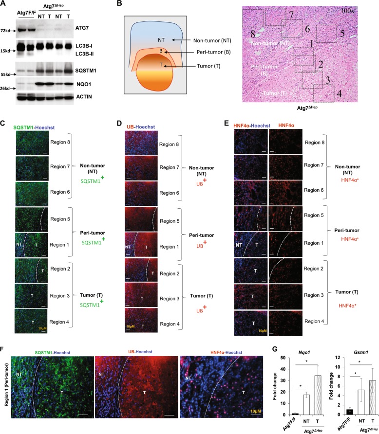
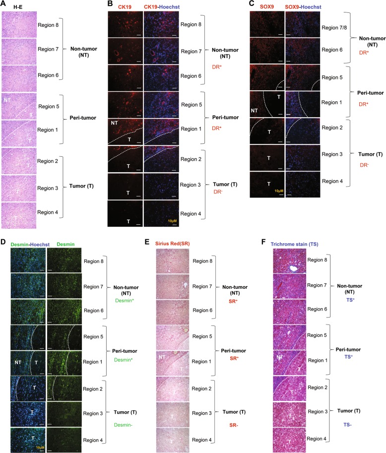
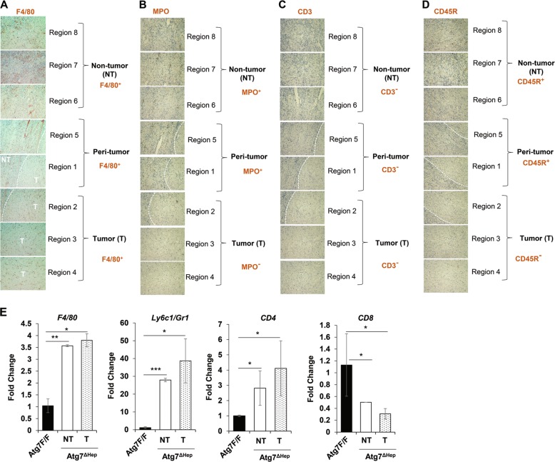
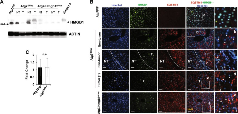
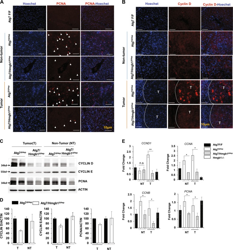
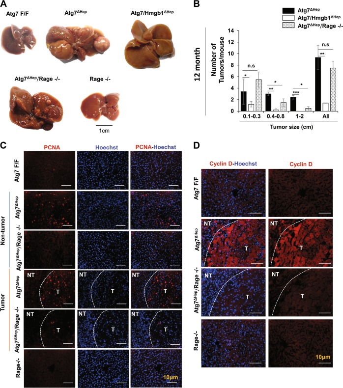
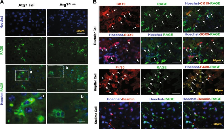
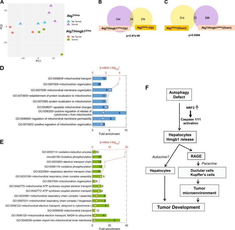
Autophagy is an intracellular lysosomal degradative pathway important for tumor surveillance. Autophagy deficiency can lead to tumorigenesis. Autophagy is also known to be important for the aggressive growth of tumors, yet the mechanism that sustains the growth of autophagy-deficient tumors is not unclear. We previously reported that progression of hepatic tumors developed in autophagy-deficient livers required high mobility group box 1 (HMGB1), which was released from autophagy-deficient hepatocytes. In this study we examined the pathological features of the hepatic tumors and the mechanism of HMGB1-mediated tumorigenesis. We found that in liver-specific autophagy-deficient (Atg7ΔHep) mice the tumors cells were still deficient in autophagy and could also release HMGB1. Histological analysis using cell-specific markers suggested that fibroblast and ductular cells were present only outside the tumor whereas macrophages were present both inside and outside the tumor. Genetic deletion of Hmgb1 or one of its receptors, receptor for advanced glycated end product (Rage), retarded liver tumor development. HMGB1 and RAGE enhanced the proliferation capability of the autophagy-deficient hepatocytes and tumors. However, RAGE expression was only found on ductual cells and Kupffer's cells but not on hepatoctyes, suggesting that HMGB1 might promote hepatic tumor growth through a paracrine mode, which altered the tumor microenvironment. Finally, RNAseq analysis of the tumors indicated that HMGB1 induced a much broad changes in tumors. In particular, genes related to mitochondrial structures or functions were enriched among those differentially expressed in tumors in the presence or absence of HMGB1, revealing a potentially important role of mitochondria in sustaining the growth of autophagy-deficient liver tumors via HMGB1 stimulation.


Conflict of interest statement

The authors declared that they have no conflict of interest.

Figures

Fig. 1
Fig. 1. Hepatic tumor in autophagy-deficient livers are derived from autophagy-deficient hepatocytes.
a Immunoblot analysis of autophagy function-related proteins (ATG7, SQSTM1, LC3B-I/II) and NRF2 pathway-related proteins (NQO1) in whole livers isolated from 15-month-old Atg7F/F, and Atg7ΔHep mice. b Schematic representation of the non-tumor, peri-tumor, and tumor region of the liver sections. Region 1 and Region 5: peri-tumor region, Region 2-Region 4: tumor region, and Region 6- Region 8: non-tumor region. ce Livers from 12-month-old mice of Atg7ΔHep genotype were sectioned and immunostained with anti-SQSTM1(C), Anti-Ubiquitin (UB) (d), or anti-HNF4α (e). Dotted lines indicate the tumor border. f Magnified image of the region 1(peri- and intra-tumor region) of (ce). g The hepatic mRNA expression level of NRF2 target genes, Nqo1 and Gstm1, in the livers of 15-month-old Atg7F/F, and in the non-tumor and tumor samples from the liver of age-matched Atg7ΔHep mice. NT, non-tumor, T, tumor. Data are reported as mean ± SE, *P < 0.05; n = 3 mice per group.
Fig. 2
Fig. 2. Hepatic progenitor cells and fibrosis are localized exclusively in peri-tumor and non-tumor regions but are absent inside the tumor.
Liver sections from 12-month-old mice of the Atg7ΔHep genotype were subjected to H-E staining (a) (original magnification, ×200) and immunostaining for CK19 (b), SOX9 (c), Desmin immunostaining (d), Sirius Red stain (e), or Trichrome stain (f) (original magnification, ×200). Dotted lines indicate the tumor border. NT, non-tumor, T, tumor.
Fig. 3
Fig. 3. Macrophages but not other immune cells are found within the tumor.
Liver sections from 12-month-old mice of Atg7ΔHep genotype were subjected to immunohistochemistry staining for F4/80 (a), Myeloperoxidase (MPO) (b), CD3 (c) and, CD45R (d) (original magnification, ×100). Dotted lines indicate the tumor border. (e) The hepatic mRNA expression level of immune cell-associated genes in 15-month-old Atg7F/F and Atg7ΔHep liver tissues. NT, non-tumor, T, tumor. Data are reported as mean ± SE, *P < 0.05, **P < 0.01, ***P < 0.001, n.s.: no significance; n = 3 mice per group.
Fig. 4
Fig. 4. Hepatic HMGB1 is absent in the tumor of autophagy-deficient livers.
a Livers of 15-month-old mice of different genotypes were examined for HMGB1 by immunoblotting assay. b Liver sections from 15-month-old mice of different genotypes were immunostained with anti-HMGB1 and anti-SQSTM1. White dotted lines indicate the tumor border. White arrowhead indicates the hepatocytes without nuclear HMGB1. c The hepatic mRNA expression level of Hmgb1 in 15-month old-Atg7F/F and Atg7ΔHep mice, determined by real-time PCR. NT, non-tumor, T, tumor. Data are reported as mean ± SE, n.s., no significance; n = 3 mice per group.
Fig. 5
Fig. 5. Loss of HMGB1 in hepatocytes correlates with reduced proliferation in the tumor.
a, b Liver sections from 15-month-old mice of different genotypes were immunostained with anti-PCNA (a), or anti-Cyclin D (b). White arrow indicated proliferating hepatocytes. White dotted lines indicate the tumor border. c Immunoblot analysis of PCNA, cyclin D1, and cyclin E proteins in the tumor or non-tumor sample of 15-month-old Atg7ΔHep and, Atg7/Hmgb1ΔHep mice. d Densitometry qualification of the indicated proteins. e The hepatic mRNA level of indicated genes were determined in the indicated tissues of 15-month-old mice of different genotypes, determined by real-time PCR. NT, non-tumor, T, tumor. Data are reported as mean ± SE, *P < 0.05, **P < 0.01, n.s., no significance; n = 3 mice per group.
Fig. 6
Fig. 6. Genetic loss of Rage inhibits tumorigenesis in autophagy-deficient livers.
a Gross images of representative livers of 12-month-old Atg7ΔHep, Atg7/Hmgb1ΔHep, Atg7ΔHepRage-/-, and Rage-/- mice. b Average number and size distribution of the tumors observed in the livers of 12-month-old mice of different genotypes. c–d Liver sections from 12-month-old mice of different genotypes were immunostained with anti-PCNA (c), or anti-Cyclin D (d). White dotted lines indicate the tumor border. NT. non-tumor, T, tumor. Data are reported as mean ± SE, *P < 0.05, **P < 0.01, ***P < 0.001, n.s., no significance; n = 3 mice per group. Size information of the tumor from Atg7/Hmgb1ΔHep livers is derived from what we has previously reported.
Fig. 7
Fig. 7. RAGE is expressed by ductular cells and Kupffer’s cells but not by hepatocytes or stellate cells.
a Immunofluorescence staining for RAGE antigen in the livers of 9-week-old mice of Atg7F/F and Atg7ΔHepgenotype. Framed areas are enlarged and shown in separate panels (a, b). b Liver sections from 9-week-old Atg7ΔHepmice were coimmunostained with anti-RAGE, together with anti-CK19 or SOX9 or F4/80 or Desmin. White arrows indicate cells with colocalized signals.
Fig. 8
Fig. 8. RNAseq analysis indicates transcriptomic differences in the hepatic tumors of Atg7ΔHepmice and Atg7/Hmgb1ΔHep mice.
a PCA of transcriptomic data based on 12 RNA-seq samples under the four indicated combinations of genotypes and tissue types. bc Numbers of DEGs that are significantly upregulated (b) or downregulated (c) (p < 0.01) in the tumor samples of Atg7/Hmgb1ΔHep and/or Atg7ΔHepmice. The p-values are indicated for the overlap between the two groups of upregulated or downregulated DEGs, respectively. d GO biological processes significantly over-represented in the non-overlapped 256 DEGs uniquely elevated in the tumor samples of the Atg7ΔHep mice. e GO biological processes and KEGG pathways significantly enriched in the non-overlapped 288 DEGs uniquely repressed in the tumor samples of the Atg7/Hmgb1ΔHep mice. For (d) and (e), the heights of bars indicate the fold enrichment compared with random selection, whereas the red dots represent the statistical significance, p-value after FDR-adjusted multiple test correction. The numbers in the bars represent the numbers of DEGs in the particular group which are associated with corresponding GO terms. f Schematic model for the role of HMGB1 in tumor development in the autophagy-deficient liver. HMGB1 is released from autophagy-deficient hepatocytes via the NRF2-inflammasome pathway. Deletion of RAGE, an HMGB1 receptor, mimicked the effect of HMGB1 deletion in delaying tumor development, suggesting that HMGB1 affects tumor development via its released form, but not its DNA-binding form. That HMGB1 may act on hepatocytes in an autocrine fashion could not be completely excluded although hepatocytes do not seem to express a detectable level of RAGE. Released HMGB1 could thus have paracrine effects on target cells that express RAGE and may affect tumor development by altering the microenvironment.

Similar articles

Cited by

References

    1. Ni HM, et al. Nrf2 promotes the development of fibrosis and tumorigenesis in mice with defective hepatic autophagy. J. Hepatol. 2014;61:617–625. doi: 10.1016/j.jhep.2014.04.043. - DOI - PMC - PubMed
    1. Khambu B, et al. HMGB1 promotes ductular reaction and tumorigenesis in autophagy-deficient livers. J. Clin. Invest. 2018;128:2419–2435. doi: 10.1172/JCI91814. - DOI - PMC - PubMed
    1. Inami Y, et al. Persistent activation of Nrf2 through p62 in hepatocellular carcinoma cells. J. Cell Biol. 2011;193:275–284. doi: 10.1083/jcb.201102031. - DOI - PMC - PubMed
    1. Takamura A, et al. Autophagy-deficient mice develop multiple liver tumors. Genes Dev. 2011;25:795–800. doi: 10.1101/gad.2016211. - DOI - PMC - PubMed
    1. Hu TH, et al. Expression and prognostic role of tumor suppressor gene PTEN/MMAC1/TEP1 in hepatocellular carcinoma. Cancer. 2003;97:1929–1940. doi: 10.1002/cncr.11266. - DOI - PubMed

Publication types

MeSH terms

Substances