. 2020 Aug;34(8):2074-2086.
doi: 10.1038/s41375-020-0826-9. Epub 2020 May 7.

High-risk additional chromosomal abnormalities at low blast counts herald death by CML

Collaborators, Affiliations

High-risk additional chromosomal abnormalities at low blast counts herald death by CML

Rüdiger Hehlmann et al. Leukemia. 2020 Aug.

Erratum in

  • Correction: High-risk additional chromosomal abnormalities at low blast counts herald death by CML.
    Hehlmann R, Voskanyan A, Lauseker M, Pfirrmann M, Kalmanti L, Rinaldetti S, Kohlbrenner K, Haferlach C, Schlegelberger B, Fabarius A, Seifarth W, Spieß B, Wuchter P, Krause S, Kolb HJ, Neubauer A, Hossfeld DK, Nerl C, Gratwohl A, Baerlocher GM, Burchert A, Brümmendorf TH, Hasford J, Hochhaus A, Saußele S, Baccarani M; SAKK and the German CML Study Group. Hehlmann R, et al. Leukemia. 2020 Oct;34(10):2823. doi: 10.1038/s41375-020-01039-7. Leukemia. 2020. PMID: 32913312 Free PMC article.

Abstract

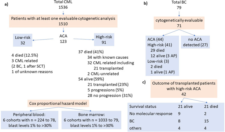
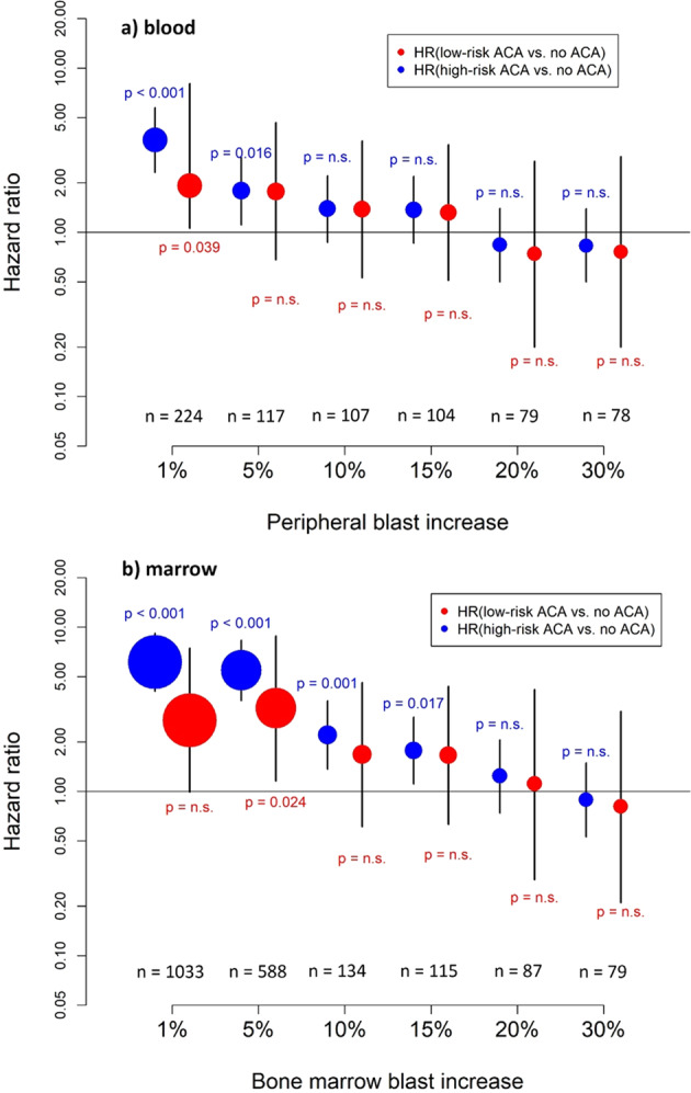
Blast crisis is one of the remaining challenges in chronic myeloid leukemia (CML). Whether additional chromosomal abnormalities (ACAs) enable an earlier recognition of imminent blastic proliferation and a timelier change of treatment is unknown. One thousand five hundred and ten imatinib-treated patients with Philadelphia-chromosome-positive (Ph+) CML randomized in CML-study IV were analyzed for ACA/Ph+ and blast increase. By impact on survival, ACAs were grouped into high risk (+8, +Ph, i(17q), +17, +19, +21, 3q26.2, 11q23, -7/7q abnormalities; complex) and low risk (all other). The presence of high- and low-risk ACAs was linked to six cohorts with different blast levels (1%, 5%, 10%, 15%, 20%, and 30%) in a Cox model. One hundred and twenty-three patients displayed ACA/Ph+ (8.1%), 91 were high risk. At low blast levels (1-15%), high-risk ACA showed an increased hazard to die compared to no ACA (ratios: 3.65 in blood; 6.12 in marrow) in contrast to low-risk ACA. No effect was observed at blast levels of 20-30%. Sixty-three patients with high-risk ACA (69%) died (n = 37) or were alive after progression or progression-related transplantation (n = 26). High-risk ACA at low blast counts identify end-phase CML earlier than current diagnostic systems. Mortality was lower with earlier treatment. Cytogenetic monitoring is indicated when signs of progression surface or response to therapy is unsatisfactory.


Conflict of interest statement

The authors declare that they have no conflict of interest.

Figures

Fig. 1
Fig. 1. Flow chart.
a Patients with ACA, b patients with BC, and c transplanted patients with high-risk ACA. BC blast crisis, AP accelerated phase, SCT stem cell transplantation.
Fig. 2
Fig. 2. Impact of high- vs. low-risk ACA on survival.
The left-side panel shows the impact of +8 (a), +Ph (b), complex ACA (c), and of chromosome 3, 7, 17, 19, and 21 aberrations combined (d) on survival in patients with primary imatinib treatment after the emergence of ACA. Suvival after emergence of low-risk ACA in imatinib-treated patients serves as control. The right-side panel shows the impact of rare high-risk ACA of chromosomes 3, 7, 17, 19, and 21 on survival (ei).
Fig. 3
Fig. 3. Hazard to die with high-risk and low-risk ACA compared to no ACA dependent on blast counts.
Hazard ratios for mortality in imatinib-treated patients with high-risk and low-risk ACA were determined in six different (but overlapping) patient groups (blast increase to 1–30%) together with 95% confidence intervals a in peripheral blood and b in bone marrow. The size of the circle correlates with the sample size. Thirty-seven patients with high-risk ACA and four patients with low-risk ACA died. In 34 patients with high-risk ACA, causes of death were known. Thirty-two of these (94%) died of progression, including progression-related transplantation in 21 patients. Two patients died of CML-unrelated causes. Causes of death were unknown in three patients. With low-risk ACA, causes of death were CML related in three patients and unknown in one patient.

References

    1. Johansson B, Fioretos T, Mitelman F. Cytogenetic and molecular genetic evolution of chronic myeloid leukemia. Acta Haematol. 2002;107:76–94. doi: 10.1159/000046636. - DOI - PubMed
    1. Mitelman F, Levan G, Nilsson PG, Brandt L. Non-random karyotypic evolution in chronic myeloid leukemia. Int J Cancer. 1976;18:24–30. doi: 10.1002/ijc.2910180105. - DOI - PubMed
    1. Sadamori N, Matsunaga M, Yao E, Ichimaru M, Sandberg AA. Chromosomal characteristics of chronic and blastic phases of Ph-positive chronic myeloid leukemia. Cancer Genet Cytogenet. 1985;15:17–24. doi: 10.1016/0165-4608(85)90127-X. - DOI - PubMed
    1. Anastasi J, Feng J, Le Beau MM, Larson RA, Rowley JD, Vardiman JW. The relationship between secondary chromosomal abnormalities and blast transformation in chronic myelogenous leukemia. Leukemia. 1995;9:628–33. - PubMed
    1. Majlis A, Smith TL, Talpaz M, O'Brien S, Rios MB, Kantarjian HM. Significance of cytogenetic clonal evolution in chronic myelogenous leukemia. J Clin Oncol. 1996;14:196–203. doi: 10.1200/JCO.1996.14.1.196. - DOI - PubMed

Publication types