. 2020 May 7;10(1):7734.
doi: 10.1038/s41598-020-64672-3.

Fractionated radiation suppresses Kruppel-like factor 2 pathway to a greater extent than by single exposure to the same total dose

Affiliations

Fractionated radiation suppresses Kruppel-like factor 2 pathway to a greater extent than by single exposure to the same total dose

Ratan Sadhukhan et al. Sci Rep. .

Abstract

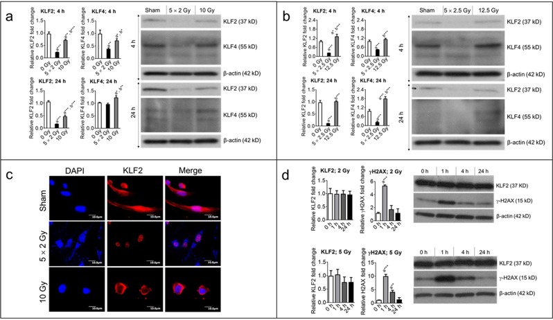
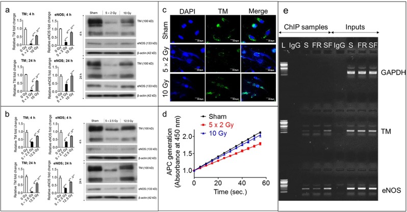
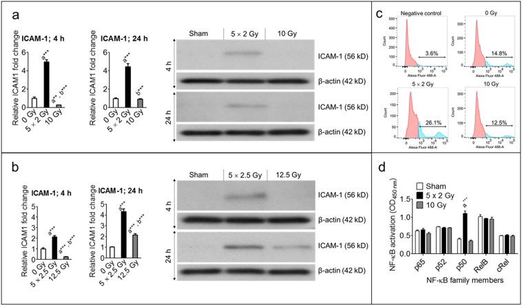
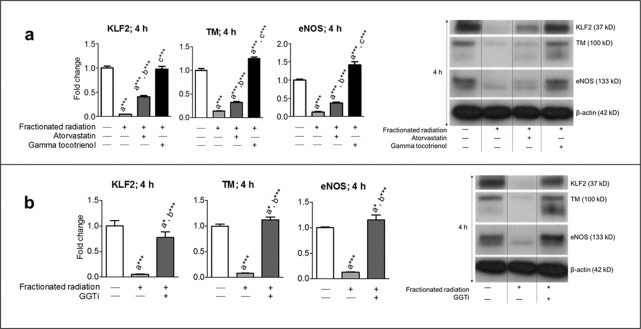
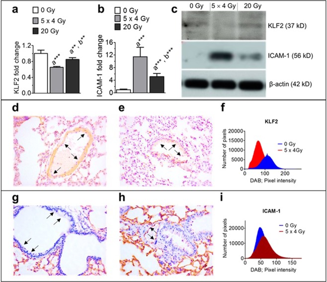
Kruppel-like factor 2 (KLF2) is a positive transcriptional regulator of several endothelial protective molecules, including thrombomodulin (TM), a surface receptor, and endothelial nitric oxide synthase (eNOS), an enzyme that generates nitric oxide (NO). Loss of TM and eNOS causes endothelial dysfunction, which results in suppressed generation of activated protein C (APC) by TM-thrombin complex and in upregulation of intercellular adhesion molecule 1 (ICAM-1). Mechanistic studies revealed that activation of extracellular signal-regulated kinase 5 (ERK5) via upregulation of myocyte enhancer factor 2 (MEF2) induces KLF2 expression. Radiation causes endothelial dysfunction, but no study has investigated radiation's effects on the KLF2 pathway. Because fractionated radiation is routinely used during cancer radiotherapy, we decided to delineate the effects of radiation dose fractionation on the KLF2 signaling cascade at early time points (up to 24 h). We exposed human primary endothelial cells to radiation as a series of fractionated or as a single exposure, with the same total dose delivered to each group. We measured the expression and activity of critical members of the KLF2 pathway at subsequent time points, and determined whether pharmacological upregulation of KLF2 can reverse the radiation effects. Compared to single exposure, fractionated radiation profoundly suppressed KLF2, TM, and eNOS levels, subdued APC generation, declined KLF2 binding ability to TM and eNOS promoters, enhanced ICAM-1 expression, and decreased expression of upstream regulators of KLF2 (ERK5 and MEF2). Pharmacological inhibitors of the mevalonate pathway prevented fractionated-radiation-induced suppression of KLF2, TM, and eNOS expression. Finally, fractionated irradiation to thoracic region more profoundly suppressed KLF2 and enhanced ICAM-1 expression than single exposure in the lung at 24 h. These data clearly indicate that radiation dose fractionation plays a critical role in modulating levels of KLF2, its upstream regulators, and its downstream target molecules in endothelial cells. Our findings will provide important insights for selecting fractionated regimens during radiotherapy and for developing strategies to alleviate radiotherapy-induced toxicity to healthy tissues.


Conflict of interest statement

The authors declare no competing interests.

Figures

Figure 1
Figure 1
Fractionated, compared to single exposure, radiation profoundly suppresses levels of KLF2 and KLF4. Representative (3–5 independent experiments) Western blot analysis and quantification of KLF2 (n = 5) and KLF4 (n = 3) levels in whole-cell lysates from nonirradiated (sham) and irradiated HUVECs 4 h and 24 h after exposure to (a) either five fractions of 2 Gy (5 × 2 Gy) or single exposure to 10 Gy and (b) either five fractions of 2.5 Gy (5 × 2.5 Gy) or single exposure to 12.5 Gy. Fractions delivered at 24-h intervals. β-actin served as a loading control. c Representative photomicrograph (20× magnification) showing immunofluorescence of KLF2 (red) in nonirradiated (sham) and irradiated HUVECs 4 h after exposure to either five fractions of 2 Gy or single exposure to 10 Gy (n = 3). Nuclei were stained with DAPI (blue). d Representative Western blot analysis and quantification of KLF2 (n = 3) and γ-H2AX (n = 3) phosphorylation levels in nonirradiated (0 h) and irradiated HUVECs at indicated time intervals after single exposure to 2 Gy and 5 Gy. β-actin served as a loading control. (n, number of independent experiments performed; a, significant statistical difference between nonirradiated and irradiated groups; b, significant statistical difference between fractionated irradiation and single exposure; *, p < 0.05; **, p < 0.01; ***, p < 0.001).
Figure 2
Figure 2
Fractionated, compared to single exposure, radiation more profoundly suppressed TM and eNOS. Representative (3 independent experiments) Western blot analysis and quantification of TM and eNOS levels in whole-cell lysates from nonirradiated (sham) and irradiated HUVECs 4 h and 24 h after exposure to (a) either five fractions of 2 Gy (5 × 2 Gy) or single exposure to 10 Gy and (b) either five fractions of 2.5 Gy (5 × 2.5 Gy) or single exposure to 12.5 Gy. Fractions were delivered at 24-h intervals. β-actin served as a loading control. c Representative photomicrograph (20× magnification) showing immunofluorescence of TM (green) in nonirradiated (sham) and irradiated HUVECs 4 h after exposure to either five fractions of 2 Gy or single exposure to 10 Gy (n = 3). Nuclei were stained with DAPI (blue). d APC generation in HUVECs 24 h after exposure to 0 Gy (sham), five fractions of 2 Gy, or single exposure to 10 Gy. Experiments were performed twice (n = 2) with four biological replicates. e ChIP assay showing KLF2’s binding ability to TM and eNOS promoter 24 h after 3 fractions of 2.5 Gy or single fraction of 7.5 Gy in HUVECs; Glyceraldehyde 3‐phosphate dehydrogenase (GAPDH) was used as control for ChIP assay. (L, ladder (100 bp); S, sham; FR, fractionated radiation; SF, single fraction).
Figure 3
Figure 3
Fractionated, compared to single exposure, radiation more profoundly enhanced ICAM-1 expression. Representative (3 independent experiments) Western blot analysis and quantification of ICAM-1 levels in whole-cell lysates from nonirradiated (sham) and irradiated HUVECs 4 h and 24 h after exposure to (a) either five fractions of 2 Gy (5 × 2 Gy) or single exposure to 10 Gy and (b) either five fractions of 2.5 Gy (5 × 2.5 Gy) or single exposure to 12.5 Gy. Fractions delivered at 24-h intervals. β-actin served as a loading control. c Ectopic expression of ICAM-1 after exposure to 0 Gy, five fractions of 2 Gy, or 10 Gy, as measured by flow cytometry (n = 3). d NF-κB activation after 4 h of exposure to 0 Gy, five fractions of 2 Gy, or 10 Gy (n = 2).
Figure 4
Figure 4
Fractionated, compared to single exposure, radiation more profoundly suppressed upstream regulators of KLF2. Representative Western blot analysis and quantification of pERK5, tERK5, and MEF2A/C levels in nonirradiated (0 Gy) and irradiated HUVECs 4 h after exposure to either five fractions of 2 Gy (5 × 2 Gy), five fractions of 2.5 Gy (5 × 2.5 Gy), single exposure to 10 Gy, or single exposure to 12.5 Gy (n = 3). β-actin served as a loading control. (n, number of independent experiments performed; a, significant statistical difference between nonirradiated and irradiated groups; b, significant statistical difference between five fractions of 2 Gy vs single exposure to 10 Gy; c, significant statistical difference between five fractions of 2.5 Gy vs single exposure to 12.5 Gy; *, p < 0.05; **, p < 0.01; ***, p < 0.001).
Figure 5
Figure 5
Mevalonate pathway inhibitors reversed fractionated-radiation–induced suppression of KLF2 and its downstream target molecules. Representative Western blot analysis and quantification of KLF2, TM, and eNOS 4 h after exposure to five fractions of 2 Gy (a) in presence or absence of atorvastatin (1 μM) or GT3 (5 μM) and (b) in presence or absence of GGTi (10 μM) (n = 3). β-actin served as a loading control. (n, number of independent experiments performed; a, significant statistical difference between nonirradiated and irradiated groups; b, significant statistical difference between fractionated irradiation and single exposure; *, p < 0.05; **, p < 0.01; ***, p < 0.001).
Figure 6
Figure 6
Fractionated thoracic irradiation suppressed KLF2 and enhanced ICAM-1 levels in the lung. Quantification of KLF2 (a) and ICAM-1 (b) protein levels and representative Western blot analysis (c) in the lung tissue of mice (n = 6) at 24 h following 5 fractions of 4 Gy at 24 h intervals or single exposure to 20 Gy. β-actin served as a loading control. KLF2 immunostaining in the lung tissue samples of sham irradiated (d), irradiated (e), and quantitation (f) at 24 h after exposure to 5 fractions of 4 Gy at 24 h intervals or single exposure to 20 Gy. ICAM-1 immunostaining in the lung tissue samples of sham irradiated (g), or irradiated (h), and quantitation (i) at 24 h after exposure to 5 fractions of 4 Gy at 24 h intervals or single exposure to 20 Gy. (n, number of independent experiments performed; a, significant statistical difference between nonirradiated and irradiated groups; b, significant statistical difference between fractionated irradiation and single exposure; *p < 0.05; **p < 0.01; ***p < 0.001).

References

    1. Galley HF, Webster NR. Physiology of the endothelium. British journal of anaesthesia. 2004;93:105–113. doi: 10.1093/bja/aeh163. - DOI - PubMed
    1. Esmon CT. Protein C anticoagulant system–anti-inflammatory effects. Seminars in immunopathology. 2012;34:127–132. doi: 10.1007/s00281-011-0284-6. - DOI - PMC - PubMed
    1. Forstermann U, Munzel T. Endothelial nitric oxide synthase in vascular disease: from marvel to menace. Circulation. 2006;113:1708–1714. doi: 10.1161/CIRCULATIONAHA.105.602532. - DOI - PubMed
    1. Ito T, Maruyama I. Thrombomodulin: protectorate God of the vasculature in thrombosis and inflammation. Journal of thrombosis and haemostasis: JTH. 2011;9(Suppl 1):168–173. doi: 10.1111/j.1538-7836.2011.04319.x. - DOI - PubMed
    1. Martin FA, Murphy RP, Cummins PM. Thrombomodulin and the vascular endothelium: insights into functional, regulatory, and therapeutic aspects. American journal of physiology. Heart and circulatory physiology. 2013;304:H1585–1597. doi: 10.1152/ajpheart.00096.2013. - DOI - PMC - PubMed

Publication types

MeSH terms

Substances