

High grade acromioclavicular injury: Comparison of arthroscopic assisted acromioclavicular joint fixation and anatomic acromioclavicular joint reconstruction
- PMID: 32382217
- PMCID: PMC7200614
- DOI: 10.1016/j.jor.2020.04.007
High grade acromioclavicular injury: Comparison of arthroscopic assisted acromioclavicular joint fixation and anatomic acromioclavicular joint reconstruction
Abstract
Background: Injuries to the acromioclavicular (AC) joint disrupting the AC and Coracoclavicular (CC) ligaments could lead to loss of clavicular strut function. A High-grade AC joint injury remain challenging because of highly variety of treatment and limited evidence exists comparing clinical outcomes and complications after surgery.
Purpose: The purpose of this study was to compare the clinical and radiological outcome after arthroscopic assisted acromioclavicular Joint fixation and anatomic acromioclavicular joint reconstruction.
Study design: Retrospective Clinical Cohort Study.
Methods: Twenty-nine patients with a high grade (Rockwood Type III-V) Acromioclavicular Joint Injury, operatively treated with arthroscopic assisted acromioclavicular joint fixation and anatomic acromioclavicular joint reconstruction from 2012 to 2018. The clinical assessment consists of Specific AC Score (SACS) and Nottingham Score. The radiographic evaluations were performed to evaluate the stability of reduction and the complications are assessed.
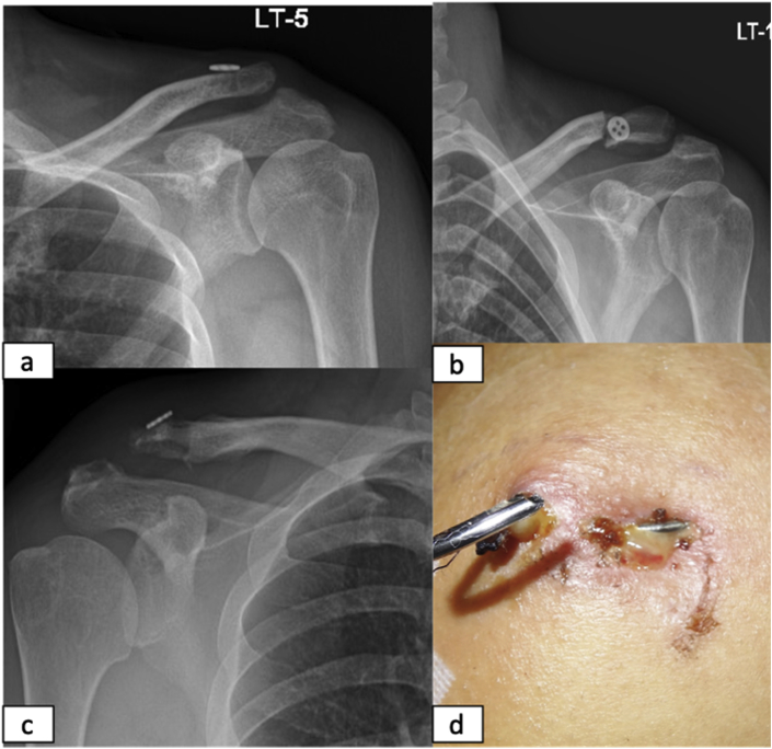
Results: There were a significant different between groups with AC joint reconstruction and fixation group (20 vs. 8, p < 0.005) and (19 vs. 10, p < 0.005) in SAC score and Nottingham score, respectively. There were no significant different in CC dstance between two groups. At the last follow up 6 patient develop loss of reduction with CC percentage >50% in AC joint fixation. Complication developed in 5 patients (17%) including 2 mild osteolysis, 1 superficial infection, 1 implant failure, and 1 clavicle fracture.
Conclusion: Anatomic AC joint reconstruction was associated with functional and radiologic benefits and better restored the stability.
Keywords: AC joint Fixation; AC joint Reconstruction; Acromioclavicular joint injury.
© 2020 Published by Elsevier B.V. on behalf of Professor P K Surendran Memorial Education Foundation.
Conflict of interest statement
Both authors declare that they do not have any conflict of interest. Both authors have nothing to disclose.
Figures


References
-
- Dyrna F., Imhoff F.B., Haller B. Primary stability of an acromioclavicular joint repair is affected by the type of additional reconstruction of the acromioclavicular capsule. Am J Sports Med. 2018;46(14):3471–3479. - PubMed
-
- Gowd A.K., Liu J.N., Cabarcas B.C. Current concepts in the operative management of acromioclavicular dislocations: a systematic review and meta-analysis of operative techniques. Am J Sports Med. 2018:1–14. - PubMed
-
- Braun S., Imhoff A.B., Martetschlaeger F. Primary fixation of acromioclavicular joint disruption. Oper Tech Sports Med. 2014;22(3):221–226. doi: 10.1053/j.otsm.2014.03.005. [Internet] Available at. - DOI
-
- Cano-Martínez J.A., Nicolás-Serrano G., Bento-Gerard J., Picazo-Marín F., Andrés-Grau J. Acute high-grade acromioclavicular dislocations treated with triple button device (MINAR): preliminary results. Injury. 2016;47(11):2512–2519. - PubMed
LinkOut - more resources
Full Text Sources
