Case Reports
. 2020 Apr 28;15(6):683-687.
doi: 10.1016/j.radcr.2020.03.004. eCollection 2020 Jun.

Anaplastic meningioma seeding of the abdominal wall following calvarial bone flap preservation

Affiliations
Case Reports

Anaplastic meningioma seeding of the abdominal wall following calvarial bone flap preservation

Melissa Ling et al. Radiol Case Rep. .

Abstract

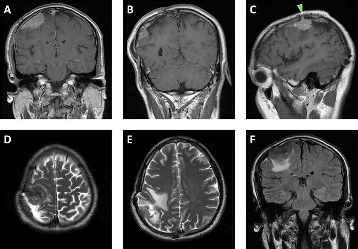
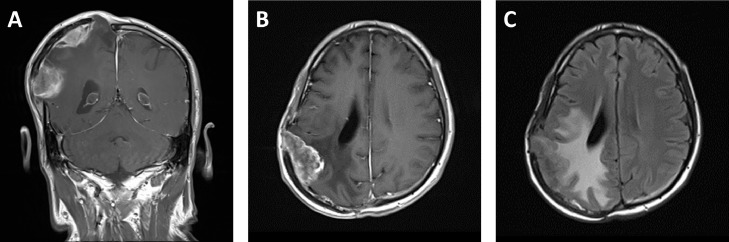
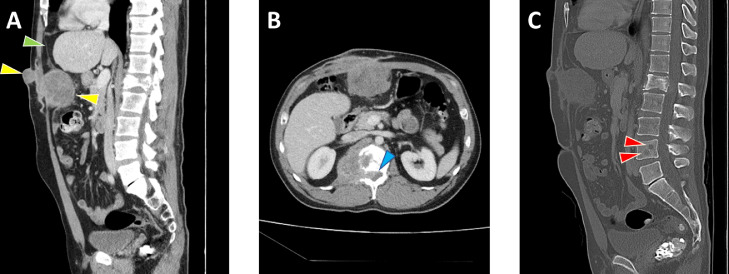
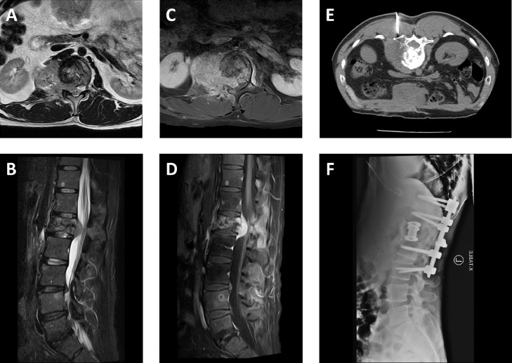
Meningiomas are common intracranial tumors that rarely metastasize. We present a highly unusual case of a 42-year-old man with direct seeding of meningioma to the abdominal wall. The patient had a history of multiple operations for a recurrent intracranial meningioma with decompressive craniectomy and preservation of the calvarial bone flap by implantation into the subcutaneous layer of the anterior abdominal wall. Following removal of the bone flap, a new abdominal wall mass was identified, consistent with iatrogenic implantation of anaplastic meningioma.

Keywords: Anaplastic meningioma; Calvarial bone flap preservation; Central nervous system neoplasms; Metastatic meningioma.


Figures

Fig. 1
Fig. 1
Initial meningioma recurrence. Coronal postcontrast T1-weighted MR images of the brain (A, B) depict 2 dural-based, enhancing masses underlying a right parietal craniotomy flap. Sagittal postcontrast T1-weighted image (C) illustrates one of these masses extending into the craniotomy kerf (green arrowhead). Axial T2-weighted images (D, E) depict areas of hypointensity within the masses and reactive edema in the underlying parietal lobe, which is also highlighted on the coronal fluid-attenuated inversion recovery image (F). The edema results in moderate elevation of the craniotomy flap (B, E). (Color version of figure is available online.)
Fig. 2
Fig. 2
Second meningioma recurrence. Coronal (A) and axial (B) postcontrast T1-weighted MR images of the brain depict multiple dural-based enhancing masses underlying the right parietal craniotomy flap. The axial fluid-attenuated inversion recovery image (C) shows significant reactive edema in the underlaying parietal lobe. Associated brain swelling markedly elevates the craniotomy flap (A).
Fig. 3
Fig. 3
Abdominal wall implantation and lumbar spine meningioma metastases. Sagittal (A) and axial (B) contrast-enhanced CT images of the abdomen and pelvis on soft-tissue windows reveal a lobulated mass (yellow arrowheads) in the anterior abdominal wall at the site of prior craniotomy flap storage extending from the skin surface to the preperitoneal fat and deflecting the parietal peritoneum (green arrowhead). Also seen is a pathologic fracture of the L1 vertebra with associated enhancing soft tissue mass extending into the spinal canal (blue arrowhead) and right psoas muscle. Sagittal CT image on bone window (C) reveals additional lytic lesions in the L4 vertebra (red arrowheads) in addition to better delineating the L1 pathologic fracture. (Color version of figure is available online.)
Fig. 4
Fig. 4
Spinal cord compression due to metastatic meningioma. Axial T2-weighted (A), sagittal short tau inversion recovery (B), and axial (C) and sagittal (D) postcontrast T1-weighted fat suppressed MR images of the lumbar spine reveal an enhancing mass associated with a pathologic fracture at the L1 level extending into the right ventrolateral epidural space and deforming the contour of the conus medullaris. Additional enhancing metastatic lesions are also seen in the L4 vertebral body. The mass at L1 underwent CT-guided biopsy (E) demonstrating anaplastic meningioma, following by decompression, corpectomy, and T11-L3 spinal fixation with associated hardware seen on lateral radiograph (F).

Similar articles

Cited by

References

    1. Thomas R.Z., Dalal I. Extracranial metastases of anaplastic meningioma. BJR Case Rep. 2017 7;3(2) doi: 10.1259/bjrcr.20150092. - DOI - PMC - PubMed
    1. Enam S.A., Abdulrauf S., Mehta B., Malik G.M., Mahmood A. Metastasis in meningioma. Acta Neurochir (Wien) 1996;138(10):1172–1177. doi: 10.1007/bf01809747. discussion 1177-8. - DOI - PubMed
    1. Kok D.L., Hendry S., Alvarez B. Iatrogenic subcutaneous metastasis from WHO Grade I intracranial meningioma. J Clin Neurosci. 2018;58:224–225. doi: 10.1016/j.jocn.2018.09.005. - DOI - PubMed
    1. Singh R.V., Yeh J.S., Campbell D.A. Implantation meningioma in temporalis muscle: case report. Br J Neurosurg. 1994;8(1):93–95. doi: 10.3109/02688699409002400. - DOI - PubMed
    1. Lüdemann W.O., Obler R., Tatagiba M., Samii M. Seeding of malignant meningioma along a surgical trajectory on the scalp. Case report and review of the literature. J Neurosurg. 2002;97(3):683–686. doi: 10.3171/jns.2002.97.3.0683. - DOI - PubMed

Publication types

LinkOut - more resources