Review
. 2020 Jun 6;10(3):20190098.
doi: 10.1098/rsfs.2019.0098. Epub 2020 Apr 17.

Sleep, circadian rhythms and health

Affiliations
Review

Sleep, circadian rhythms and health

Russell G Foster. Interface Focus. .

Abstract

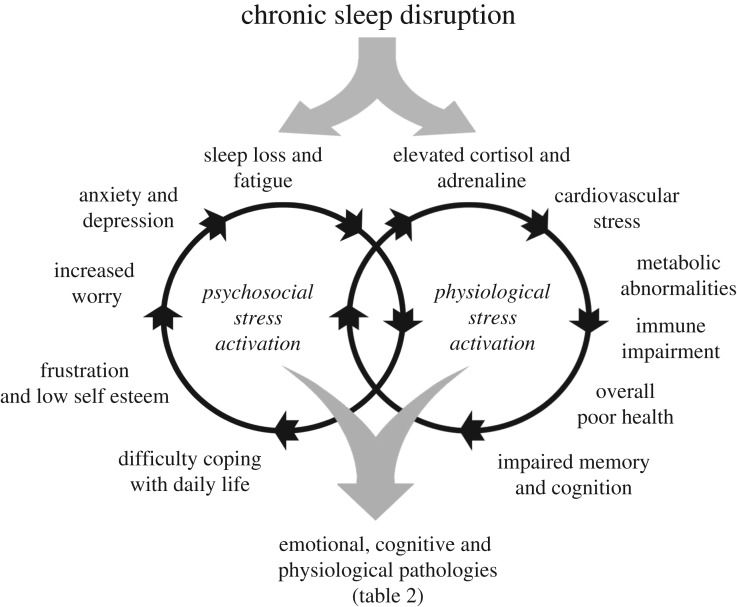
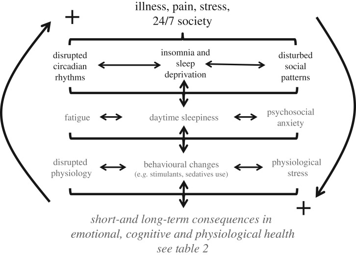
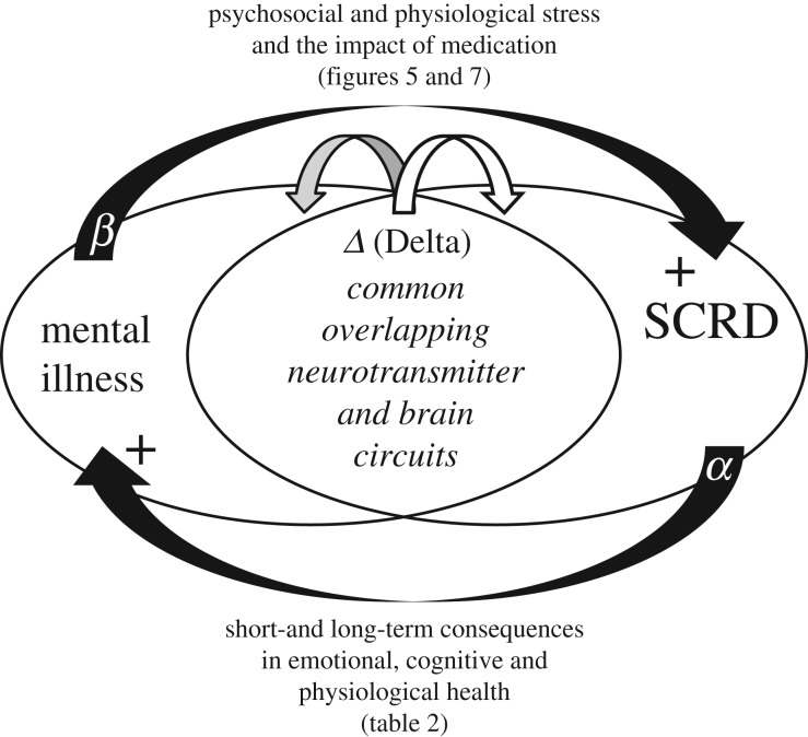
At the core of human thought, for the majority of individuals in the developed nations at least, there is the tacit assumption that as a species we are unfettered by the demands imposed by our biology and that we can do what we want, at whatever time we choose, whereas in reality every aspect of our physiology and behaviour is constrained by a 24 h beat arising from deep within our evolution. Our daily circadian rhythms and sleep/wake cycle allow us to function optimally in a dynamic world, adjusting our biology to the demands imposed by the day/night cycle. The themes developed in this review focus upon the growing realization that we ignore the circadian and sleep systems at our peril, and this paper considers the mechanisms that generate and regulate circadian and sleep systems; what happens mechanistically when these systems collapse as a result of societal pressures and disease; how sleep disruption and stress are linked; why sleep disruption and mental illness invariably occur together; and how individuals and employers can attempt to mitigate some of the problems associated with working against our internal temporal biology. While some of the health costs of sleep disruption can be reduced, in the short-term at least, there will always be significant negative consequences associated with shift work and sleep loss. With this in mind, society needs to address this issue and decide when the consequences of sleep disruption are justified in the workplace.

Keywords: CBTi; circadian; health; insomnia; sleep; stress.


Conflict of interest statement

I declare I have no competing interests.

Figures

Figure 1.
Figure 1.
The mammalian suprachiasmatic nuclei (SCN) and retina. (i) The mouse brain from the side showing the suprachiasmatic nuclei (SCN) which contains the master circadian pacemaker of mammals. The SCN receive a dedicated projection from the retina called the retino-hypothalamic tract (RHT); (ii) the frontal view of the brain shows the small-paired SCN which are located either side of the third ventricle and sit on top of the optic chiasm (where the optic nerves combine). In mice, the SCN comprises approximately 20 000 neurons, and in humans 50 000 neurons. See text for details. (iii) Retinal rods and cones convey visual information to the retinal ganglion cells (RGCs) via the second order neurons of the inner retina—the bipolar (B), horizontal (H) and amacrine (A) neurons. The optic nerve is formed from the axons of all the ganglion cells and this large nerve takes light information to the brain. A subset of photosensitive retinal ganglion cells (pRGC—shown in dark grey) can also detect light directly. The pRGCs use the blue light-sensitive photopigment, melanopsin or OPN4. Thus photodetection in the retina occurs in three types of cell: the rods, cones and pRGCs. The pRGCs also receive signals from the rods and cones, and, although not required, they can help drive light responses by the pRGCs.
Figure 2.
Figure 2.
The mammalian molecular clock. The driving force of the mammalian molecular clockwork is transcriptional/translational feedback loop (TTFL) the transcriptional drive is provided by two proteins named ‘Circadian Locomotor Output Cycles Kaput’, or less torturously CLOCK (CLK), which links with ‘Brain muscle arnt-like 1’ or BMAL1. The CLK–BMAL1 complex binds to E-box promoters driving transcription of five core clock genes, three Period genes (Per) giving rise to the proteins PER1, PER2 and PER3, and two Cryptochrome genes (Cry) which encode the CRY1 and CRY2 proteins. The PER proteins combine with the kinase CK1 (Casein kinase 1) and are phosphorylated. The PER–CK1 complex then binds to the CRYs to form a CRY–PER–CK1 complex. Within the complex of CRY–PER–CK1, CRY and PER are phosphorylated by other kinases which then allows the CRY–PER–CK1 complex to move into the nucleus and inhibit CLK–BMAL1 transcription of the Per and Cry genes forming the negative limb of the TTFL. The CRY–PER–CK1 protein complex levels rise throughout the day, peak at dusk, are then degraded and decline to their lowest level the following dawn. The net result is a TTFL, whereby the Per and Cry genes and their protein products interact and feedback to inhibit their own transcription, generating a 24 h cycle of protein production and degradation. Note, multiple other genes and their proteins, generate additional feedback loops to provide further stability to the circadian oscillation [8]. Significantly, polymorphisms in several of these clock genes have been associated with human ‘morning types’ (larks) and ‘evening types’ (owls) [9].
Figure 3.
Figure 3.
Sleep/wake states arise from mutually excitatory and inhibitory circuits that result in two distinct behavioural states of wake (consciousness) and sleep. The diagram illustrated here represents a greatly simplified version of the interactions associated with the wake/sleep switch. During wake orexin (also known as hypocretin) neurons in the lateral hypothalamus project to and excite (+) different populations of wake-promoting neurons within the hind- and mid-brain including monoaminergic neurons which release histamine, dopamine, noradrenaline and serotonin; cholinergic neurons in the hind-brain which release acetylcholine; and an important group of broadly distributed neurons that release glutamate. These neurotransmitters drive wakefulness and consciousness within the cortex. In addition, acute activation of the stress axis (figure 5) will also contribute to sleep/wake regulation, acting to promote wake and inhibit sleep. During wake, the monoaminergic neurons project to (dotted line) and inhibit (−) the ventrolateral preoptic nuclei (VLPO). During sleep, circadian and homeostatic sleep drivers (figure 4) activate the VLPO which releases the neurotransmitters gamma-aminobutyric acid (GABA) and galanin to inhibit the orexin neurons in the lateral hypothalamus, and the monoamninergic, cholinergic and glutamatergic neuronal populations (−) directly. Further, a subpopulation of interneurons in the cortex project long distances to the cerebral cortex and release the inhibitory neurotransmitter GABA during sleep. These neurons are activated during sleep in a manner proportional to the homeostatic sleep drive for sleep (figure 4). The primary measures used to define sleep in mammals is the electroencephalogram (EEG) which characterizes sleep as either rapid eye movement (REM) or non-rapid eye movement (NREM) states. The NREM–REM switch occurs every approximately 60–90 min and is driven by a network of neurons within the mid- and hind-brain. During REM sleep monoaminergic neurons remain inhibited, but cholinergic neurons are activated (+). REM-on neurons project to the spinal cord and drive muscle paralysis (atonia) [38]. If the atonia pathway fails to activate, conditions termed REM sleep behaviour disorder (RBD) can arise. Further, the level of loss of atonia can predict the development of Parkinson's disease [39]. It is worth emphasizing that we have only a rudimentary understanding of the real function of REM versus NREM sleep [40].
Figure 4.
Figure 4.
A depiction of the two-process model of sleep regulation [41]. A 24 h circadian timer (process C) and a homeostatic driver (process S, dotted line) interact to determine the timing, duration and structure of sleep. A circadian driven rhythm of sleep promotion during the night and wake during the day is opposed by a homeostatic driver which increasingly promotes sleep (S) during the day, and then during sleep, homeostatic sleep pressure is dissipated towards the end of the sleep episode. The time of day most suitable for sleep—the ‘sleep window’ occurs as a result of the combined effects of the circadian and homeostatic drivers. Sleep pressure within the sleep window will be highest during the first part of the night but increasingly reduced as the homeostatic drive for sleep dissipates towards the end of the night. During sleep most humans experience 4–5 cycles of NREM/REM sleep and, without the influence of an alarm clock, we wake naturally from REM sleep [42].
Figure 5.
Figure 5.
The impact of chronic sleep disruption and reduced sleep on the promotion and interaction of physiological stress via the hypothalamic-pituitary-adrenal (HPA) and sympatho-adreno-medullary (SAM) axes and psychosocial stress whereby sleep loss and fatigue result in an imbalance between the demands placed upon an individual and an inability of the individual to manage these demands. Ultimately, the combined and interlocking effects of physiological and psychosocial stress lead to emotional, cognitive and physiological pathologies (table 2).
Figure 6.
Figure 6.
Illustration of altered sleep patterns arising from multiple causes. Filled horizontal bars represent periods of sleep on consecutive work days and at the weekend. Advanced sleep phase disorder (ASPD) is characterized by difficulty staying awake in the evening and difficulty staying asleep in the early morning. Typically, individuals go to bed and rise about 3 or more hours earlier than the societal norm; delayed sleep phase disorder (DSPD) is characterized by 3 h delay or more in sleep onset and offset. This often leads to greatly reduced sleep duration during the working week and extended sleep on free days. ASPD and DSPD can be considered as pathological extremes of morning (lark) or evening (owl) preferences. It is important to stress that ASPD and DSPD are not merely shifted sleep/wake patterns, but conditions that cause distress or impairment because they conflict with the schedules demanded by societal pressures or personal preferences; Free-running or non-24-h sleep/wake disorder describes a condition where an individual's sleep occurs later and later each day. This has been observed in individuals with complete eye loss or other conditions such as schizophrenia; irregular or completely fragmented sleep is typically observed in individuals who lack a circadian clock. Note: ASPD, DSPS, free-running and irregular sleep/wake patterns are most often, but not exclusively, linked to circadian rhythm abnormalities. Insomnia can be used to describe both a symptom or a disorder, and if a disorder describes a condition that leads to difficulty falling asleep or staying asleep, even when a person has the chance to do so. Insomnia is frequently associated with reduced sleep (hyposomnia) and can arise from multiple causes [68].
Figure 7.
Figure 7.
The drivers of emotional, cognitive and physiological poor health. Factors such as Illness, illness resulting in pain, stressful situations and/or the impact of shift work and the 24/7 society can all lead to disrupted circadian rhythms, insomnia, sleep deprivation and abnormal patterns in social behaviour. Collectively these problems can give rise to fatigue, daytime sleepiness and psychosocial anxiety arising from altered patterns of social interaction. These altered behaviours will, in turn, disrupt physiology (e.g. metabolic abnormalities), drive abnormal patterns of behaviour (promote the use of stimulants and sedatives) and stimulate physiological stress (chronic release of cortisol and adrenaline). Collectively, this cascade of events underpins short- and long-term somatic and mental illness (table 2). Furthermore, it is important to appreciate that many of these interactions are bi-directional, acting to reinforce each other via multiple positive feedback loops.
Figure 8.
Figure 8.
Diagram illustrating the possible relationship between mental illness and sleep and circadian rhythm disruption (SCRD). The diagram illustrates the hypothesis that mental illness and SCRD share common and overlapping pathways within the brain. As a result, an altered pattern of neurotransmitter release (shown as Δ Delta) that predispose an individual to mental illness will result in a parallel impact upon the sleep/circadian systems. Disruption of sleep (shown as α) will, likewise, impact upon multiple aspects of brain function with both short- and long-term consequences in emotional, cognitive and physiological health (figures 5 and 7; and table 2), and in the young may even have developmental consequences. The consequences of mental illness (shown as β), giving rise to psychosocial (e.g. social isolation) and physiological stress (figures 5 and 7), along with the impact of medication, will impinge upon the sleep and circadian systems. A positive feedback loop could rapidly be established whereby a small change in neurotransmitter release could be amplified via positive feedback loops into more pronounced SCRD and poorer mental health.

References

    1. Foster RG, Kreitzman L. 2014. The rhythms of life: what your body clock means to you! Exp. Physiol. 99, 599–606. (10.1113/expphysiol.2012.071118) - DOI - PubMed
    1. Foster RG, Kreitzman L. 2004. Rhythms of life: the biological clocks that control the daily lives of every living thing, xii, 276 pp London, UK: Profile Books.
    1. Lockley SW, Foster RG. 2012. Sleep: a very short introduction, xvi, 146 pp Oxford, NY: Oxford University Press.
    1. Moore RY, Lenn NJ. 1972. A retinohypothalamic projection in the rat. J. Comp. Neurol. 146, 1–14. (10.1002/cne.901460102) - DOI - PubMed
    1. Stephan FK, Zucker I. 1972. Circadian rhythms in drinking behavior and locomotor activity of rats are eliminated by hypothalamic lesions. Proc. Natl Acad. Sci. USA 69, 1583–1586. (10.1073/pnas.69.6.1583) - DOI - PMC - PubMed

LinkOut - more resources