. 2020 Apr 9:7:100878.
doi: 10.1016/j.mex.2020.100878. eCollection 2020.

A protocol for the analysis of DTI data collected from young children

Affiliations

A protocol for the analysis of DTI data collected from young children

Maksym Tokariev et al. MethodsX. .

Abstract

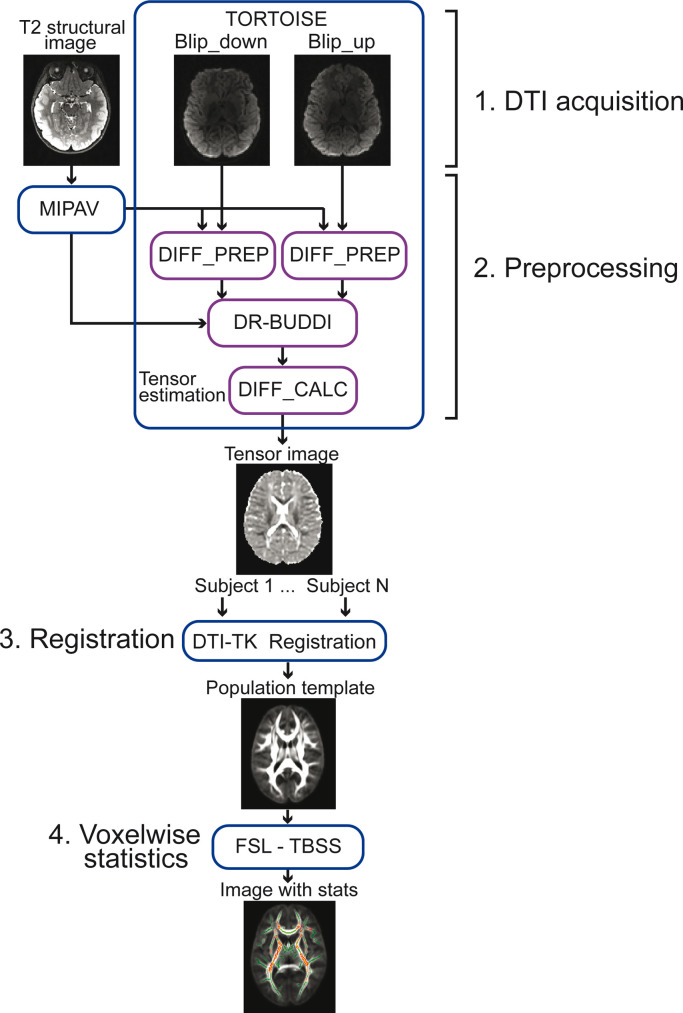
Analysis of scalar maps obtained by diffusion tensor imaging (DTI) produce valuable information about the microstructure of the brain white matter. The DTI scanning of child populations, compared with adult groups, requires specifically designed data acquisition protocols that take into consideration the trade-off between the scanning time, diffusion strength, number of diffusion directions, and the applied analysis techniques. Furthermore, inadequate normalization of DTI images and non-robust tensor reconstruction have profound effects on data analyses and may produce biased statistical results. Here, we present an acquisition sequence that was specifically designed for pediatric populations, and describe the analysis steps of the DTI data collected from extremely preterm-born young school-aged children and their age- and gender-matched controls. The protocol utilizes multiple software packages to address the effects of artifacts and to produce robust tensor estimation. The computation of a population-specific template and the nonlinear registration of tensorial images with this template were implemented to improve alignment of brain images from the children.

Keywords: Diffusion tensor imaging (DTI); Nonlinear registration; Pediatric; Tract-based spatial statistics (TBSS).


Conflict of interest statement

The authors declare no conflict of interest.

Figures

Image, graphical abstract
Graphical abstract
Fig 1
Fig. 1
Illustration of the protocol that was designed for the analysis of diffusion tensor imaging (DTI) data collected from young school-aged children. The protocol has four major parts: (1) The acquisition of diffusion weighted images (DWIs), (2) preprocessing of DWIs, (3) nonlinear registration of the images to a population-specific template, and (4) voxelwise statistical analysis. MIPAV = Medical Image Processing, Analysis, and Visualization; TBSS = tract-based spatial statistics.
Fig 2
Fig. 2
The DTI masks (cyan) are shown on directionally encoded color (DEC) map of the mean FA template and on the mean FA skeleton (red) obtained from TBSS analysis. The DEC map shows the preferred orientation of fibers using the red (left-right), green (anterior-posterior) and blue (superior-inferior) color scheme. The horizontal lines in the sagittal brain slice of the population template indicate the level of axial slice planes that are presented in the frames with the corresponding colors. CC = corpus callosum; CST = corticospinal tract; SLF = superior longitudinal tract; ROI = region of interest; R = right.
Fig 3
Fig. 3
Results of correlation analyses between the fractional anisotropy (FA) and response times of 1-back tasks in normally developing, young school-aged control children (n = 15) born at term-age. The images illustrate significant correlations (P < 0.05, shown in red-yellow) between the FA and response times when the tract-based spatial statistical (TBSS) analysis (P < 0.05, TFCE-corrected) was performed (A) using the DTI-TK nonlinear registration to the population template, and (B) when TBSS analysis was performed using the FSL registration. The horizontal lines in the sagittal brain slices of the population template (A), and the most representative subject's template (B), indicate the level of the selected axial slice planes that are presented in the frames with the corresponding colors. CC = corpus callosum; CST = corticospinal tract; MCP = middle cerebellar peduncle; R = right.

References

    1. Vanderwal T., Kelly C., Eilbott J., Mayes L.C., Castellanos F.X. Inscapes: a movie paradigm to improve compliance in functional magnetic resonance imaging. Neuroimage. 2015;122:222–232. - PMC - PubMed
    1. Poldrack R.A., Pare-Blagoev E.J., Grant P.E. Pediatric functional magnetic resonance imaging: progress and challenges. Top. Magn. Reson. Imaging. 2002;13:61–70. - PubMed
    1. Tamnes C.K., Roalf D.R., Goddings A.L., Lebel C. Diffusion MRI of white matter microstructure development in childhood and adolescence: methods, challenges and progress. Dev. Cogn. Neurosci. 2018;33:161–175. - PMC - PubMed
    1. Le Bihan D., Poupon C., Amadon A., Lethimonnier F. Artifacts and pitfalls in diffusion MRI. J. Magn. Reson. Imaging. 2006;24(3):478–488. - PubMed
    1. Alexander A.L., Lee J.E., Lazar M., Field A.S. Diffusion tensor imaging of the brain. Neurotherapeutics. 2007;4(3):316–329. - PMC - PubMed

LinkOut - more resources