. 2020 Apr 8:30:105468.
doi: 10.1016/j.dib.2020.105468. eCollection 2020 Jun.

DNA-PAINT super-resolution imaging data of surface exposed active sites on particles

Affiliations

DNA-PAINT super-resolution imaging data of surface exposed active sites on particles

Pietro Delcanale et al. Data Brief. .

Abstract

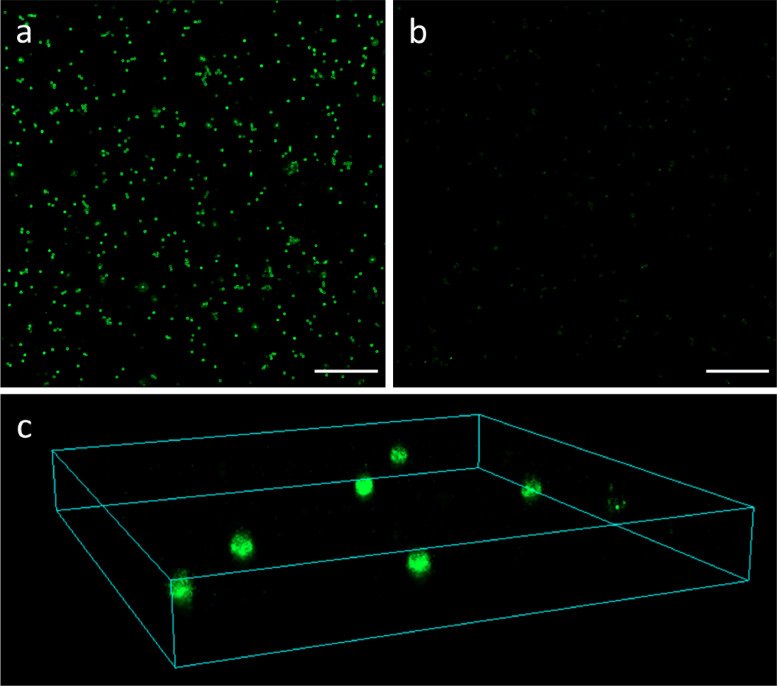
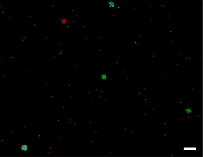
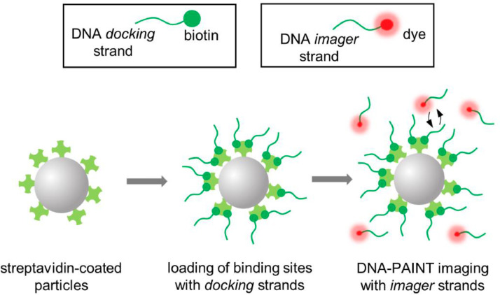
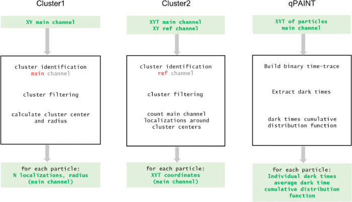
Surface functionalization with targeting ligands confers to nanomaterials the ability of selectively recognize a biological target. Therefore, a quantitative characterization of surface functional molecules is critical for the rational development of nanomaterials-based applications, especially in nanomedicine research. Single-molecule localization microscopy can provide visualization of surface molecules at the level of individual particles, preserving the integrity of the material and overcoming the limitations of analytical methods based on ensemble averaging. Here we provide single-molecule localization data obtained on streptavidin-coated polystyrene particles, which can be exploited as a model system for surface-functionalized materials. After loading of the active sites of streptavidin molecules with a biotin-conjugated probe, they were imaged with a DNA-PAINT imaging approach, which can provide single-molecule imaging at subdiffraction resolution and molecule counting. Both raw records and analysed data, consisting in a list of space-time single-molecule coordinates, are shared. Additionally, Matlab functions are provided that analyse the single-molecule coordinates in order to quantify features of individual particles. These data might constitute a valuable reference for applications of similar quantitative imaging methodologies to other types of functionalized nanomaterials.

Keywords: DNA-PAINT; Functional materials; Nanoparticles; Single-molecule localization microscopy; Super-resolution microscopy.


Figures

Fig 1
Fig. 1
Representative examples of DNA-PAINT images of particles reconstructed from provided data. (a-b): Two-dimensional DNA-PAINT images obtained using a complementary (a) and a non-complementary (b) docking-imager strands pair. Scale bars 5 µM. (c): Three-dimensional DNA-PAINT image of a selected region of interest, obtained with complementary docking-imager pair. Size 8 × 8 × 1 µm.
Fig 2
Fig. 2
Representative example of a multi-colour DNA-PAINT image of particles loaded with different docking strands, reconstructed from provided data. Different false colours are assigned to results obtained with imager strand 1 (cyan), 2 (green) and 3 (red), complementary to docking1, docking2 and docking3, respectively. Scale bar 0.6 µM. (For interpretation of the references to color in this figure legend, the reader is referred to the web version of this article.)
Fig 3
Fig. 3
Schematic representation of the procedure followed for sample preparation and DNA-PAINT imaging of sites exposed on streptavidin coated particles.
Fig 4
Fig. 4
Schematic workflow of the operations implemented in the provided codes for analysis.

References

    1. Delcanale P., Miret-Ontiveros B., Arista-Romero M., Pujals S., Albertazzi L. Nanoscale mapping functional sites on nanoparticles by points accumulation for imaging in nanoscale topography (PAINT) ACS Nano. 2018;12:7629–7637. doi: 10.1021/acsnano.7b09063. - DOI - PubMed
    1. Schnitzbauer J., Strauss M.T., Schlichthaerle T., Schueder F., Jungmann R. Super-resolution microscopy with DNA-PAINT. Nat. Protoc. 2017;12:1198–1228. doi: 10.1038/nprot.2017.024. - DOI - PubMed
    1. Jungmann R., Avendaño M.S., Dai M., Woehrstein J.B., Agasti S.S., Feiger Z., Rodal A., Yin P. Quantitative super-resolution imaging with qPAINT. Nat. Methods. 2016;13:439–442. doi: 10.1038/nmeth.3804. - DOI - PMC - PubMed
    1. van der Meel R., Sulheim E., Shi Y., Kiessling F., Mulder W.J.M., Lammers T. Smart cancer nanomedicine. Nat. Nanotechnol. 2019;14:1007–1017. doi: 10.1038/s41565-019-0567-y. - DOI - PMC - PubMed
    1. Rabanel J.-.M., Adibnia V., Tehrani S.F., Sanche S., Hildgen P., Banquy X., Ramassamy C. Nanoparticle heterogeneity: an emerging structural parameter influencing particle fate in biological media? Nanoscale. 2019;11:383–406. doi: 10.1039/C8NR04916E. - DOI - PubMed

LinkOut - more resources