. 2020 Mar;6(3):10.18103/imr.v6i3.852.
doi: 10.18103/imr.v6i3.852.

Etiologies of Proptosis: A review

Affiliations

Etiologies of Proptosis: A review

Nicole J Topilow et al. Intern Med Rev (Wash D C). 2020 Mar.

Abstract

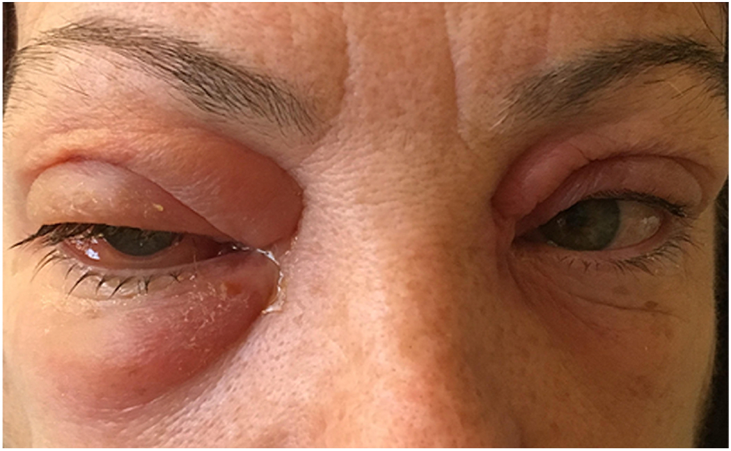
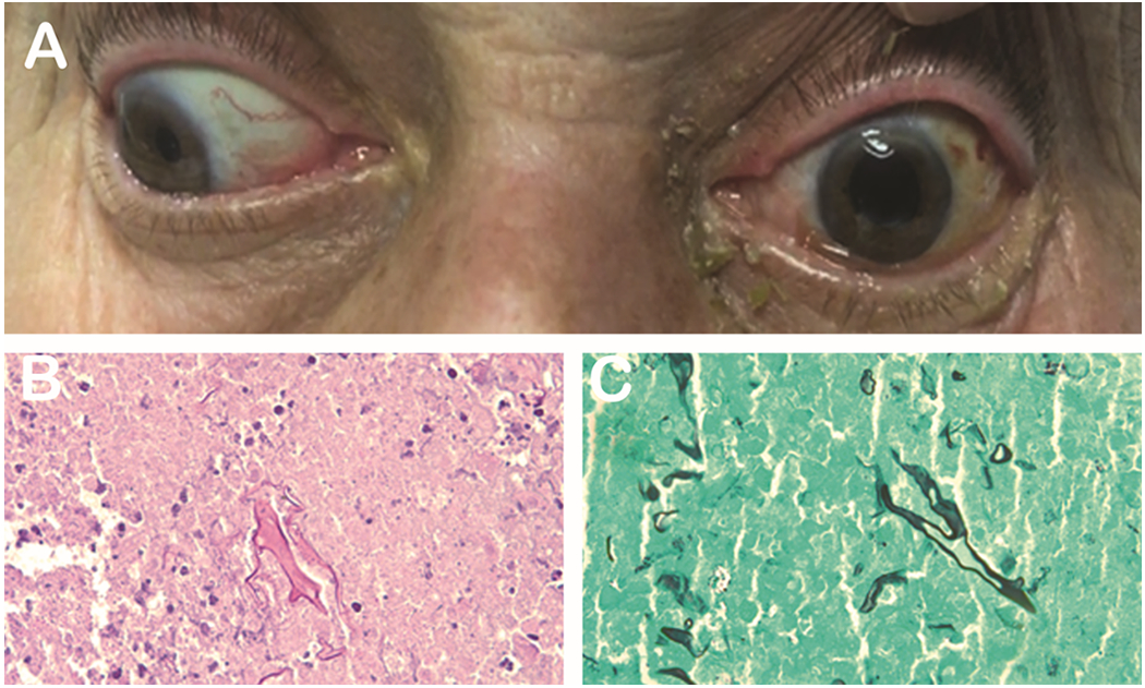
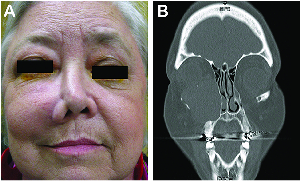
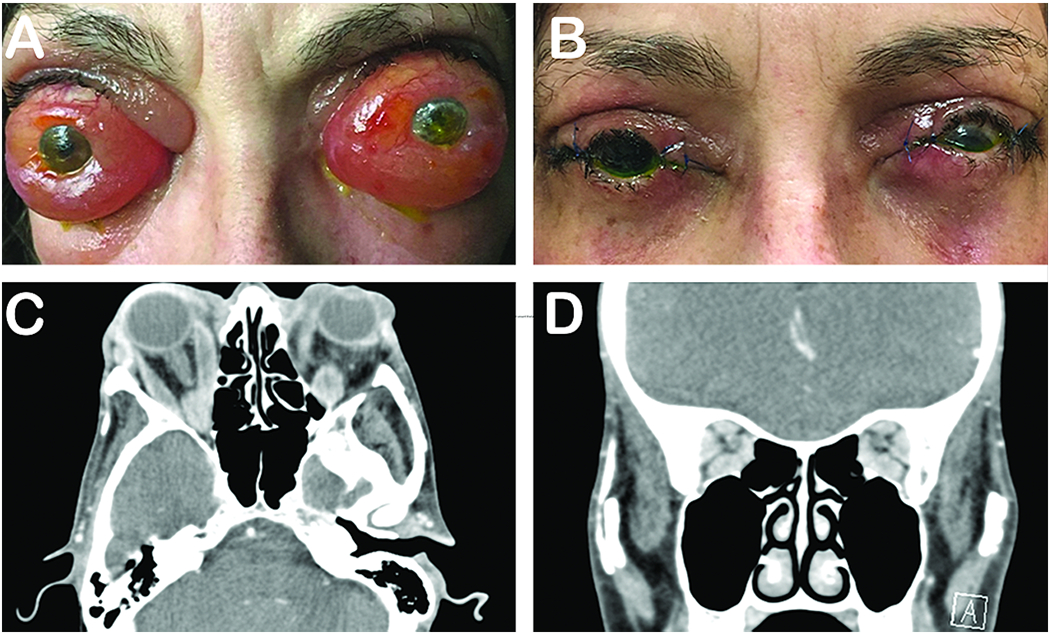
Proptosis, the protrusion of the eyeball from the orbit, results from a wide variety of pathologies that can be vision- or life-threatening. Clinical history, associated physical exam findings, and imaging features are all crucial in establishing the underlying etiology. The differential diagnosis is broad, and includes infectious, inflammatory, vascular, and neoplastic entities that range from benign and indolent, to malignant and aggressive. While treatment varies significantly based on the disease process, all are aimed at preserving vision, salvaging the globe, preventing disfigurement, and reducing mortality. Both internists and general ophthalmologists should be familiar with the causes of proptosis in order to initiate the work-up for, and appropriately triage, affected patients.

Keywords: Proptosis; orbital infection; orbital inflammation; orbital tumors; orbital vascular anomalies.


Figures

Figure 1.
Figure 1.. Orbital cellulitis.
64-year-old healthcare worker found to be MRSA positive who presented with 4-day history of periorbital edema, lid swelling and chemosis.
Figure 2.
Figure 2.. Mucormycosis.
72-year-old man with a past medical history of diabetes and COPD on steroids presented with 3 day history of mild proptosis, ophthalmoplegia and decrease in vision (A). H&E (B) and GMS (C) show growth of mucormycosis. Photo courtesy of Kenneth Fan, MD, MBA.
Figure 3.
Figure 3.. Mucocele.
A 63-year-old woman who presented with a slowly progressing mass on the check with diplopia and upward displacement of the left globe (A). CT scan revealed an enlarged encapsulated left maxillary sinus mucocele (B).
Figure 4.
Figure 4.. Thyroid eye disease.
57-year-old woman presented with a long-standing history of proptosis and chemosis with acute decrease in vision (A). The patient underwent a bilateral 3-wall orbital decompression (B). Pre-operative CT axial (C) and coronal (D) illustrate substantial orbital apex crowding. Photo courtesy of Alexandra Levitt, MD.
Figure 5.
Figure 5.. Non-specific orbital inflammatory syndrome.
26-year-old Haitian woman who presented with 1-year history of loss of vision and proptosis. Patient did not respond to intravenous steroids. Biopsy revealed non-specific inflammation, negative for IgG4.
Figure 6.
Figure 6.. Carotid-cavernous fistulas.
An 83-year-old woman who presented with bilateral orbital congestion syndrome and prominent corkscrew episcleral vessels was found to have a carotid cavernous fistula and underwent coil embolization.
Figure 7.
Figure 7.. Orbital varix.
63-year-old woman presents with left bluish upper eyelid lesion (A) that increases in size with Valsalva (B). MRI shows isointense vascular lesion (C) and H&E shows thrombosed orbital varix (D).
Figure 8.
Figure 8.. Orbital lymphangioma.
25-year-old man presents with intermittent proptosis and subconjunctival hemorrhage (A). MRI showsw an intraconal multi-cystic lesion (B). Posterior exam shows optic nerve edema and tortuous vessels (C) and retinal folds (D).
Figure 9.
Figure 9.. Capillary hemangioma.
4-year-old boy presents with gradual right orbital proptosis over past few months (A). MRI imaging reveals a large homogenous hyperintense bright lesion that enhances with gadolinium (B). An encapsulated vascular lesion was removed (C). H&E illustrates a tumor composed of capillary sized blood vessels (D) (Photo courtesy of David Tse and Sara Wester).
Figure 10.
Figure 10.. Rhabdomyosarcoma.
9-year-old girl presents with an 8-day history of progressive periorbital swelling and decreased vision (A). MRI demonstrates a large, enhancing, infiltrative intraconal mass (B). Orbital biopsy shows a small blue cell tumor staining for Myod1, desmin, and myogenin consistent with embryonal rhabdomyosarcoma (C).
Figure 11.
Figure 11.. Orbital lymphoma.
86-year-old man with multiple myeloma status post 3 weeks of chemotherapy presents with 2 months worsening right eye pain, vision loss, proptosis and complete ophthalmoplegia. Photo courtesy of Zubair Ansari, MD.
Figure 12.
Figure 12.. Adenoid cystic carcinoma.
53-year-old woman presents with a painful supratemporal mass (A). Imaging reveals lacrimal gland mass with irregular margins and bony erosion (B). Histopathological analysis shows cribriform “Swiss cheese” pattern (C) (photo courtesy of David Tse).
Figure 13.
Figure 13.. Invasive squamous cell into the orbit.
51-year-old man with prior history of squamous cell carcinoma of the brow status post excision who presents with 2 month history of worsening proptosis of the left eye.

References

    1. Rootman J Diseases of the Orbit: A Multidisciplinary Approach. 2nd ed. Philadelphia: Lippincott Williams & Wilkins; 2003.
    1. Dutton JJ. Atlas of Clinical and Surgical Orbital Anatomy. 2nd ed. Philadelphia: Elsevier Saunders; 2011.
    1. Aviv RI, Casselman J. Orbital imaging: Part 1. Normal anatomy Clin Radiol. 2005. March;60(3):279–87. - PubMed
    1. Conneely MF, Hacein-Bey L, Jay WM. Magnetic resonance imaging of the orbit. Semin Ophthalmol. 2008. May-Jun;23(3): 179–89. - PubMed
    1. Hallman JT, Pillay P, Koh LH, Goh KY, Yu WY. Eye Globe Abnormalities on MR and CT in adults: An Anatomical Approach. Korean J Radiol. 2016. Sep-Oct; 17(5): 664–673. - PMC - PubMed

LinkOut - more resources