. 2021 Mar;52(1):33-39.
doi: 10.1007/s42770-020-00276-3. Epub 2020 May 7.

Human serum proteins bind to Sporothrix schenckii conidia with differential effects on phagocytosis

Affiliations

Human serum proteins bind to Sporothrix schenckii conidia with differential effects on phagocytosis

Silvia Guzman Beltrán et al. Braz J Microbiol. 2021 Mar.

Abstract

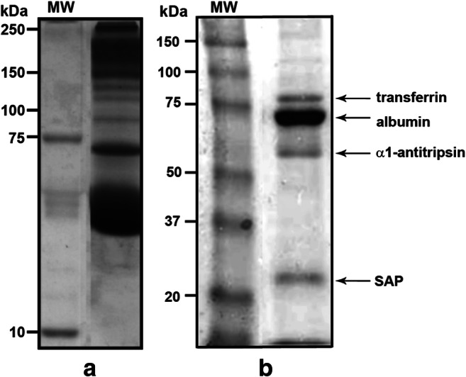
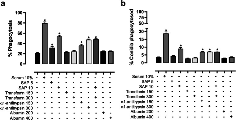
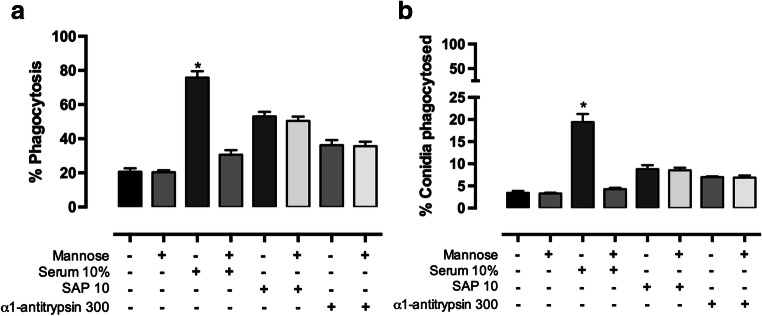
Serum is an important source of proteins that interact with pathogens. Once bound to the cell surface, serum proteins can stimulate the innate immune system. The phagocytosis of Sporothrix schenckii conidia by human macrophages is activated through human serum opsonisation. In this study, we have attempted to characterise human blood serum proteins that bind to the cell wall of S. schenckii conidia. We systematically observed the same four proteins independent of the plasma donor: albumin, serum amyloid protein (SAP), α-1 antitrypsin (AAT), and transferrin were identified with the help of tandem mass spectrometry. Phagocytosis depended on the concentration of the SAP or α-1 antitrypsin that was used to opsonise the conidia; however, transferrin or albumin did not have any effect on conidia internalisation. The presence of mannose did not affect macrophage phagocytosis of the conidia opsonised with SAP or α-1 antitrypsin, which suggests that these proteins are not recognised by the mannose receptor.

Keywords: Conidia; Macrophage; Phagocytosis; Serum amyloid protein; Sporotrichosis; α-1 antitrypsin.


Figures

Fig. 1
Fig. 1
Isolation of S. schenckii-binding proteins from human serum. a Protein profile of total human serum resolved on a 10% SDS-PAGE. b Bound human serum proteins to conidia, separated on 12% SDS-PAGE. Proteins were identified as transferrin (80 kDa), albumin (66.5 kDa), α1-antitripsin (54 kDa), and serum amyloid P component (25 kDa) by tandem mass spectrometry. The gels were stained with Coomassie Brilliant Blue
Fig. 2
Fig. 2
Phagocytosis of S. schenckii conidia by monocyte-derived Mϕs. Mϕs were incubated with non-opsonised (a) or 10% HHS opsonised conidia (b) for 60 min 37 °C, at a ratio Mϕ:fungi of 1:10. After incubation, cells were stained with Giemsa
Fig. 3
Fig. 3
Phagocytosis of S. schenckii conidia opsonised with 10% human serum or with bound proteins by human monocyte-derived macrophages. Opsonised conidia with 10% HHS or with two concentrations of the serum proteins identified by LC/ESI–MS/MS were incubated with Mϕs at a ratio Mϕ:fungi of 1:10 at 37 °C for 60 min. After incubation, cells were stained with Giemsa. a Phagocytosis was assessed by counting the number of macrophages that internalised at least one conidium. b Percentage of fungi that were phagocytised and were obtained by counting the conidia observed in vesicles in the macrophages. Data for phagocytic assay were collected in at least four independent experiments. Data are expressed as mean ± SD; n = 5. *P < 0.05 versus control (non-opsonised)
Fig. 4
Fig. 4
Effect of mannose on conidia phagocytosis by human monocyte-derived macrophages. Opsonised conidia with 10% human serum, 10 μg of SAP, or 300 μg of ΑΑΤ were incubated with macrophages in the absence or presence of 30 mM of mannose. a Phagocytosis was assessed by counting the number of macrophages that internalised at least one conidium. b Percentage of fungi that were phagocytised and were obtained by counting the conidia observed in vesicles in the macrophages. Data for phagocytic assay were collected in at least four independent experiments. Data are expressed as mean ± SD; n = 5. *P < 0.05 versus control (non-opsonised)

References

    1. Conceição-Silva F, Morgado F. Immunopathogenesis of human sporotrichosis: what we already know. J Fungi (Basel) 2018;4:89. doi: 10.3390/jof4030089. - DOI - PMC - PubMed
    1. Zhang Y, Hagen F, Stielow B, Rodrigues AM, Samerpitak K, Zhou X, Feng P, Yang L, Chen M, Deng S, Li S, Liao W, Li R, Li F, Meis JF, Guarro J, Teixeira M, al-Zahrani HS, de Camargo ZP, Zhang L, de Hoog GS. Phylogeography and evolutionary patterns in Sporothrix spanning more than 14 000 human and animal case reports. Persoonia. 2015;35:1–20. doi: 10.3767/003158515x687416. - DOI - PMC - PubMed
    1. Barros MB, Paes RD, Schubach AO. Sporothrix schenckii and Sporotrichosis. Clin Microbiol Rev. 2011;24:633–654. doi: 10.1128/cmr.00007-11. - DOI - PMC - PubMed
    1. Schubach A, Schubach TMP, Barros MBDL, Wanke B. Cat-transmitted Sporotrichosis, Rio de Janeiro, Brazil. Emerg Infect Dis. 2005;11:1952–1954. doi: 10.3201/eid1112.040891. - DOI - PMC - PubMed
    1. Morgado FN, Schubach AO, Pimentel MI, Lyra MR, Vasconcellos ÉCF, Valete-Rosalino CM, Conceição-Silva F. Is there any difference between the in situ and systemic IL-10 and IFN-γ production when clinical forms of cutaneous sporotrichosis are compared? PLoS One. 2016;11:e0162764. doi: 10.1371/journal.pone.0162764. - DOI - PMC - PubMed

Supplementary concepts

Grants and funding