Review
. 2020 Jun;24(12):6510-6522.
doi: 10.1111/jcmm.15279. Epub 2020 May 8.

The role of mitochondrial reactive oxygen species, NO and H2 S in ischaemia/reperfusion injury and cardioprotection

Affiliations
Review

The role of mitochondrial reactive oxygen species, NO and H2 S in ischaemia/reperfusion injury and cardioprotection

Ioanna Andreadou et al. J Cell Mol Med. 2020 Jun.

Abstract

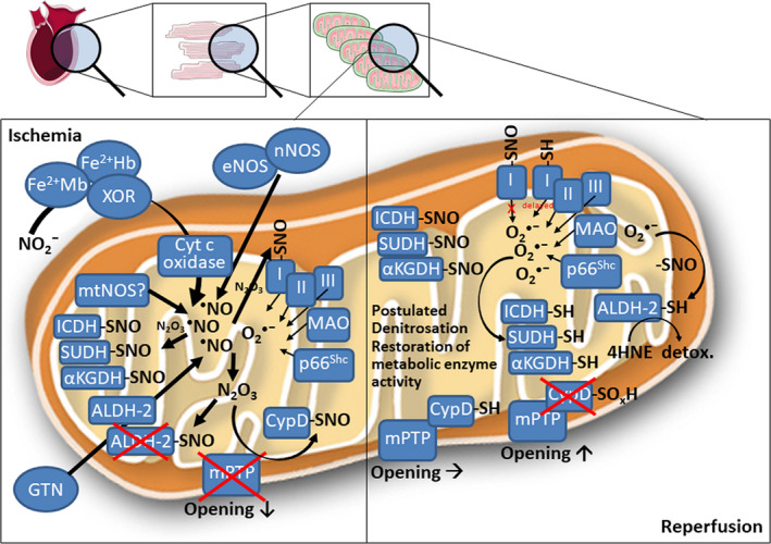
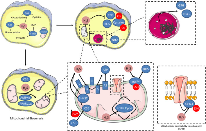
Redox signalling in mitochondria plays an important role in myocardial ischaemia/reperfusion (I/R) injury and in cardioprotection. Reactive oxygen and nitrogen species (ROS/RNS) modify cellular structures and functions by means of covalent changes in proteins including among others S-nitros(yl)ation by nitric oxide (NO) and its derivatives, and S-sulphydration by hydrogen sulphide (H2 S). Many enzymes are involved in the mitochondrial formation and handling of ROS, NO and H2 S under physiological and pathological conditions. In particular, the balance between formation and removal of reactive species is impaired during I/R favouring their accumulation. Therefore, various interventions aimed at decreasing mitochondrial ROS accumulation have been developed and have shown cardioprotective effects in experimental settings. However, ROS, NO and H2 S play also a role in endogenous cardioprotection, as in the case of ischaemic pre-conditioning, so that preventing their increase might hamper self-defence mechanisms. The aim of the present review was to provide a critical analysis of formation and role of reactive species, NO and H2 S in mitochondria, with a special emphasis on mechanisms of injury and protection that determine the fate of hearts subjected to I/R. The elucidation of the signalling pathways of ROS, NO and H2 S is likely to reveal novel molecular targets for cardioprotection that could be modulated by pharmacological agents to prevent I/R injury.

Keywords: cardioprotection; heart; hydrogen sulphide; ischaemia; mitochondria; nitric oxide; reactive oxygen species; reperfusion.


Conflict of interest statement

PF is a founder and CEO of Pharmahungary Group, a group of R&D companies.

Figures

FIGURE 1
FIGURE 1
Proposed cardioprotective mechanisms of nitric oxide in cardiac mitochondria during ischaemia/reperfusion. During ischaemia, endogenous nitric oxide formation is potentiated from several sources. Mild nitrosative stress from inorganic nitrite conversion into nitric oxide, high activity of nitric oxide synthases (NOS) or pharmacological nitric oxide formation from nitroglycerin (GTN) combine with low superoxide levels from various mitochondrial sources to generate the potent nitrosating species N2O3. This leads to widespread nitros(yl)ation of mitochondrial enzymes involved in energy metabolism, as well as cyclophilin D (CypD). Nitros(yl)ated CypD cannot bind properly to the mitochondrial permeability transition pore (mPTP), thereby decreasing its open probability. Nitros(yl)ated enzymes involved in energy metabolism are inactive, yet nitros(yl)ation partially protects against irreversible oxidative damage. For instance, nitros(yl)ated aldehyde dehydrogenase 2 (ALDH‐2) limits GTN‐dependent NO formation in mitochondria thereby preventing severe nitrosative stress but also partially protects this important antioxidant enzyme against irreversible oxidative damage. Nitros(yl)ation of complex I limit infarct I/R injury by reducing/delaying superoxide formation at the onset of reperfusion. During reperfusion, superoxide formation from mentioned sources is increased and may lead to the postulated superoxide‐dependent denitrosation of enzymes involved in energy metabolism, thus restoring their activity for the required energy supply after an ischaemia/reperfusion episode. Superoxide‐dependent denitrosation of CypD restores its regulatory effect on mPTP favouring its opening. Superoxide‐dependent denitrosation of ALDH‐2 supports detoxification of cardiac damage by excessive formation of the detrimental 4‐hydroxynonenal (4HNE). αKGDH, α‐ketoglutarate dehydrogenase; ALDH‐2, mitochondrial aldehyde dehydrogenase; Fe2+Hb/Mb, ferrous haemoglobin/myoglobin; ICDH, isocitrate dehydrogenase; mtNOS, mitochondrial nitric oxide synthase; SUDH, succinate dehydrogenase; XOR, xanthine oxidoreductase. This scheme contains images from Servier Medical Art by Servier, licensed under a Creative Commons Attribution 3.0 Unported License
FIGURE 2
FIGURE 2
Proposed sources and targets for mitochondrial H2S generation involved in cardioprotection. H2S can be generated from 3‐mercaptopyruvate sulphurtransferase (3‐MST), that has been found in both cytosol and mitochondria and from the translocation of cystathionine γ‐lyase (CSE) from the cytosol to mitochondria after prolonged elevation of Ca2+ levels. H2S induces cardioprotection by preservation of mitochondrial function: H2S can inhibit ROS and RNS formation preventing irreversible cysteine overoxidation and preserving protein functions. H2S activates the master‐regulator of antioxidant responses Nrf2, increases glutathione (GSH) synthesis and up‐regulates the expression of thioredoxin. H2S may act as an endogenous antioxidant mediator by inhibition of p66Shc‐mediated mitochondrial ROS production. Another possible mechanism of action for H2S is based on its ability to modulate cellular respiration during reperfusion. Under physiological H2S concentrations, cytochrome c oxidase remains functional, whereas sulphide oxidation likely contributes to mitochondrial ATP production. Additionally, H2S regulates mitochondrial biogenesis by activation of AMP‐activated protein kinase and peroxisome proliferator‐activated receptor γ coactivator 1α. H2S modulates cellular signalling by sulfhydration, and among the proteins confirmed to undergo sulfhydration upon exposure to H2S, several are involved in cardioprotection including the pore forming subunit of ATP‐sensitive potassium channels (Kir 6.1)

References

    1. Sies H.Oxidative stress: introduction In: Sies H. ed. Oxidative Stress: Oxidants and Antioxidants. London: Academic Press, 1991:xv‐xxii.
    1. Brandes RP, Rezende F, Schroder K. Redox regulation beyond ROS: why ROS should not be measured as often. Circ Res. 2018;123:326‐328. - PMC - PubMed
    1. Griendling KK, Touyz RM, Zweier JL, et al. Measurement of reactive oxygen species, reactive nitrogen species, and redox‐dependent signaling in the cardiovascular system: a scientific statement from the american heart association. Circ Res. 2016;119:e39‐e75. - PMC - PubMed
    1. Dalle‐Donne I, Rossi R, Colombo R, et al. Biomarkers of oxidative damage in human disease. Clin Chem. 2006;52:601‐623. - PubMed
    1. Steinhubl SR. Why have antioxidants failed in clinical trials? Am J Cardiol. 2008;101:14D‐9D. - PubMed

Publication types

MeSH terms