Characterization of the immune profile of oral tongue squamous cell carcinomas with advancing disease
- PMID: 32383556
- PMCID: PMC7333861
- DOI: 10.1002/cam4.3106
Characterization of the immune profile of oral tongue squamous cell carcinomas with advancing disease
Abstract
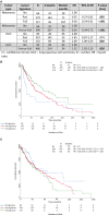
We investigated whether a unique immune response was instigated with the development of oral tongue squamous cell carcinomas (OTSCC), with/without nodal involvement, with/without recurrent metastatic disease, or within tumor involved nodes. One hundred and ten formalin-fixed paraffin-embedded samples were collected from a retrospective cohort of 67 OTSCC patients and 10 non-cancerous tongue samples. Targets including CD4, CD8, FOXP3, PD-L1, and PD-1 were analyzed by immunohistochemistry. The Nanostring PanCancer Immune Profiling Panel was used for gene expression profiling. Data were externally validated in the The Cancer Genome Atlas (TCGA) head and neck (HNSCC), melanoma and lung squamous cell carcinoma (LSCC) cohorts. A 24-immune gene signature was identified that discriminated more aggressive OTSCC cases, and although not prognostic in HNSCC was associated with survival in other TCGA cohorts (improved survival for melanoma, P < .001 and worse survival for LSCC, P = .038). OTSCC exhibited concordant gene and immunohistochemical (IHC) features characterized by a TH-2 biased, proinflammatory profile with upregulated B cell and neutrophil gene activity and increased CD4, FOXP3, and PD-L1 expression (P < .001 for all by IHC). Compared to less advanced disease, nodal involvement and recurrent OTSCC did not induce a different immune response although recurrent disease was characterized by significantly higher PD-L1 expression (P = .004 by SP263, P = .013 by 22C3, P = .004 for gene expression). Identification of a gene signature associated with different prognostic effects in other cancers highlights common pathways of immune dysregulation that are impacted by the tumor origin. The significant immunosuppressive signaling in OTSCC indicates primary failure of immune system to control carcinogenesis emphasizing the need for early, combination therapeutic approaches.
Keywords: FOXP3; Immune signature; PD-L1; oral tongue squamous cell carcinoma.
© 2020 The Authors. Cancer Medicine published by John Wiley & Sons Ltd.
Conflict of interest statement
AM Lim ‐ uncompensated advisory board from Merck Sharp & Dohme and Bristol‐Myers Squibb with travel and accommodation expenses. The other authors declare that there is no potential conflict of interest.
Figures


References
-
- Burtness B, Harrington KJ, Greil R, et al. Pembrolizumab alone or with chemotherapy versus cetuximab with chemotherapy for recurrent or metastatic squamous cell carcinoma of the head and neck (KEYNOTE‐048): a randomised, open‐label, phase 3 study. Lancet. 2019;394:1915‐1928. - PubMed
-
- Varilla V, Atienza J, Dasanu CA. Immune alterations and immunotherapy prospects in head and neck cancer. Expert Opin Biol Ther. 2013;13:1241‐1256. - PubMed
-
- Strome SE, Dong H, Tamura H, et al. B7–H1 blockade augments adoptive T‐cell immunotherapy for squamous cell carcinoma. Cancer Res. 2003;63:6501‐6505. - PubMed
-
- Chung CH, Parker JS, Karaca G, et al. Molecular classification of head and neck squamous cell carcinomas using patterns of gene expression. Cancer Cell. 2004;5:489‐500. - PubMed
MeSH terms
Substances
LinkOut - more resources
Full Text Sources
Molecular Biology Databases
Research Materials

