. 2020 Jun 2;92(11):7596-7603.
doi: 10.1021/acs.analchem.0c00174. Epub 2020 May 21.

In Vivo Fast Photochemical Oxidation of Proteins Using Enhanced Multiplexing Proteomics

Affiliations

In Vivo Fast Photochemical Oxidation of Proteins Using Enhanced Multiplexing Proteomics

Jessica A Espino et al. Anal Chem. .

Abstract

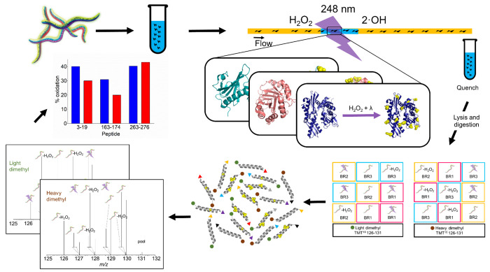
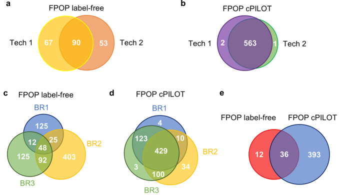
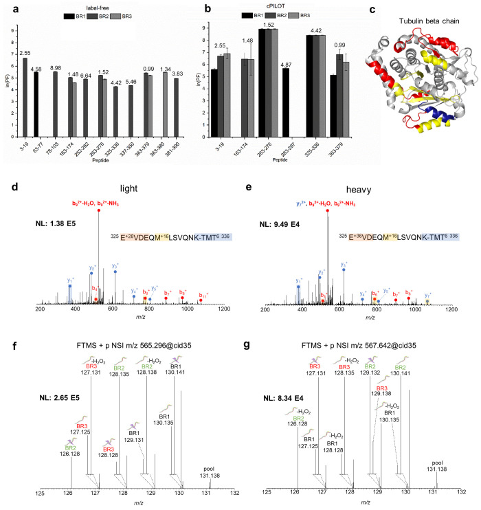
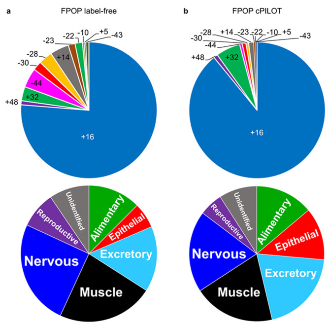
In vivo fast photochemical oxidation of proteins (IV-FPOP) is a hydroxyl radical protein footprinting method used to study protein structure and protein-protein interactions. Oxidatively modified proteins by IV-FPOP are analyzed by mass spectrometry (MS), and the extent of oxidation is quantified by label-free MS. Peptide oxidation changes yield useful information about protein structure, due to changes in solvent accessibility. However, the sample size necessary for animal studies requires increased sample preparation and instrument time. Here, we report the combined application of IV-FPOP and the enhanced multiplexing strategy combined precursor isotopic labeling and isobaric tagging (cPILOT) for higher-throughput analysis of oxidative modifications in C. elegans. Key differences in the performance of label-free MS and cPILOT were identified. The addition of oxygen (+16) was the most abundant modification identified among all known possible FPOP modifications. This study presents IV-FPOP coupled with enhanced multiplexing strategies such as cPILOT to increase throughput of studies seeking to examine oxidative protein modifications.


Conflict of interest statement

The authors declare no competing financial interest.

Figures

Figure 1
Figure 1
Experimental workflow for IV-FPOP cPILOT. C. elegans (N = 20,000) is grown to its fourth larva stage (L4). Three conditions (control, control oxidation, and FPOP) have three biological replicates. Worms in the presence of hydrogen peroxide are flown through a 250 μm i.d. fused silica and irradiated using a KrF excimer at 248 nm wavelength. Each biological replicate has two workflow replicates, resulting in 18 samples. Following oxidative labeling, samples are lysed and digested, and peptides are further labeled by cPILOT. Specifically, peptides (50 μg) are labeled by either light- or heavy dimethylation at the N-terminus and isobarically tagged by TMT 10-plex at lysine residues. Finally, peptides are analyzed using LC-MS, MS/MS, and MS3 on an Orbitrap Fusion Lumos, and the extent of modification of each protein of interest for every condition is calculated.
Figure 2
Figure 2
Protein oxidation quantification across biological and technical replicates. (a) IV-FPOP representative biological replicate of oxidatively modified proteins identified by label-free MS across two technical (tech) replicates: one (yellow, N = 157) and two (orange, N = 143). (b) IV-FPOP oxidatively modified proteins identified by cPILOT across technical replicates one (purple, N = 565) and two (green, N = 564). Venn diagrams of oxidatively modified proteins by IV-FPOP across three biological replicates (BR) identified by (c) label-free MS (N = 830) and (d) cPILOT (N = 703). (e) Venn diagram of common oxidatively modified proteins among label-free (red, N = 48) and cPILOT (blue, N = 429).
Figure 3
Figure 3
Protein oxidation for tubulin beta chain. Calculated ln(PF) for tubulin beta chain oxidatively modified peptides identified by (a) label-free MS (N = 11) and (b) cPILOT (N = 6) across three biological replicates. SASA calculated values using the Homo sapiens tubulin beta chain cryo-EM structure (PDB: 5N5N(31)) are displayed on top of each bar. (c) Oxidatively modified peptides identified by label-free MS only (red), cPILOT only (blue), and both methods only (yellow) mapped on the cryo-EM structure of the human tubulin beta chain. CID-MS/MS spectra of the tubulin beta chain peptide 325–336 showing b- and y-ions for (d) light and (e) heavy dimethylation plus a +16 FPOP modification for residue M330 and isobaric-tag. HCD-MS3 spectra generated from the 10 most intense fragment ions (SPS-10) of the (f) light and (g) heavy CID-MS/MS ion.
Figure 4
Figure 4
Oxidative mass shift modifications and body systems identified by IV-FPOP. (a) Oxidative mass shift modifications observed by HCD-MS/MS fragmentation (top) and body systems (bottom) identified by IV-FPOP label-free MS. (b) Oxidative mass shift modifications observed by CID-MS/MS fragmentation (top) and body systems (bottom) identified IV-FPOP-cPILOT.

Similar articles

Cited by

References

    1. Shi Y. A glimpse of structural biology through X-ray crystallography. Cell 2014, 159 (5), 995–1014. 10.1016/j.cell.2014.10.051. - DOI - PubMed
    1. Huang C.; Kalodimos C. G. Structures of Large Protein Complexes Determined by Nuclear Magnetic Resonance Spectroscopy. Annu. Rev. Biophys. 2017, 46, 317–336. 10.1146/annurev-biophys-070816-033701. - DOI - PubMed
    1. Adrian M.; Dubochet J.; Lepault J.; McDowall A. W. Cryo-electron microscopy of viruses. Nature 1984, 308 (5954), 32–6. 10.1038/308032a0. - DOI - PubMed
    1. Kaur U.; Johnson D. T.; Chea E. E.; Deredge D. J.; Espino J. A.; Jones L. M. Evolution of Structural Biology through the Lens of Mass Spectrometry. Anal. Chem. 2019, 91 (1), 142–155. 10.1021/acs.analchem.8b05014. - DOI - PMC - PubMed
    1. Chea E. E.; Jones L. M. Analyzing the structure of macromolecules in their native cellular environment using hydroxyl radical footprinting. Analyst 2018, 143 (4), 798–807. 10.1039/C7AN01323J. - DOI - PubMed

Publication types

MeSH terms

Substances