. 2020 Jun 19;48(11):e63.
doi: 10.1093/nar/gkaa270.

Split & mix assembly of DNA libraries for ultrahigh throughput on-bead screening of functional proteins

Affiliations

Split & mix assembly of DNA libraries for ultrahigh throughput on-bead screening of functional proteins

Laurens Lindenburg et al. Nucleic Acids Res. .

Abstract

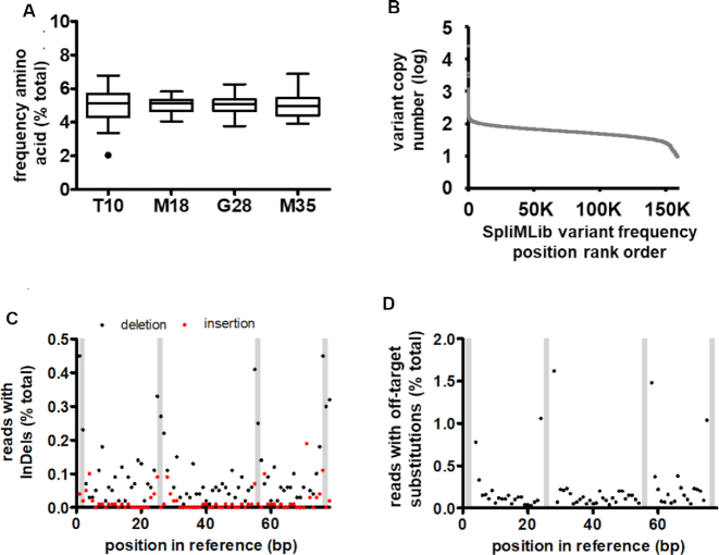
Site-saturation libraries reduce protein screening effort in directed evolution campaigns by focusing on a limited number of rationally chosen residues. However, uneven library synthesis efficiency leads to amino acid bias, remedied at high cost by expensive custom synthesis of oligonucleotides, or through use of proprietary library synthesis platforms. To address these shortcomings, we have devised a method where DNA libraries are constructed on the surface of microbeads by ligating dsDNA fragments onto growing, surface-immobilised DNA, in iterative split-and-mix cycles. This method-termed SpliMLiB for Split-and-Mix Library on Beads-was applied towards the directed evolution of an anti-IgE Affibody (ZIgE), generating a 160,000-membered, 4-site, saturation library on the surface of 8 million monoclonal beads. Deep sequencing confirmed excellent library balance (5.1% ± 0.77 per amino acid) and coverage (99.3%). As SpliMLiB beads are monoclonal, they were amenable to direct functional screening in water-in-oil emulsion droplets with cell-free expression. A FACS-based sorting of the library beads allowed recovery of hits improved in Kd over wild-type ZIgE by up to 3.5-fold, while a consensus mutant of the best hits provided a 10-fold improvement. With SpliMLiB, directed evolution workflows are accelerated by integrating high-quality DNA library generation with an ultra-high throughput protein screening platform.


Figures

Figure 1.
Figure 1.
Design of SpliMLiB for solid-phase cloning of site saturation libraries. (A) SpliMLiB consists of a number of DNA attachment-rounds, where DNA is immobilised to the bead surface (first attachment-round) or immobilised DNA is extended by ligation (subsequent attachment-rounds). Beads are split into different tubes, with the number of vessels corresponding to the desired number of different amino acid variants at a position of interest within the encoded protein. Beads are mixed between DNA additions, ensuring all combinations of positional variants are achieved. This process may be continued for several attachment-rounds, resulting in a final diversity of nm where n is the number of splits per attachment-round and m is the number of attachment-rounds. Each tube within a split receives a DNA fragment carrying a single codon variant, as indicated by the lower dash-lined box shown for the second SpliMLiB round only. (B) SpliMLiB results in a site saturation library represented by beads each densely coated in identical DNA.
Figure 2.
Figure 2.
Design of bead surface and solid-phase manipulations of DNA. (A) Beads were designed to display both azide (labelled ‘N3’) and SpyTag (labelled ‘ST’) moieties (surface modification described in Supplementary Figure S1). (B) Flow cytometric analysis of beads for fluorescein-derived fluorescence intensity before (grey) and after (black) immobilisation of fluorescein and DBCO-functionalised DNA (top histogram), after Esp3I treatment (2 hours at 37°C) of the DNA-coated beads (middle histogram) and after exposure of Esp3I-treated beads to a fluorescein-labelled DNA duplex that had a 5′-overhang complementary to the 5′-overhang of bead-immobilised DNA, in T4 DNA ligase buffer, with (black) or without (grey) T4 DNA ligase (bottom histogram). Details of the DNA sequences used for the generation of this panel are set out in Supplementary Figure S4. (C) Schematic overview of on-bead assembly allowing potential saturation of three codons in close proximity. The final, bead-attached DNA assembly is shown at the top of the panel, with the three DNA fragments used in the construction are shown below. Restriction sites are depicted in red, target codons in green and sequences used for hybridisation during ligation in blue. The first, PCR-generated amplicon (frag3) was attached to bead (via copper-free click chemistry) and digested by Esp3I. DNA on the bead was extended using an oligonucleotide duplex (frag2) carrying a 5′-phosphorylated cohesive end; the sequence used to ensure stability of the duplex (stability stuffer) prior to ligation is indicated in a diagonal pattern. Once this duplex had been appended to the bead by ligation, a new cohesive end was generated (and stability stuffer removed) through BspQI digestion. Finally, another PCR amplicon (frag1), separately prepared with a cohesive end (using BspQI) was ligated to the bead-immobilised DNA. Details of the DNA sequences used for the generation of this panel are set out in Supplementary Figure S5. (D) Flow cytometric analysis of untreated beads (top trace), beads carrying full length starting template (i.e. with FAM at one end and DBCO at the other, middle trace) and beads having gone through the 3-codon SpliMLiB process described in C. (E) Sanger sequencing chromatogram (templated by a PCR amplicon obtained directly from beads) of the exemplary bead-surface assembled construct shown in panel C where codons to be mutated were designed to be in close proximity (bottom). As in panel C, the green coloring refers to mutated positions, while the blue coloring refers to sequences used for ligations.
Figure 3.
Figure 3.
Design and workflow of a SpliMLiB library for ZIgE. (A) Model structure for ZIgE (modelled by Swissmodel (118), based on a template with PDB ID 2m5a (119), indicating the locations of the four positions targeted in the SpliMLiB library. (B) Schematic overview of the final ZIgE expression construct that was assembled in four SpliMLiB attachment-rounds. The ZIgE sequence was divided into four sets of fragments, each of which carried one of the targeted positions. These SpliMLiB input fragments were generated either by PCR (fragment sets fragT10 & fragM35) or through annealing of partially complementary oligonucleotides (fragment sets fragM18 & fragG28). The first set of fragments to be immobilised, fragM35, was functionalised with DBCO, allowing immobilisation of fragments through copper-free click chemistry to azide-functionalised beads. The last set of fragments to be ligated, fragT10, was functionalised with FAM, allowing monitoring of the efficiency of total SpliMLiB library assembly efficiency. The Esp3I type IIs sites included on the ends of the PCR-generated fragments supported seamless ligations to the oligonucleotide duplexes which had 5′-overhangs by design and which had been enzymatically 5′-phosphorylated. (C) The SpliMLiB workflow is schematically depicted. In a first attachment-round, DNA was immobilised on split populations of beads using copper-free click chemistry (i), before beads were mixed (ii) and subjected to an on-bead restriction reaction (iii) in order to generate a 5′-overhang. Next, beads were split again and 5′-phosphorylated synthetic duplex DNA with a 5′-overhang complementary to the 5′-overhang (generated in step iii) was ligated to the bead-immobilised DNA. After subsequent mixing (v) and splitting of the beads, the bead-bound DNA was ready for extension by yet another 5′-phosphorylated synthetic duplex DNA fragment (vi). Beads were then mixed (vii) and split for the final ligation (viii) to add a PCR fragment carrying a 5′-overhang (generated by off-bead type IIs restriction), complementary to the penultimate fragment, the 5′-phosphorylated synthetic duplex DNA. Each PCR amplicon from this last set of fragments was labelled with a 5′-FAM at the far end, for flow cytometric analysis of the mixed final library (ix). (D) The efficiency of SpliMLiB library construction was analysed by flow cytometry. The positive control (PC) was prepared by immobilising the full length ZIgE DNA fragment by click chemistry on the beads (identically end-labelled with fluorescein as the library bead DNA). Untreated beads that did not contain any DNA served as the negative control (NC).
Figure 4.
Figure 4.
Analysis of ZIgE SpliMLiB library by NGS. (A) Box and whiskers plots for the frequency of all 20 amino acids at each of the four target sites. As per convention, the Tukey whiskers are extended along 1.5 times the interquartile distance or to the highest/lowest point, whichever is shorter. The sole data point outside the range of the whiskers (for T10P) is indicated by a black dot. (B) Frequency distribution of all theoretical library variants arranged in order of frequency with which they were observed in NGS. (C) Frequency of insertions and deletions occurring at each position of the sequenced fragment from the SpliMLiB library. (D) Frequency of off-target substitutions occurring at each position of the sequenced fragment from the SpliMLiB library. In panels C & D, shaded bars represent the positions of the four targeted codons (from left to right, T10, M18, G28 and M35).
Figure 5.
Figure 5.
Microemulsion-based bead display screening of the ZIgE SpliMLiB library. (A) Schematic overview of a round of SpliMLiB-enabled directed evolution of ZIgE. SpliMLiB beads (i) were singly encapsulated in emulsion IVTT at 37°C for 1 h (ii), sufficient time to allow for both ZIgE-SpyCatcher variants’ expression as well as for their SpyTag-SpyCatcher-mediated immobilisation on the bead surface, after which the emulsion was broken, and the washed beads were exposed to Cy5-labelled IgE (iii), followed by flow cytometric sorting of beads based on Cy5 signal (iv). (B) Representative histogram recorded during the flow cytometric sorting of SpliMLiB ZIgE library beads. The range of fluorescence intensity used for each of the sorting gates 1–4 is indicated. (C) Analysis of pooled, recovered and subcloned DNA from the sorting gates set out in panel B. DNA was used to express protein in IVTT under bulk, i.e. non-emulsion conditions, in the presence of SpyTag-functionalised microbeads. The microbeads, having captured the SpyCatcher fusion proteins, were then incubated with 200 nM IgE-Cy5 and analysed by flow cytometry. Cy5 fluorescence intensity was normalised to a sample prepared from beads exposed to purified ZIgEwild-type-SpyCatcher protein (WT, grey bar). Negative control (NC) was beads not exposed to any ZIgE-SpyCatcher protein. (D) Analysis of bacterially expressed & purified variants derived from the stringently sorted library output from FACS sorting gate 4. Beads that had been bound with ZIgE-SpyCatcher variants were incubated with 200 nM IgE-Cy5 and analysed by flow cytometry. ZIgEwild-type-SpyCatcher (labelled WT) was included as control and was used to normalise all fluorescent values. The variant showing the highest Cy5 median signal (variant 33, marked by a single asterisk) and second highest (variant 44, marked by a double asterisk) signal were taken forward for further analysis. (E) As panel D, except for 48 randomly picked clones derived from the unsorted SpliMLiB input library beads. (F) Frequencies of amino acids encountered in selected variants displaying a higher binding signal than ZIgEwild-type-SpyCatcher (17 in total). The most frequent amino acid at each position is indicated in bold to emphasise it.

References

    1. Currin A., Swainston N., Day P.J., Kell D.B.. Synthetic biology for the directed evolution of protein biocatalysts: navigating sequence space intelligently. Chem. Soc. Rev. 2015; 44:1172–1239. - PMC - PubMed
    1. Acevedo-Rocha C.G., Hoebenreich S., Reetz M.T.. Iterative saturation mutagenesis: a powerful approach to engineer proteins by systematically simulating Darwinian evolution. Methods Mol. Biol. 2014; 1179:103–128. - PubMed
    1. van den Bergh T., Tamo G., Nobili A., Tao Y., Tan T., Bornscheuer U.T., Kuipers R.K.P., Vroling B., de Jong R.M., Subramanian K. et al. .. CorNet: Assigning function to networks of co-evolving residues by automated literature mining. PLoS One. 2017; 12:e0176427. - PMC - PubMed
    1. Kelly R.M., Leemhuis H., Dijkhuizen L.. Conversion of a cyclodextrin glucanotransferase into an alpha-amylase: assessment of directed evolution strategies. Biochemistry. 2007; 46:11216–11222. - PubMed
    1. Parra L.P., Agudo R., Reetz M.T.. Directed evolution by using iterative saturation mutagenesis based on multiresidue sites. ChemBioChem. 2013; 14:2301–2309. - PubMed

Publication types