. 2020 May 6;11(5):511.
doi: 10.3390/genes11050511.

Efficient Generation and Correction of Mutations in Human iPS Cells Utilizing mRNAs of CRISPR Base Editors and Prime Editors

Affiliations

Efficient Generation and Correction of Mutations in Human iPS Cells Utilizing mRNAs of CRISPR Base Editors and Prime Editors

Duran Sürün et al. Genes (Basel). .

Abstract

In contrast to CRISPR/Cas9 nucleases, CRISPR base editors (BE) and prime editors (PE) enable predefined nucleotide exchanges in genomic sequences without generating DNA double strand breaks. Here, we employed BE and PE mRNAs in conjunction with chemically synthesized sgRNAs and pegRNAs for efficient editing of human induced pluripotent stem cells (iPSC). Whereas we were unable to correct a disease-causing mutation in patient derived iPSCs using a CRISPR/Cas9 nuclease approach, we corrected the mutation back to wild type with high efficiency utilizing an adenine BE. We also used adenine and cytosine BEs to introduce nine different cancer associated TP53 mutations into human iPSCs with up to 90% efficiency, generating a panel of cell lines to investigate the biology of these mutations in an isogenic background. Finally, we pioneered the use of prime editing in human iPSCs, opening this important cell type for the precise modification of nucleotides not addressable by BEs and to multiple nucleotide exchanges. These approaches eliminate the necessity of deriving disease specific iPSCs from human donors and allows the comparison of different disease-causing mutations in isogenic genetic backgrounds.

Keywords: CRISPR/Cas9; base editors; human induced pluripotent stem cells; mRNA; prime editors.


Conflict of interest statement

The authors declare no conflict of interest. The funders had no role in the design of the study; in the collection, analyses, or interpretation of data; in the writing of the manuscript, or in the decision to publish the results.

Figures
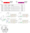
Figure 1
Figure 1
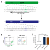
Base editing in HEK293T cells via mRNA transfection. (A) Schematic illustration of adenine and cytosine base editor (ABE and CBE) mRNAs. (B) GFP targeting sgRNAs. The introduced changes on DNA and protein levels, as well as the gRNA sequences and the employed BE are shown. (C) FACS profiles of HEK293T cells 10 days post co-transfection of indicated BE mRNAs and sgRNAs. The corresponding sequence reads recovered from sorted cells are shown to the right, and the green arrow indicates the expected change. Note the bystander mutations (indicated as red arrows) detected in the CBE-M1* fraction and in the ABE-Y66H GFP negative (neg) fraction. (D) Quantification of GFP edited cells after treatment. Results are represented as means ± SD of three independent experiments. NTC, non-targeting control. *** p < 0.001.
Figure 2
Figure 2
mRNA delivery into hiPS cells. (A) Schematic illustration of EGFP mRNA. (B) Fluorescent microscopy images of hiPS cells 24h post transfection with EGFP mRNA in the DEF-CS culture system with Lipofectamine MessengerMAXTM transfection reagent. The upper panels show brightfield images and the lower panels show fluorescent recordings. (C) FACS profiles of hiPS cells 24h post transfection or nucleofection with EGFP mRNA. Grey = control cells (non-transfected).
Figure 3
Figure 3
ABE conversion of GFP-to-BFP in hiPS cells. (A) Schematic illustration of the GFP-to-BFP conversion. The employed sgRNA is high-lighted by an arrow, with the protospacer-adjacent motif (PAM) sequence shown in bold italicized and the codon to be altered shown in bold green. The changed amino acids are depicted from green to blue, respectively. (B) FACS profiles of AAVS1-eGFP hiPS cells 10 days post co-transfection with ABE mRNA alone (NTC) or in combination with the GFP-sgRNA-Y66H. Note that AAVS1-eGFP hiPS cells harbor two copies of the GFP gene, explaining cells that are green and blue fluorescent (shown in orange). (C) Quantification of GFP edited cells after transfection. Results are represented as means ± SD of three independent experiments. *** p < 0.001.
Figure 4
Figure 4
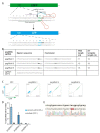
Repair of a SAMHD1 mutation in patient-derived hiPS cells. (A) Schematic illustration of the relevant region of the SAMHD1 locus. The sgRNA-targeting sequence is shown as an arrow, with the protospacer-adjacent motif (PAM) sequence indicated in bold italicized. The mutated codon is indicated in bold with the mutated nucleotide shown in red. (B) Editing rate of the SAMHD1 gene utilizing the ABE system. Absolute numbers and percentages of investigated clones are provided. The percentage of correctly repaired clones is shown in bold. Mixed clones have still a residual pick of initial nucleotide in the Sanger sequencing. Almost 30% of the clones were unedited and no indels were detected. (C) Sanger sequencing data showing the heterozygous c.1642C > T (Q548X) mutation in the patient hiPSC line (top) and the ABE-corrected locus (bottom). The “Y” boxed in yellow highlights the nucleotide that was changed.
Figure 5
Figure 5
BE-mediated generation of isogenic hiPSCs carrying TP53 mutations. (A) Schematic illustration of the TP53 gene with its addressed mutations labeled. TAD, transcriptional repression domain; PRD, proline- rich domain; TET, tetramerization domain; REG, basic C-terminal regulatory domain. (B) Important features of the selected TP53 mutations. The introduced mutations, frequencies of the mutations in the cosmic database and the sequence of the employed sgRNAs are shown with nucleotides in bold highlighting the positions to be changed). (C) Quantification of editing efficiencies based on deep sequencing results of the TP53 gene. (D) Quantification of cells in G2 after Nutlin-3 treatment in indicated cell lines. Results are represented as mean ± SEM of three independent experiments. *** p < 0.001.
Figure 6
Figure 6
Conversion of GFP-to-CFP in hiPS cells with prime editing. (A) Schematic illustration of the GFP-to-CFP conversion. The employed pegRNA is high-lighted by a line with important features highlighted. PBS, primer binding site; RT, reverse transcriptase. The PAM sequence is shown in bold italicized. The position where Cas9 introduces a nick is indicated by an arrow. The changed codon and amino acids are depicted in green and blue, respectively. (B) Features of the employed pegRNAs and sgRNA for the GFP-to-CFP conversion. Nucleotides to be changed are depicted in bold. PBS, primer binding site; RT, reverse transcriptase. (C) FACS profiles of hiPS cells 14 days post transfection with indicated PEs. Percentages of the gated fractions are shown. (D) Quantification of editing efficiencies based on the FACS results. Results are represented as mean ± SEM of three independent experiments. (E) Sanger sequencing data from CFP positive sorted cells. Converted nucleotides are shown in a red box.

References

    1. Takahashi K., Yamanaka S. Induction of pluripotent stem cells from mouse embryonic and adult fibroblast cultures by defined factors. Cell. 2006;126:663–676. doi: 10.1016/j.cell.2006.07.024. - DOI - PubMed
    1. Drews K., Jozefczuk J., Prigione A., Adjaye J. Human induced pluripotent stem cells-from mechanisms to clinical applications. J. Mol. Med. 2012;90:735–745. doi: 10.1007/s00109-012-0913-0. - DOI - PubMed
    1. Hu B.Y., Weick J.P., Yu J., Ma L.X., Zhang X.Q., Thomson J.A., Zhang S.C. Neural differentiation of human induced pluripotent stem cells follows developmental principles but with variable potency. Proc. Natl. Acad. Sci. USA. 2010;107:4335–4340. doi: 10.1073/pnas.0910012107. - DOI - PMC - PubMed
    1. Choi K.-D., Yu J., Smuga-Otto K., Salvagiotto G., Rehrauer W., Vodyanik M., Thomson J., Slukvin I. Hematopoietic and Endothelial Differentiation of Human Induced Pluripotent Stem Cells. Stem Cells. 2009;27:559–567. doi: 10.1002/stem.20080922. - DOI - PMC - PubMed
    1. Zwi L., Caspi O., Arbel G., Huber I., Gepstein A., Park I.-H., Gepstein L. Cardiomyocyte differentiation of human induced pluripotent stem cells. Circulation. 2009;120:1513–1523. doi: 10.1161/CIRCULATIONAHA.109.868885. - DOI - PubMed

Publication types

MeSH terms

Supplementary concepts