. 2020 May 6;9(5):1144.
doi: 10.3390/cells9051144.

Integrative Cluster Analysis of Whole Hearts Reveals Proliferative Cardiomyocytes in Adult Mice

Affiliations

Integrative Cluster Analysis of Whole Hearts Reveals Proliferative Cardiomyocytes in Adult Mice

Anne-Marie Galow et al. Cells. .

Abstract

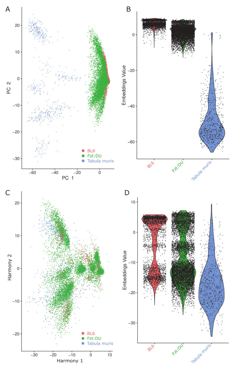
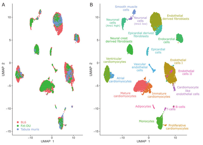
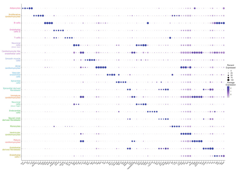
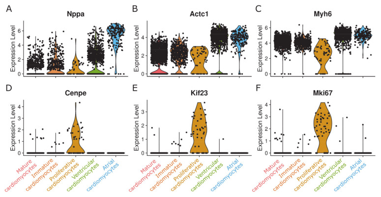
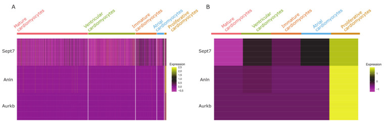
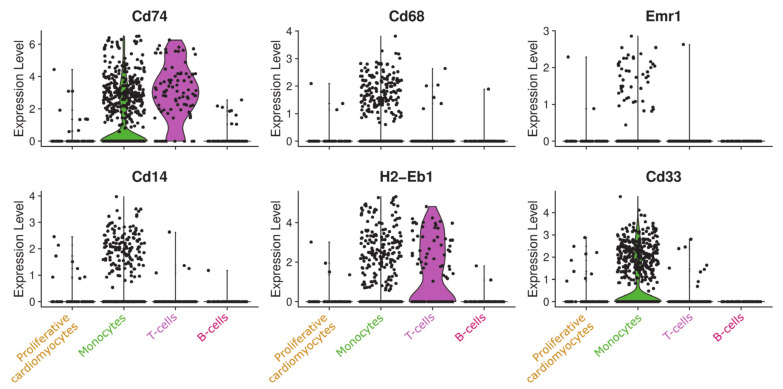
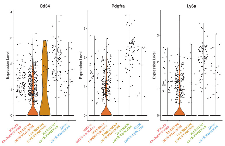
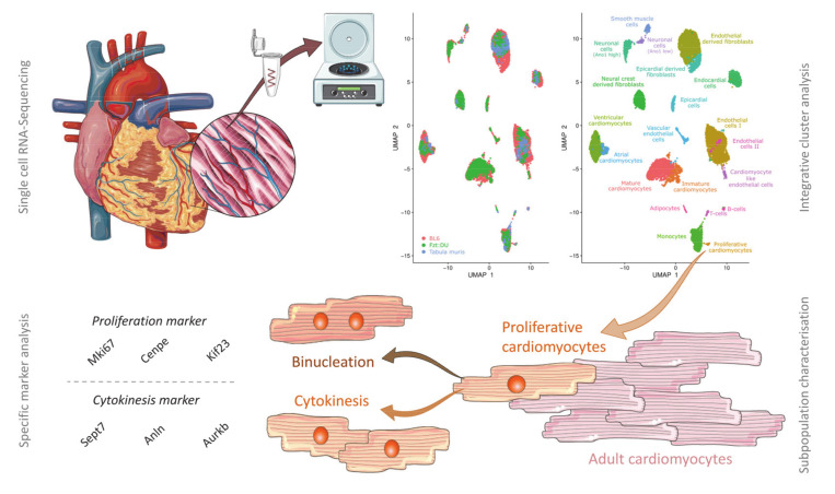
The recent development and broad application of sequencing techniques at the single-cell level is generating an unprecedented amount of data. The different techniques have their individual limits, but the datasets also offer unexpected possibilities when utilized collectively. Here, we applied snRNA-seq in whole adult murine hearts from an inbred (C57BL/6NRj) and an outbred (Fzt:DU) mouse strain to directly compare the data with the publicly available scRNA-seq data of the tabula muris project. Explicitly choosing a single-nucleus approach allowed us to pin down the typical heart tissue-specific technical bias, coming up with novel insights on the mammalian heart cell composition. For our integrated dataset, cardiomyocytes, fibroblasts, and endothelial cells constituted the three main cell populations accounting for about 75% of all cells. However, their numbers severely differed between the individual datasets, with cardiomyocyte proportions ranging from about 9% in the tabula muris data to around 23% for our BL6 data, representing the prime example for cell capture technique related bias when using a conventional single-cell approach for these large cells. Most strikingly in our comparison was the discovery of a minor population of cardiomyocytes characterized by proliferation markers that could not be identified by analyzing the datasets individually. It is now widely accepted that the heart has an, albeit very restricted, regenerative potential. However there is still an ongoing debate where new cardiomyocytes arise from. Our findings support the idea that the renewal of the cardiomyocyte pool is driven by cytokinesis of resident cardiomyocytes rather than differentiation of progenitor cells. We thus provide data that can contribute to an understanding of heart cell regeneration, which is a prerequisite for future applications to enhance the process of heart repair.

Keywords: cytokinesis; harmony; heart regeneration; proliferation; single-cell RNA-Seq; single-nucleus RNA-Seq.


Conflict of interest statement

The authors declare no conflict of interest. The funders were not involved in study design, data collection and interpretation, and manuscript preparation.

Figures

Figure 1
Figure 1
Clustering effects of the data integration with and without utilizing Harmony on the embedding in the first two principal components (PC) on the indicated datasets (BL6, red; Fzt:DU, green; Tabula muris, blue). (A) PCA-plot of the three investigated datasets without Harmony processing. (B) Violin plot of the investigated datasets without Harmony processing. (C) PCA-plot of the three investigated datasets with Harmony processing. (D) Violin plot of the three investigated datasets with Harmony processing.
Figure 2
Figure 2
Uniform Manifold Approximation and Projection (UMAP) clustering of the integrated datasets representing the identified cellular clusters that are colored by data origin and cell type. (A) snRNA-seq data from whole Fzt:DU (green, 8533 nuclei) and BL6 (red, 3139 nuclei) mouse hearts were integrated with scRNA-seq data from hearts of BL6 mice of the tabula muris project (blue, 628 cells) to attain well-mixed clusters for a common downstream analysis. (B) 21 clusters were identified and annotated to specific cell types.
Figure 3
Figure 3
Dot plot of the top five markers for each identified cluster. For each cluster, the relative gene expression in percent is represented by the size of dots, e.g., a value of 100 means that each cell within this cell type expressed this gene. The average expression level for the listed genes is indicated by the color.
Figure 4
Figure 4
Violin plot of cardiomyocyte cell markers and markers for proliferation. (AC) Three typical cardiomyocyte markers are expressed in comparable levels in all clusters considered as cardiomyocyte populations. (DF) Markers for proliferation and cytokinesis are only expressed in one of the clusters, resulting in the annotation as “proliferative cardiomyocytes”.
Figure 5
Figure 5
Heatmap of cytokinesis marker expression in cardiomyocyte clusters. (A) Expression levels of three factors involved in cleavage furrow assembly during cytokinesis are shown for each individual cell in the cardiomyocyte clusters. (B) Expression of cytokinesis markers averaged for each cardiomyocyte cluster shows the result more clearly, demonstrating the notable expression of cytokinesis markers exclusively in the “proliferative cardiomyocyte cluster”.
Figure 6
Figure 6
Violin plot of monoblast markers in the proliferating cardiomyocyte population and immune cells. Proliferative cardiomyocytes show no significant expression of the indicated typical monoblast markers.
Figure 7
Figure 7
Violin plot of stem cell markers in the cardiomyocyte populations. Typical stem cell markers are expressed in low levels in immature cardiomyocytes, but not in “proliferative cardiomyocytes” except for Cd34, which showed moderate expression in several cell clusters of the integrated dataset.
Figure 8
Figure 8
Graphical illustration of the workflow for identification of bona fide proliferative cardiomyocytes. Single-cell and single-nucleus sequencing data of whole adult murine hearts from inbred (C57BL/6NRj) and outbred (Fzt:DU) mouse strains were computationally integrated via Harmony. The integrated dataset allowed for novel insights on the mammalian heart cell composition revealing a minor population of cardiomyocytes characterized by proliferation markers and therefore referred to as “proliferative cardiomyocytes.” During in-depth gene expression analysis, we found additional markers specific for cytokinesis to be upregulated, suggesting that proliferative cardiomyocytes not only undergo binucleation but also complete cytokinesis. We did not detect relevant expression of any stem cell marker, thus supporting the idea that the renewal of the cardiomyocyte pool is driven by cytokinesis of resident cardiomyocytes rather than differentiation of stem cells.

References

    1. Hu P., Liu J., Zhao J., Wilkins B.J., Lupino K., Wu H., Pei L. Single-nucleus transcriptomic survey of cell diversity and functional maturation in postnatal mammalian hearts. Genes Dev. 2018;32:1344–1357. doi: 10.1101/gad.316802.118. - DOI - PMC - PubMed
    1. Macosko E.Z., Basu A., Satija R., Nemesh J., Shekhar K., Goldman M., Tirosh I., Bialas A.R., Kamitaki N., Martersteck E.M., et al. Highly parallel genome-wide expression profiling of individual cells using nanoliter droplets. Cell. 2015;161:1202–1214. doi: 10.1016/j.cell.2015.05.002. - DOI - PMC - PubMed
    1. Paige S.L., Plonowska K., Xu A., Wu S.M. Molecular Regulation of Cardiomyocyte Differentiation. Circ. Res. 2015;116:341. doi: 10.1161/CIRCRESAHA.116.302752. - DOI - PMC - PubMed
    1. Potente M., Mäkinen T. Vascular heterogeneity and specialization in development and disease. Nat. Rev. Mol. Cell Biol. 2017;18:477–494. doi: 10.1038/nrm.2017.36. - DOI - PubMed
    1. Tallquist M.D., Molkentin J.D. Redefining the identity of cardiac fibroblasts. Nat. Rev. Cardiol. 2017;14:484. doi: 10.1038/nrcardio.2017.57. - DOI - PMC - PubMed

Publication types

LinkOut - more resources