Review
. 2020 May 6;12(5):512.
doi: 10.3390/v12050512.

Conserved and Diverse Traits of Adhesion Devices from Siphoviridae Recognizing Proteinaceous or Saccharidic Receptors

Affiliations
Review

Conserved and Diverse Traits of Adhesion Devices from Siphoviridae Recognizing Proteinaceous or Saccharidic Receptors

Adeline Goulet et al. Viruses. .

Abstract

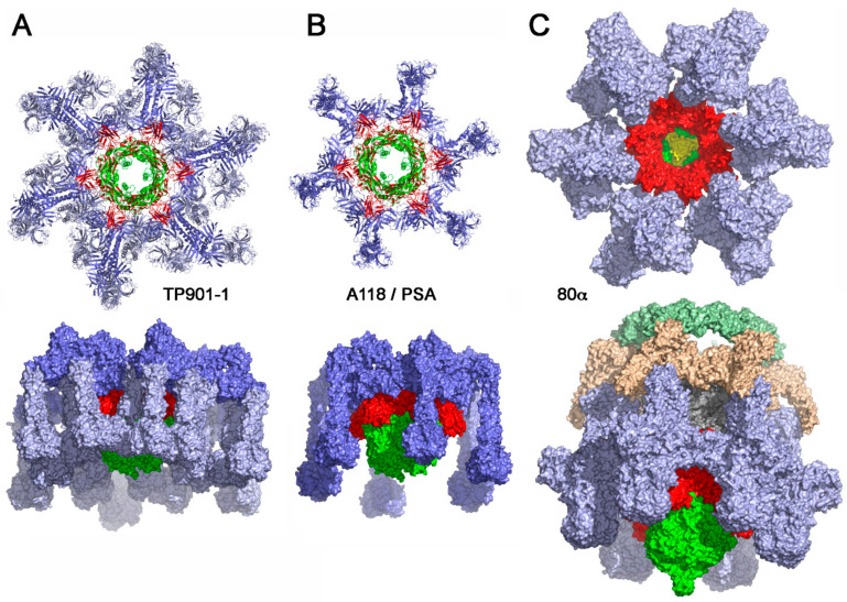
Bacteriophages can play beneficial roles in phage therapy and destruction of food pathogens. Conversely, they play negative roles as they infect bacteria involved in fermentation, resulting in serious industrial losses. Siphoviridae phages possess a long non-contractile tail and use a mechanism of infection whose first step is host recognition and binding. They have evolved adhesion devices at their tails' distal end, tuned to recognize specific proteinaceous or saccharidic receptors on the host's surface that span a large spectrum of shapes. In this review, we aimed to identify common patterns beyond this apparent diversity. To this end, we analyzed siphophage tail tips or baseplates, evaluating their known structures, where available, and uncovering patterns with bioinformatics tools when they were not. It was thereby identified that a triad formed by three proteins in complex, i.e., the tape measure protein (TMP), the distal tail protein (Dit), and the tail-associated lysozyme (Tal), is conserved in all phages. This common scaffold may harbor various functional extensions internally while it also serves as a platform for plug-in ancillary or receptor-binding proteins (RBPs). Finally, a group of siphophage baseplates involved in saccharidic receptor recognition exhibits an activation mechanism reminiscent of that observed in Myoviridae.

Keywords: Bacteriophage; Siphoviridae; baseplate; phage–host interactions; receptor-binding protein.


Conflict of interest statement

The authors declare no conflict of interest.

Figures

Figure 1
Figure 1
Examples of siphophages infecting Gram-positive and Gram-negative bacteria. Phages are classified between those binding to host-encoded proteins (green, left) or cell wall polysaccharides (green, right) and those with a ready to bind adhesion device (orange, left) and those with an activable adhesion device (orange right).
Figure 2
Figure 2
The adhesion device of siphophages. (A) Schematic representation of genes coding for the adhesion device of representative phages: lambda [7], T5 [9], SPP1 [9], c2 [23], p2 [24], 1358 [25], TP901-1 [21,26], Tuc2009 [27], PSA [13], STP1 [28], 80α [15]. Tape measure proteins (TMPs) are color-coded grey; distal tail proteins (Dits) are color-coded red; tail-associated lysozymes (Tals) are color-coded dark green (Nt-structural domain), light green (Tal extension), light blue for Tal extension tip bearing a receptor-binding domain; receptor-binding proteins (RBPs) are color-coded light blue; ancillary proteins and tail fibers are color-coded dark blue. White genes are non-structural. The c2 phage Dit and Tal are shaded grey as their annotation is highly hypothetic; (B) Schematic topology of the TMP-Dit-Tal triad (same color code as in (A)); (C) Example of a TMP-Dit-Tal triad from phage 80α baseplate [15] (same color code as in (A) and (B). The TMP is dark grey); (D) 90° rotation with respect to C.
Figure 3
Figure 3
X-ray structures of RBPs from siphophages binding to host cell wall saccharidic receptors. (A) Lactococcal phage 1358 [55]; (B) Lactococcal phage p2 [51]; (C) Lactococcal phage TP901-1 [53]; (D) Lactococcal phage Tuc2009 [54]; (E) Receptor-binding domain and stem of listerial phage PSA [13]; (F) Staphylococcal phage 80α [15]; (G) Staphylococcal phage ϕ11 [57]. The various domains are identified: shoulders and stem, binding to the rest of the baseplate, the neck joining the shoulders/stem to the head. The head bearing the receptor-binding crevice. The stem N-terminus, involved in baseplate integration of the RBP, has been boxed in red.
Figure 4
Figure 4
Architecture of the phage lambda, T5, and SPP1 host adhesion devices targeting membrane proteins. (A) Right. The lambda host adhesion device is formed by the Dit hexameric ring (gpM) and the long trimeric Tal / RBP (gpJ) central fiber. The positions of gpK, gpL, and gpI proteins in the lambda tail spike are not known. Ur-lambda also contains long tail fibers (LTFs) (Stf and Tfa). Left. Structural models, based on HHpred analyses, of gpM, gpJ middle domains (residues 326-449 and 570-811), Stf C-terminal domains (residues 578-774), and Tfa (residues 62-192). The Dit ring is a tilted view. The crystal structures used to produce these representations are PDB ID 4JMQ [31] for gpM, PDB IDs 3D37, and 5UTK for gpJ, PDB ID 2XGF for Stf [70], and PDB ID 5YVQ for Tfa [72]. (B) Right. The T5 host adhesion device is formed by the Dit hexameric ring (pb9), the trimeric Tal (pb3), and central fiber (pb4), and the RBP (pb5). LTFs (pb1) are anchored to the Dit ring via p132. Left. Ribbon representations of the pb9 (PDB ID 4JMQ) and pb1 (PDB ID 4UW8) [73] crystal structures. The Dit ring is a tilted view. Structural models, based on HHpred analyses, of pb3 (residues 528-709 and 735-849), pb4 (residues 6-319), and p132. The crystal structures used to produce these representations are PDB IDs 3D37 and 5UTK for pb3, PDB ID 5UTK for pb4, and PDB ID 3UH8 [30] for p132. (C) Right. The SPP1 host adhesion device is formed by the Dit hexameric ring (gp19.1) and the long trimeric Tal / RBP (gp21). The positions of gp22, gp23, gp23.1, and gp24 proteins in the SPP1 tail spike are not known. Left. Ribbon representations of the gp19.1 (PDB ID 2X8K), gp22 (PDB ID 2XC8), and gp23.1 (PDB ID 2XF7) crystal structures [74,75]. Structural models, based on HHpred analyses, of the gp21 N-terminal domains (residues 3-404 and 422-518). The crystal structure used to produce these representations are PDB IDs 3GS9 and 5E53 [76].
Figure 5
Figure 5
Structures of binding-ready baseplates from siphophages binding to a host cell wall saccharidic receptor. (A) X-ray structure of the baseplate from phage TP901-1; the Tal trimer has been modeled by addition of phage p2 Tal to the X-ray structure. (B) Model structure of the RBP from phages PSA or A118. (C) -cryoEM structure of phage 80α baseplate [15]. The color-coding is identical to that in Figure 2: Dit: red; tal: green: RBP: pale blue; ancillary proteins dark blue; in (C) Fiber proteins are colored beige and pale green (80α).
Figure 6
Figure 6
Structure of the activatable baseplates from siphophage p2. (A) negative staining EM (nsEM) structure of the phage p2 virion [92]. (B) Zoom on the baseplate with two RBPs fitted in the nsEM map. (C) Slicing of the nsEM map exhibiting the baseplate (Dits, Tals, and RBPs) fitted inside. (D) nsEM structure of the expressed baseplate; note the extra volume compared to the X-ray structure obtained in the presence of VHHs [24]. (E) Representation of phage p2 with the non-activated baseplate. (F) Representation of phage p2 with the X-ray determined activated baseplate localized at the tail extremity. The red arrows show the open Tal channel and the red dots the position of the receptor binding crevices. (G,H) sliced views of the structures shown in (E)- and (F)-, respectively. A model of dsDNA helix shown in the central closed or open channel. (I,J) schematic representation of the phenomenon associated with baseplate activation.
Figure 7
Figure 7
Structure of the activatable baseplates from siphophage 1358 [25]. (A) The nsEM structure of the non-activated baseplate viewed from the phage bottom. Tal and Dit are colored blue. The RBP is colored red. Note the "arm and hand" of Dits interacting with base of the RBPs (white arrow). (B) Lateral view of the baseplate showing the interactions of the Dit with the RBP N-terminal domain (shoulders) (1), the interaction of the MTP lateral extension with the RBP head domain (2) and lateral RBP contacts (3). (C) Sliced lateral view of the baseplate showing the non-assigned volume that can be attributed to a second Dit hexamer (white arrows). Tal and Dit are colored blue, RBP red, and MTPs green. (D) Lateral view of the nsEM maps of the baseplate in the non-activated state (green) and the activated state (red) resulting from a rotation of the RBPs attached to the Dits arms and hands.

References

    1. Guenther S., Herzig O., Fieseler L., Klumpp J., Loessner M.J. Biocontrol of Salmonella Typhimurium in RTE foods with the virulent bacteriophage FO1-E2. Int. J. Food Microbiol. 2012;154:66–72. doi: 10.1016/j.ijfoodmicro.2011.12.023. - DOI - PubMed
    1. Guenther S., Huwyler D., Richard S., Loessner M.J. Virulent bacteriophage for efficient biocontrol of Listeria monocytogenes in ready-to-eat foods. Appl. Environ. Microbiol. 2009;75:93–100. doi: 10.1128/AEM.01711-08. - DOI - PMC - PubMed
    1. Dion M.B., Oechslin F., Moineau S. Phage diversity, genomics and phylogeny. Nat. Rev. Microbiol. 2020;18:125–138. doi: 10.1038/s41579-019-0311-5. - DOI - PubMed
    1. Mahony J., Cambillau C., van Sinderen D. Host recognition by lactic acid bacterial phages. FEMS Microbiol. Rev. 2017;41:S16–S26. doi: 10.1093/femsre/fux019. - DOI - PubMed
    1. Kanamaru S., Leiman P.G., Kostyuchenko V.A., Chipman P.R., Mesyanzhinov V.V., Arisaka F., Rossmann M.G. Structure of the cell-puncturing device of bacteriophage T4. Nature. 2002;415:553–557. doi: 10.1038/415553a. - DOI - PubMed

MeSH terms

LinkOut - more resources