. 2020 Aug;69(8):1843-1853.
doi: 10.2337/db19-1070. Epub 2020 May 8.

Circulating Protein Signatures and Causal Candidates for Type 2 Diabetes

Affiliations

Circulating Protein Signatures and Causal Candidates for Type 2 Diabetes

Valborg Gudmundsdottir et al. Diabetes. 2020 Aug.

Abstract

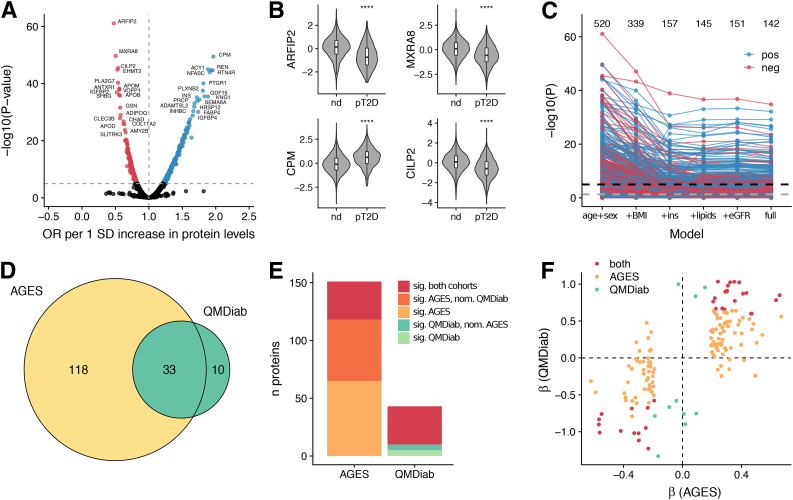
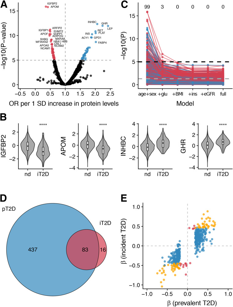
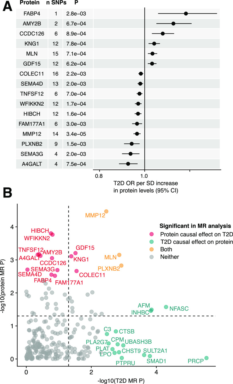
The increasing prevalence of type 2 diabetes poses a major challenge to societies worldwide. Blood-based factors like serum proteins are in contact with every organ in the body to mediate global homeostasis and may thus directly regulate complex processes such as aging and the development of common chronic diseases. We applied a data-driven proteomics approach, measuring serum levels of 4,137 proteins in 5,438 elderly Icelanders, and identified 536 proteins associated with prevalent and/or incident type 2 diabetes. We validated a subset of the observed associations in an independent case-control study of type 2 diabetes. These protein associations provide novel biological insights into the molecular mechanisms that are dysregulated prior to and following the onset of type 2 diabetes and can be detected in serum. A bidirectional two-sample Mendelian randomization analysis indicated that serum changes of at least 23 proteins are downstream of the disease or its genetic liability, while 15 proteins were supported as having a causal role in type 2 diabetes.


Figures

Figure 1
Figure 1
Serum protein associations for prevalent type 2 diabetes. A: Volcano plot demonstrating positive (blue) and negative (red) serum protein associations with prevalent type 2 diabetes; points are colored where P < 0.05/4,782. B: Violin and box plots showing serum protein levels for the AGES cohort (n = 5,438) stratified by prevalent type 2 diabetes status for the top four most significant proteins. Box plots indicate median value, 25th and 75th percentile. Whiskers extend to smallest/largest value no further than 1.5× interquartile range. Outliers not shown. ****P < 0.0001. C: Spaghetti plot providing an overview of the P values (−log10 [y-axis]) for SOMAmer associations with prevalent type 2 diabetes, using six different models (x-axis). The full model includes age, sex, BMI, fasting insulin, TG, HDL, SBP, eGFR, abdominal circumference, and parental history of diabetes. The black and gray dashed lines denote the Bonferroni-corrected (P < 0.05/4,782) and nominal (P < 0.05) significance thresholds, respectively. The numbers above denote how many unique proteins are significant at a Bonferroni-corrected threshold in each model. The color of the points/lines indicates positive (blue) or negative (red) associations. D: Venn diagram illustrating the overlap between 161 proteins that were significantly (Padjusted < 0.05) associated with type 2 diabetes in either the AGES or the QMDiab cohort and measured in both. E: For each cohort, the proportion of significant proteins that were also significant (Padjusted < 0.05) in the other cohort are shown, as well as the proportion of proteins that were nominally significant (P < 0.05) and directionally consistent in the other cohort. F: Comparison of β-coefficients for prevalent type 2 diabetes between the two cohorts. The colors indicate significant (Padjusted < 0.05) associations in either or both cohorts. ins, fasting insulin; nd, nondiabetic; neg, negative; nom, nominally significant; OR, odds ratio; pos, positive; pT2D, prevalent type 2 diabetes; sig, significant.
Figure 2
Figure 2
Serum protein associations for incident type 2 diabetes. A: Volcano plot demonstrating positive (blue) and negative (red) serum protein associations with incident type 2 diabetes; points are colored where P < 0.05/4,782. B: Violin and box plots showing serum protein levels for the AGES cohort with follow-up data (n = 2,940) stratified by incident type 2 diabetes status for the top four most significant proteins. Box plots indicate median value, 25th and 75th percentile. Whiskers extend to smallest/largest value no further than 1.5× interquartile range. Outliers not shown. ****P < 0.0001. C: Spaghetti plot providing an overview of the P values (−log10 [y-axis]) for SOMAmer associations with incident type 2 diabetes, using six different models (x-axis). The full model includes age, sex, fasting glucose, BMI, fasting insulin, TG, HDL, SBP, eGFR, abdominal circumference, and parental history of diabetes. The black and gray dashed lines denote the Bonferroni-corrected (P < 0.05/4,782) and nominal (P < 0.05) significance thresholds, respectively. The numbers above denote how many unique proteins are significant at a Bonferroni-corrected threshold in each model. The color of the points/lines indicates positive (blue) or negative (red) associations. D: Venn diagram showing the overlap between unique proteins associated with prevalent (blue) and incident (red) type 2 diabetes. E: β-Coefficients for associations between proteins and prevalent or incident type 2 diabetes. The colors denote significant associations with prevalent type 2 diabetes (blue), incident type 2 diabetes (red), or both (yellow). glu, fasting glucose; ins, fasting insulin; iT2D, incident type 2 diabetes; nd, nondiabetic; OR, odds ratio; pT2D, prevalent type 2 diabetes; T2D, type 2 diabetes.
Figure 3
Figure 3
Bidirectional Mendelian randomization analysis supports causal associations for proteins on type 2 diabetes and vice versa. A: Forest plot for the 16 proteins supported as causal (FDR <0.05) for type 2 diabetes in the two-sample MR analysis, together with the number of SNPs used as instruments and the MR P value. MR estimates were obtained using the inverse variance–weighted method when more than one SNP was available for a given protein but otherwise with the Wald ratio. B: Comparison of bidirectional two-sample MR P values (−log10) for the 246 proteins that could be investigated in both directions, where the x-axis indicates the P value for a causal effect of type 2 diabetes on protein levels and the y-axis indicates the P value for a causal effect of protein levels on type 2 diabetes. Proteins with FDR <0.05 are colored as indicated in the legend, while the dashed lines indicate P = 0.05. OR, odds ratio.

References

    1. Morris AP, Voight BF, Teslovich TM, et al. .; Wellcome Trust Case Control Consortium; Meta-Analyses of Glucose and Insulin-related traits Consortium (MAGIC) Investigators; Genetic Investigation of ANthropometric Traits (GIANT) Consortium; Asian Genetic Epidemiology Network–Type 2 Diabetes (AGEN-T2D) Consortium; South Asian Type 2 Diabetes (SAT2D) Consortium; DIAbetes Genetics Replication And Meta-analysis (DIAGRAM) Consortium . Large-scale association analysis provides insights into the genetic architecture and pathophysiology of type 2 diabetes. Nat Genet 2012;44:981–990 - PMC - PubMed
    1. Mahajan A, Go MJ, Zhang W, et al. .; DIAbetes Genetics Replication And Meta-analysis (DIAGRAM) Consortium; Asian Genetic Epidemiology Network Type 2 Diabetes (AGEN-T2D) Consortium; South Asian Type 2 Diabetes (SAT2D) Consortium; Mexican American Type 2 Diabetes (MAT2D) Consortium; Type 2 Diabetes Genetic Exploration by Next-generation sequencing in multi-Ethnic Samples (T2D-GENES) Consortium . Genome-wide trans-ancestry meta-analysis provides insight into the genetic architecture of type 2 diabetes susceptibility. Nat Genet 2014;46:234–244 - PMC - PubMed
    1. Fuchsberger C, Flannick J, Teslovich TM, et al. . The genetic architecture of type 2 diabetes. Nature 2016;536:41–47 - PMC - PubMed
    1. Scott RA, Scott LJ, Mägi R, et al. ; DIAbetes Genetics Replication And Meta-analysis (DIAGRAM) Consortium . An expanded genome-wide association study of type 2 diabetes in Europeans. Diabetes 2017;66:2888–2902 - PMC - PubMed
    1. Mahajan A, Taliun D, Thurner M, et al. . Fine-mapping type 2 diabetes loci to single-variant resolution using high-density imputation and islet-specific epigenome maps. Nat Genet 2018;50:1505–1513 - PMC - PubMed

Publication types