. 2020 May 8;11(1):2320.
doi: 10.1038/s41467-020-16134-7.

The landscape of chromothripsis across adult cancer types

Affiliations

The landscape of chromothripsis across adult cancer types

Natalia Voronina et al. Nat Commun. .

Abstract

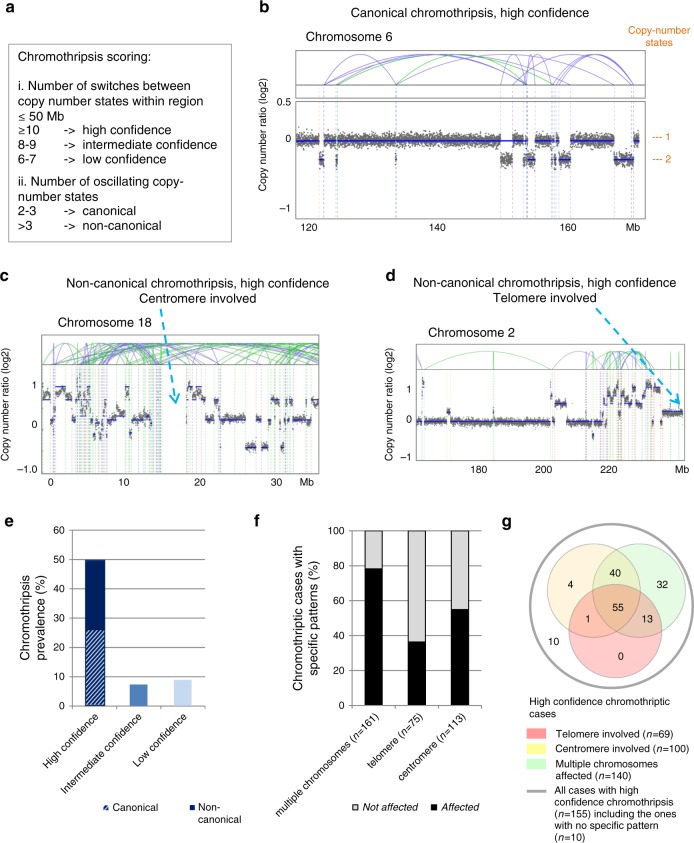
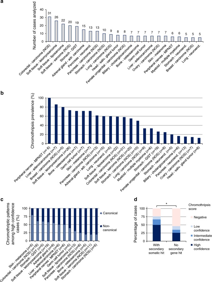
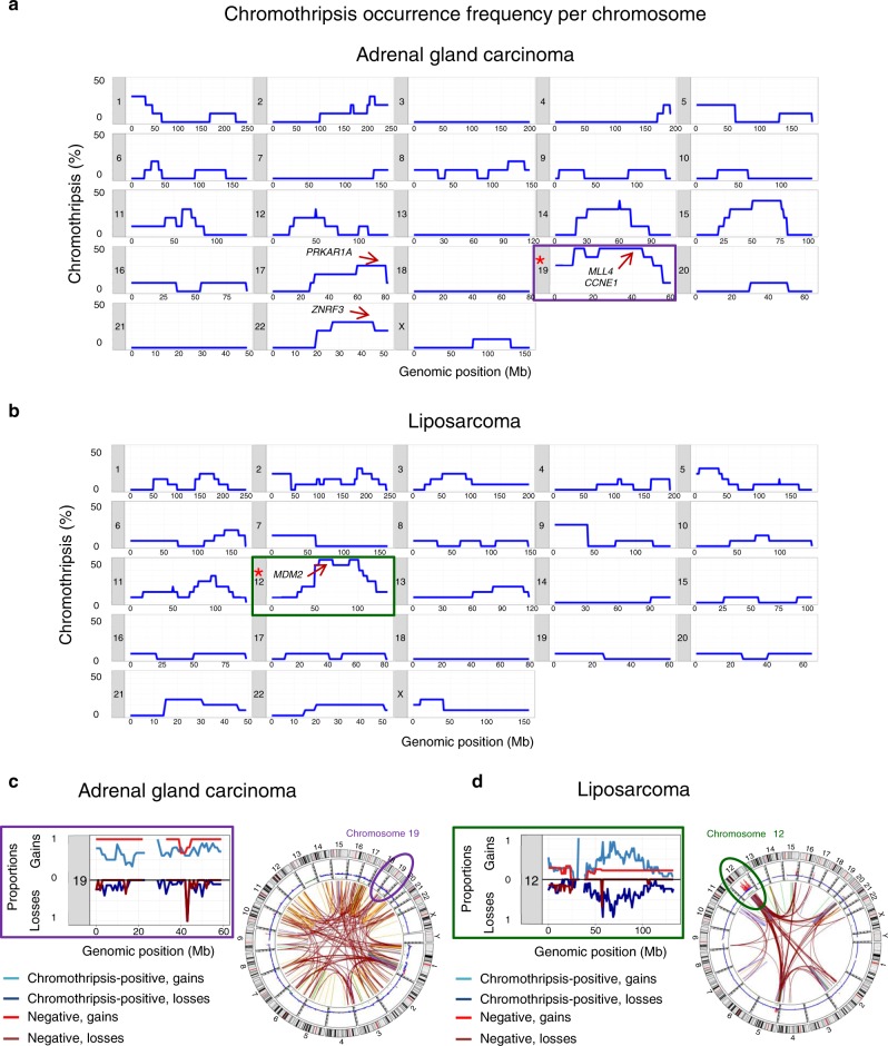
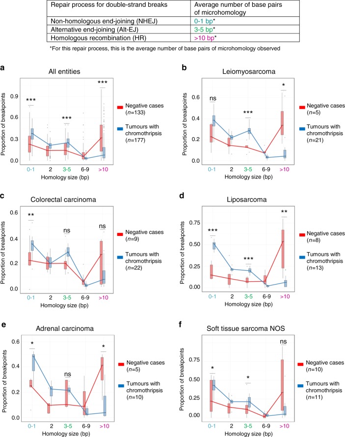
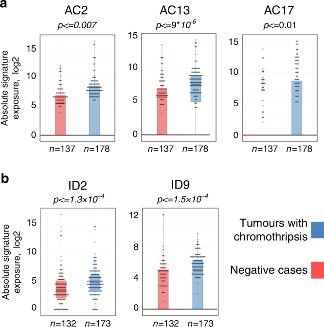
Chromothripsis is a recently identified mutational phenomenon, by which a presumably single catastrophic event generates extensive genomic rearrangements of one or a few chromosome(s). Considered as an early event in tumour development, this form of genome instability plays a prominent role in tumour onset. Chromothripsis prevalence might have been underestimated when using low-resolution methods, and pan-cancer studies based on sequencing are rare. Here we analyse chromothripsis in 28 tumour types covering all major adult cancers (634 tumours, 316 whole-genome and 318 whole-exome sequences). We show that chromothripsis affects a substantial proportion of human cancers, with a prevalence of 49% across all cases. Chromothripsis generates entity-specific genomic alterations driving tumour development, including clinically relevant druggable fusions. Chromothripsis is linked with specific telomere patterns and univocal mutational signatures in distinct tumour entities. Longitudinal analysis of chromothriptic patterns in 24 matched tumour pairs reveals insights in the clonal evolution of tumours with chromothripsis.


Conflict of interest statement

The authors declare no competing interests.

Figures

Fig. 1
Fig. 1. Chromothriptic patterns and prevalence in 316 whole-genome sequences.
a Chromothripsis scoring: criteria to determine the confidence of the scoring and to define canonical versus non-canonical chromothripsis. b Representative example of canonical chromothripsis. c Representative example of non-canonical chromothripsis. In this case, the centromere is included in the chromothriptic region. d Representative example of chromothriptic chromosome for which the telomere region is involved. e Chromothripsis prevalence shown as percentage of cases (n = 316, whole-genome sequencing) including cases with high confidence, intermediate confidence and low confidence chromothripsis. For tumours with high confidence chromothripsis, we distinguish between canonical and non-canonical chromothriptic patterns. f Chromothripsis cases with multiple versus single chromosomes affected; with or without telomere involvement (illustrated in d), as well as with or without centromere involvement (illustrated in c) from all 316 cases. High confidence, intermediate confidence and low confidence chromothriptic cases are shown. g Venn diagram showing the overlapping fractions of high confidence chromothriptic cases with multiple chromosomes affected, with or without involvement of telomeres and/or centromeres. Analyses based on whole-exome sequences and examples showing the counting of switches between copy-number states are shown in Supplementary Figs. 1–2. On the copy-number plots, blue lines indicate inversions, green lines indicate break ends, brown lines indicate translocations and orange lines indicate copy-number variation.
Fig. 2
Fig. 2. Tumour entities represented in the cohort and chromothripsis prevalence across entities.
a Number of analysed cases for each tumour entity (n = 316 whole-genome sequences in total, with 263 cases for entities with at least 5 cases per entity). Tumour entities with less than 5 cases per entity are shown in Supplementary data 1. b Chromothripsis prevalence (high confidence) per tumour entity for all entities with at least 5 cases (n = 263). c Distribution of canonical versus non-canonical chromothripsis, from all cases with high confidence chromothripsis scoring (n indicates the number of high confidence chromothriptic cases per tumour entity for all entities with at least 5 high confidence cases). d Chromothripsis prevalence among cases with germline mutations in cancer predisposition genes (n = 74 cases, whole-genome and whole-exome sequences). Statistical significance was tested using Fisher exact test (*p < 0.05, two-sided). MPNST malignant peripheral nerve sheath tumour; GIST gastrointestinal stromal tumour; NOS not otherwise specified.
Fig. 3
Fig. 3. Frequency of chromothriptic events across chromosomes for two representative tumour entities.
a adrenal gland adenocarcinoma, n = 15 cases; b liposarcoma, n = 13 cases, total for high and intermediate confidence scoring. The Y axis shows the percentage of chromothriptic events affecting each chromosomal fragment from all chromothriptic cases. Location of known driver genes frequently affected by chromothriptic events is indicated by arrows. Stars indicate chromosomes that are significantly enriched for chromothriptic events in these tumour entities (permutation test, see also Supplementary data 3). For the frequencies of chromothriptic events on all chromosomes in other tumour entities, please refer to Supplementary Fig. 3. c,d Proportions of gains (upper panels) and losses (lower panels) in tumours with chromothripsis (blue) or without chromothripsis (red) for representative chromosomes frequently affected by chromothripsis, with one illustrative CIRCOS plot for each tumour entity. Lines on CIRCOS plots show deletions in green, translocations in brown, duplications in orange and inversions in blue.
Fig. 4
Fig. 4. Gene fusions generated by chromothriptic events.
a Tumours with chromothripsis have more gene fusions per structural variant. Regression analysis for the number of fusions depending on the number of structural variants in tumours with or without chromothripsis (see Supplementary data 4 for all parameters of the regression analyses). b Chromothripsis generates clinically relevant gene fusions, as illustrated with the MYBNFIB fusion, which drives tumour development in adenoid cystic carcinoma. Blue lines indicate inversions, green lines indicate break ends, brown lines indicate translocations and orange lines indicate copy-number variation.
Fig. 5
Fig. 5. Chromothripsis is associated with specific telomere features.
Cases with gains of TERT (n = 80) or with truncating mutations in ATRX (n = 24, ATRX being strongly linked with activation of the Alternative Lengthening of Telomeres pathway) are enriched in chromothripsis-positive tumours as compared to tumours without TERT gain and without ATRX mutation (n = 212). Statistical analysis was tested using chi squared tests to compare the proportions of tumours with chromothripsis (high-confidence or intermediate-confidence). ATRX trunc, tumours with truncating mutations in ATRX.
Fig. 6
Fig. 6. Major DNA repair processes involved in the rejoining of the breakpoints after chromothripsis.
Based on the number of base pairs of homology at the breakpoint sites, we can infer the prevailing repair processes involved in the rejoining of the DNA fragments (af). Statistical significance was tested using beta-regression analyses. Family-wise correction of p values was performed according to Bonferroni for all tumour entities with at least 15 cases in total, of which at least 5 tumours showed chromothripsis and at least 5 were negative. The comparisons shown here were performed case wise (tumours with versus without chromothripsis). Comparisons performed region wise (chromothriptic chromosomes versus non-chromothriptic chromosomes) are shown in Supplementary Fig. 6. Centre lines show median values, bounds of boxes show 75th percentiles and whiskers show maximum and minimum. (*p < 0.05; **p < 0.01; ***p < 0.001).
Fig. 7
Fig. 7. Chromothripsis is linked with specific mutational signatures.
Representative examples of base substitution signatures (a) and indel signatures (b) in chromothripsis-positive versus chromothripsis-negative tumours across all entities. Significance was tested using Wilcoxon tests (two-sided). Family-wise correction of p values was performed according to Bonferroni. Supplementary Fig. 7 shows representative examples of mutational signatures in specific tumour entities. Bars show median values, dots show individual values and whiskers show maximum and minimum.
Fig. 8
Fig. 8. Longitudinal analysis of chromothriptic patterns in matched primary-relapse pairs and different metastases from the same patients.
Representative examples of the evolution of chromothriptic patterns are shown. Analyses of the evolution of chromothriptic patterns for all matched pairs (n = 41, with 24 pairs showing chromothripsis) are shown in Supplementary Data 5. Example 2 is from a different patient cohort because examples for this specific scenario with available whole-genome sequences for both time points were lacking. Blue lines indicate inversions, green lines indicate break ends, brown lines indicate translocations and orange lines indicate copy-number variation.

References

    1. Stephens PJ, et al. Massive genomic rearrangement acquired in a single catastrophic event during cancer development. Cell. 2011;144:27–40. doi: 10.1016/j.cell.2010.11.055. - DOI - PMC - PubMed
    1. Rausch T, et al. Genome sequencing of pediatric medulloblastoma links catastrophic DNA rearrangements with TP53 mutations. Cell. 2012;148:59–71. doi: 10.1016/j.cell.2011.12.013. - DOI - PMC - PubMed
    1. Korbel JO, Campbell PJ. Criteria for inference of chromothripsis in cancer genomes. Cell. 2013;152:1226–1236. doi: 10.1016/j.cell.2013.02.023. - DOI - PubMed
    1. Fontana MC, et al. Chromothripsis in acute myeloid leukemia: biological features and impact on survival. Leukemia. 2018;32:1609–1620. doi: 10.1038/s41375-018-0035-y. - DOI - PMC - PubMed
    1. Rucker FG, et al. Chromothripsis is linked to TP53 alteration, cell cycle impairment, and dismal outcome in acute myeloid leukemia with complex karyotype. Haematologica. 2018;103:e17–e20. doi: 10.3324/haematol.2017.180497. - DOI - PMC - PubMed

Publication types